HMI105 Identification and Positioning Errors
1/68
There's no tags or description
Looks like no tags are added yet.
Name | Mastery | Learn | Test | Matching | Spaced |
|---|
No study sessions yet.
69 Terms
Patient is ROTATED to the RIGHT
and TILTED to the LEFT
The Right iliac wing is elongated, and the right obturator foramen is small (ROTATION TO RIGHT)
The Left iliac wing is higher than the right (TILTED TO LEFT)
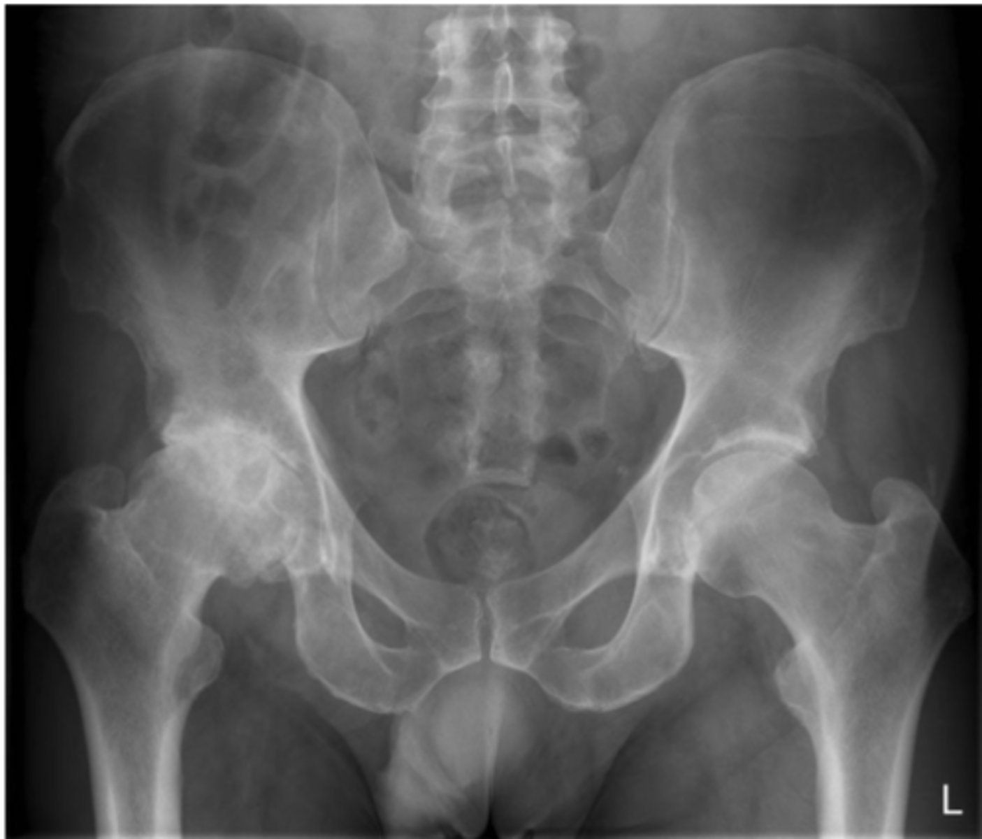
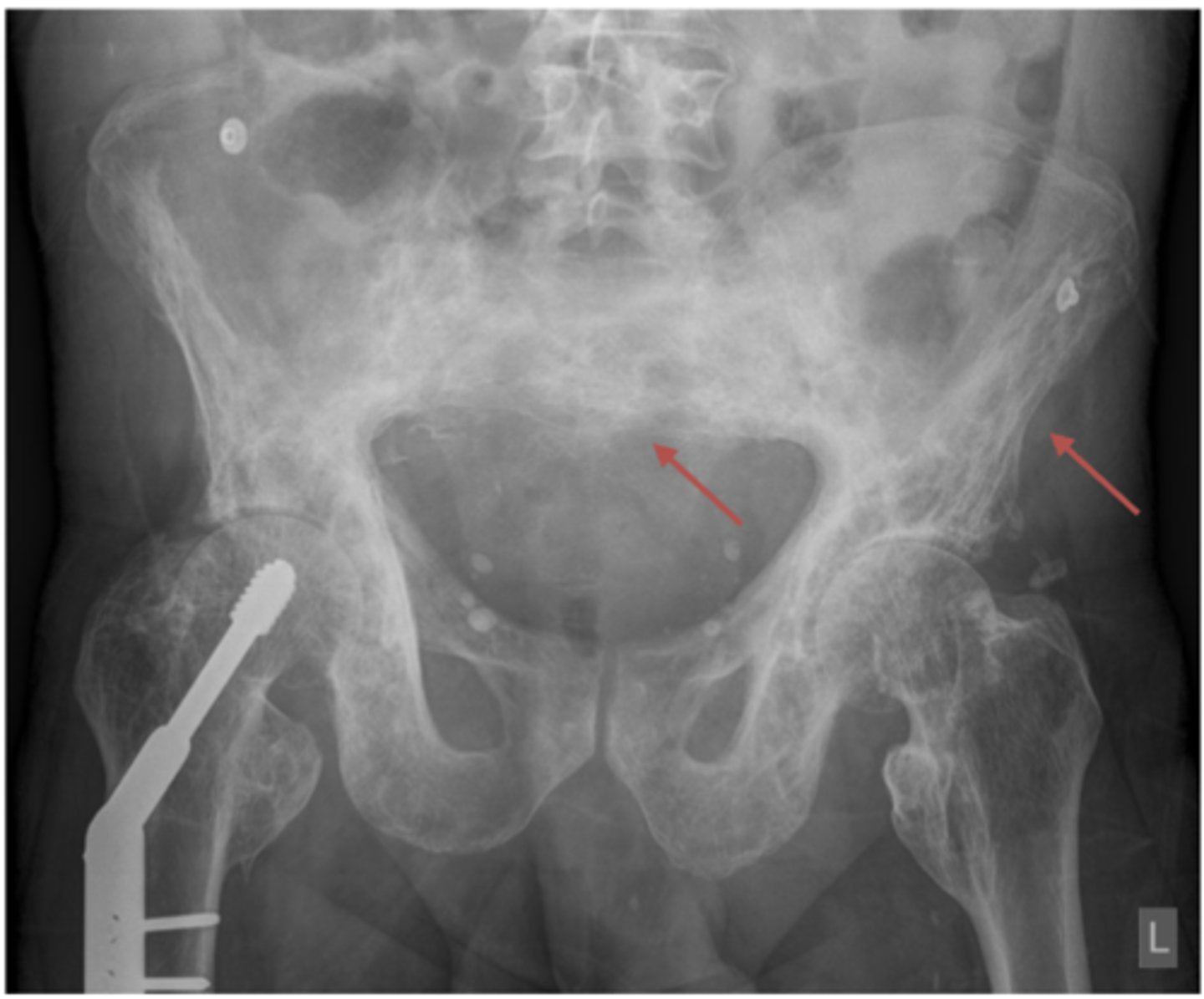
Identify the positioning errors
Left iliac wing is wider, and left obturator foramen is smaller - Patient is ROTATED to the LEFT
Patient also tilted to left (left iliac crest higher than right)
Identify the positioning errors
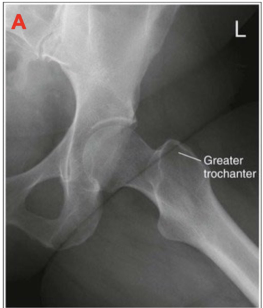
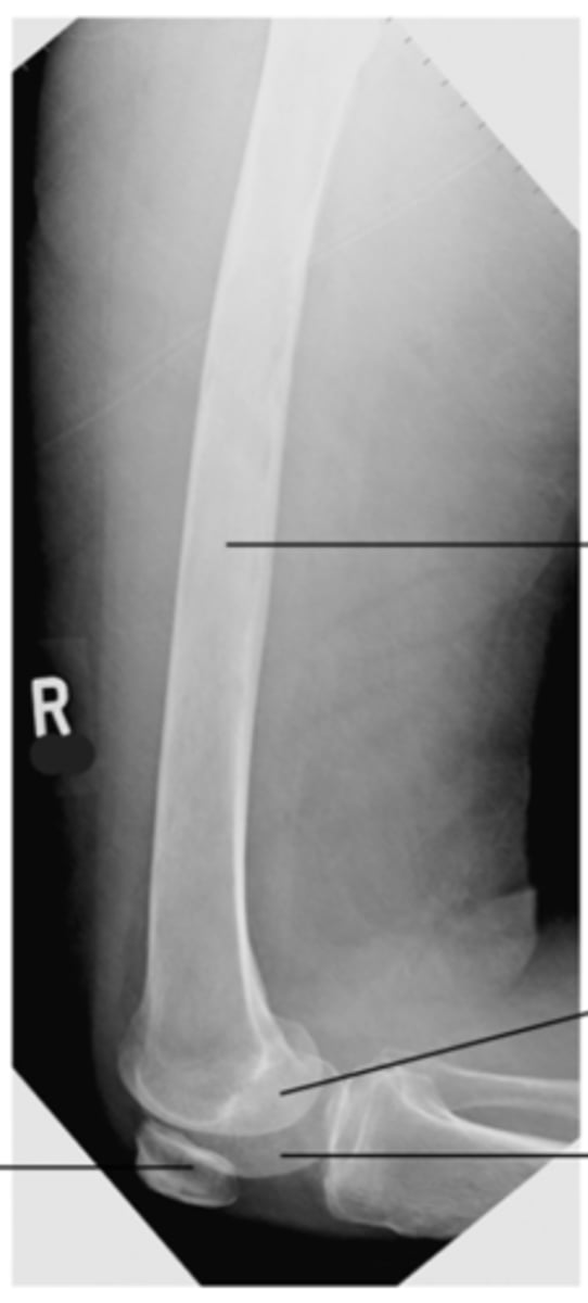
Under rotated hip and knee as greater trochanter is not superimposed with the femoral neck
Identify the positioning error
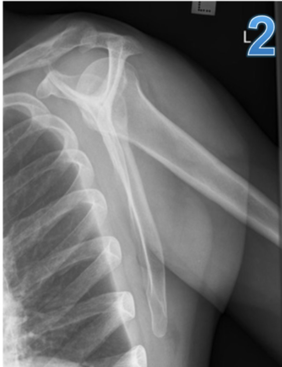
The clavicle is sitting below the superior angle of the scapula - the patient is TILTED forward.
Less than half the scapula is superimposed with the thoracic region - patient is roated away from the bucky
Name the positioning errors
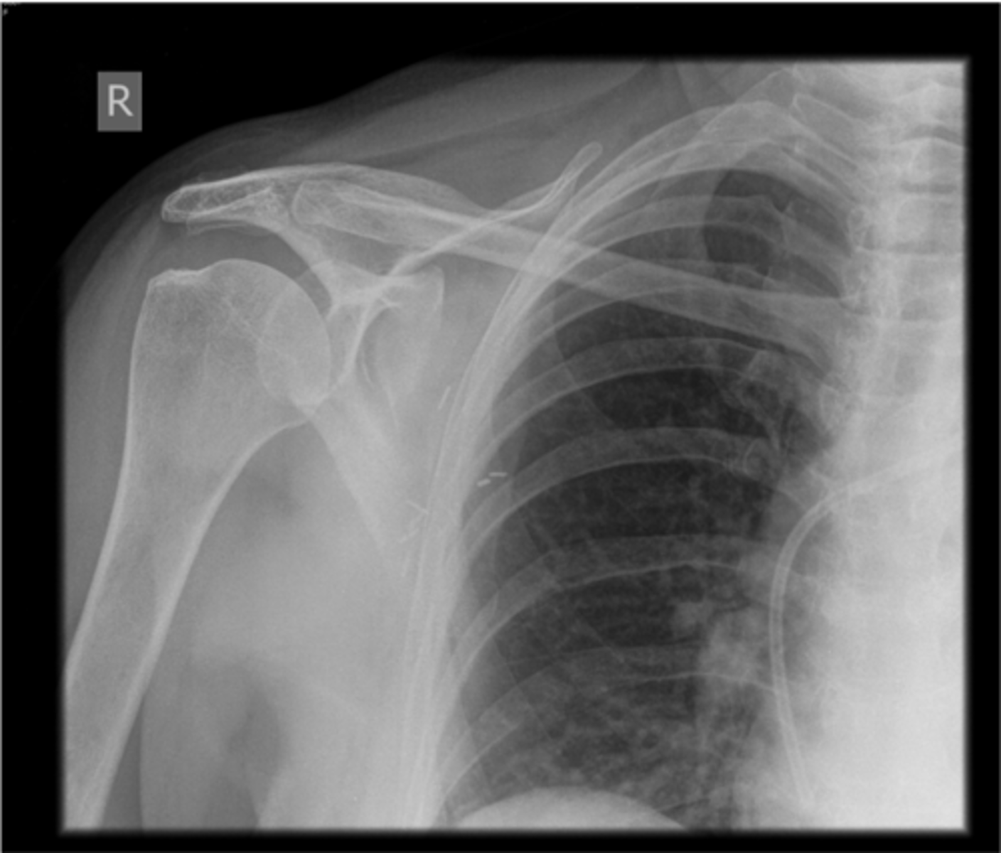
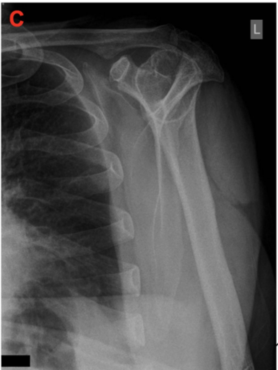
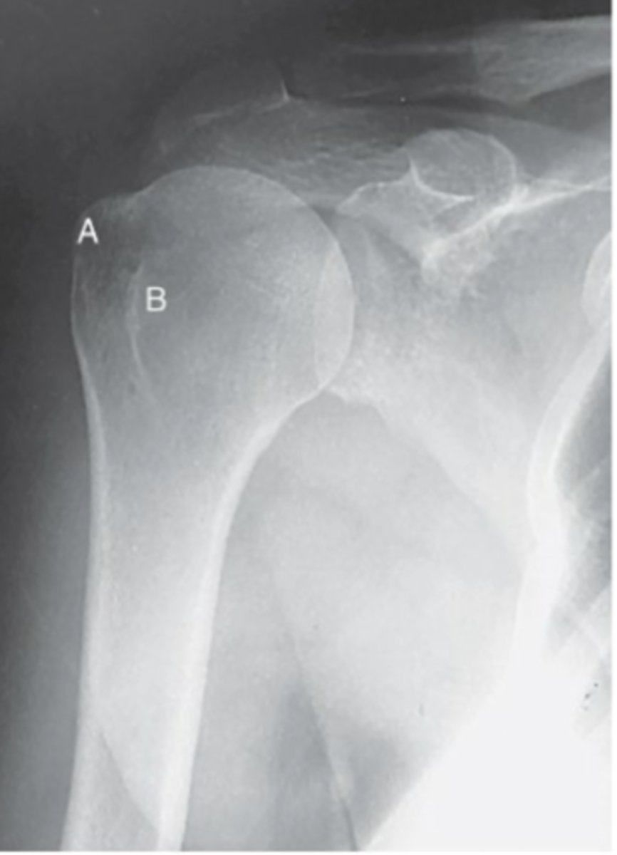
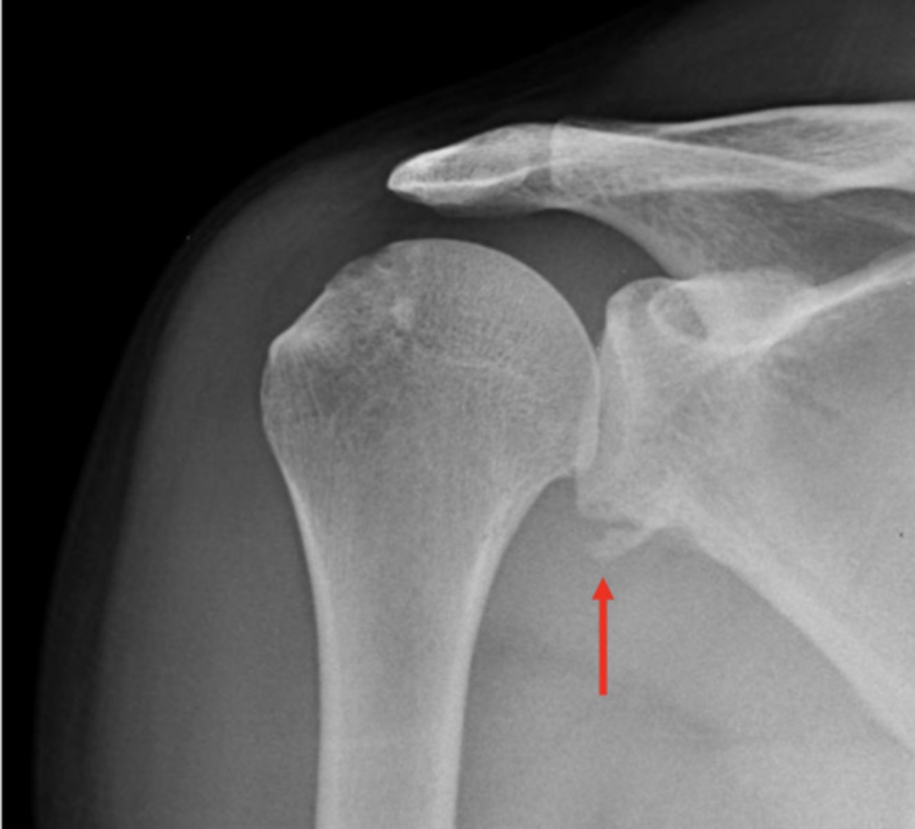
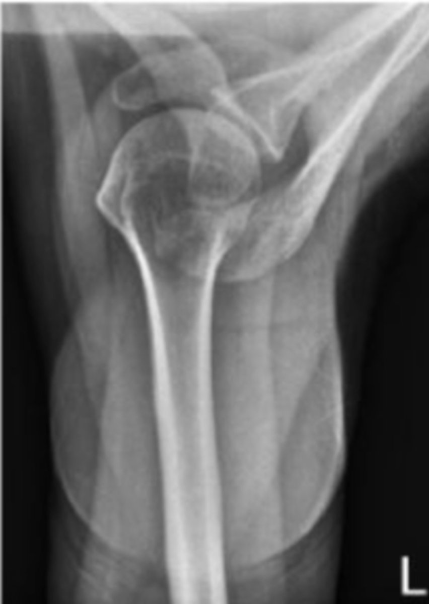
Patient is under-rotated for the glenohumeral view as the joint space is not open, and less than 1/3 of the coracoid process is superimposed by the head of the humerus
Name the positioning error
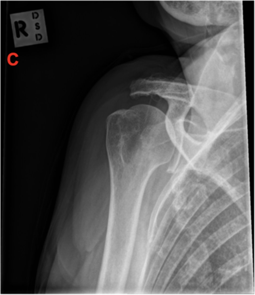
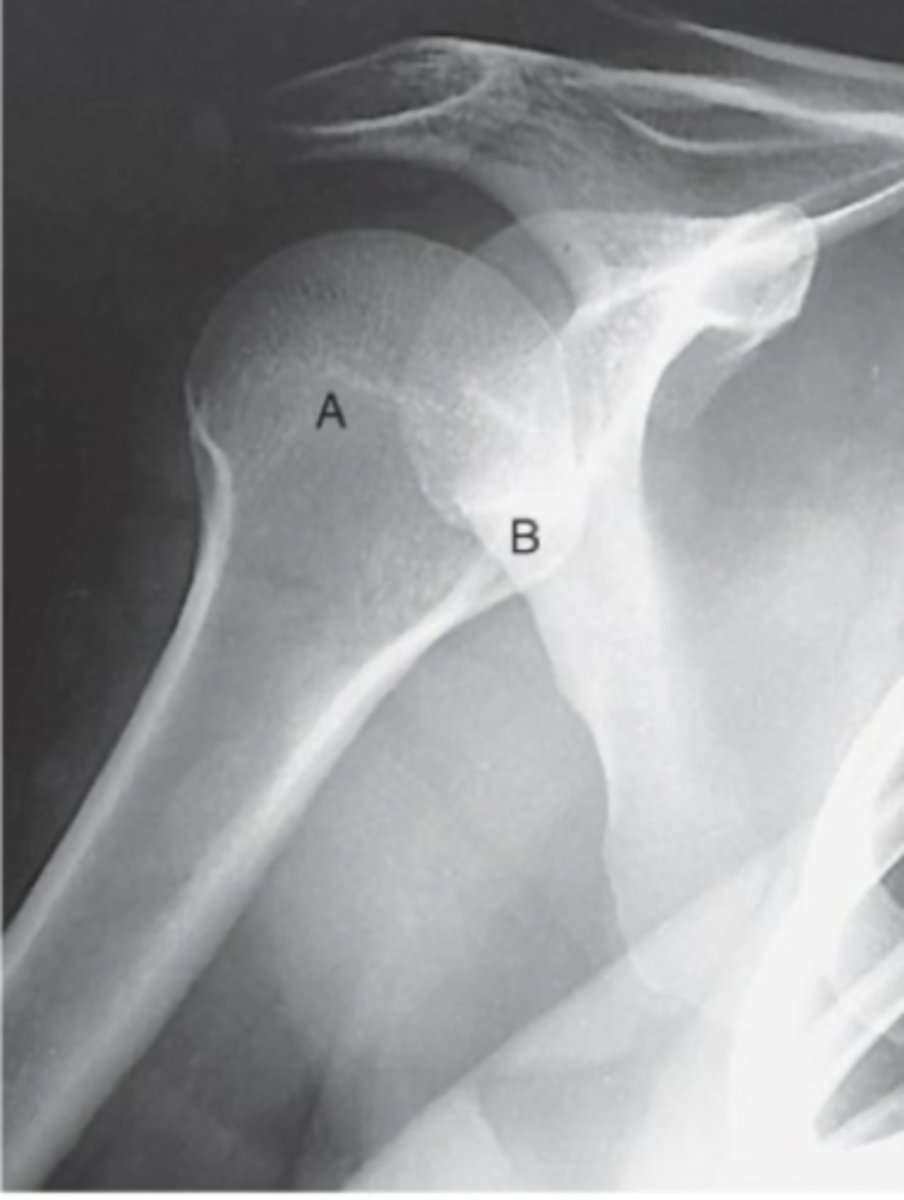
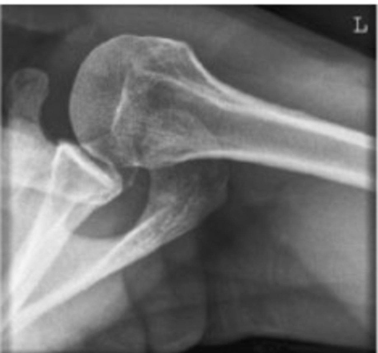
Patient is over-rotated for the glenohumeral joint, as the joint space is closed and more than 1/3 of the coracoid process is superimposed by the humeral head
Name the positioning error
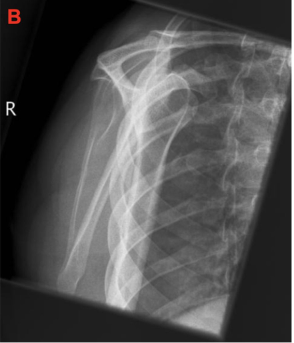
Patient is over-rotated. The humerus is superimposing the thoracic region rather than the scapula, and the lateral border of the scapula (side with 2 margins) is sitting more medial (towards the thoracic cavity)
Identify the Positioning Error
Patient is under-rotated. The lateral border of the scapula (border with 2 margins) is more lateral (further from the thoracic cavity).
Identify the Positioning Error
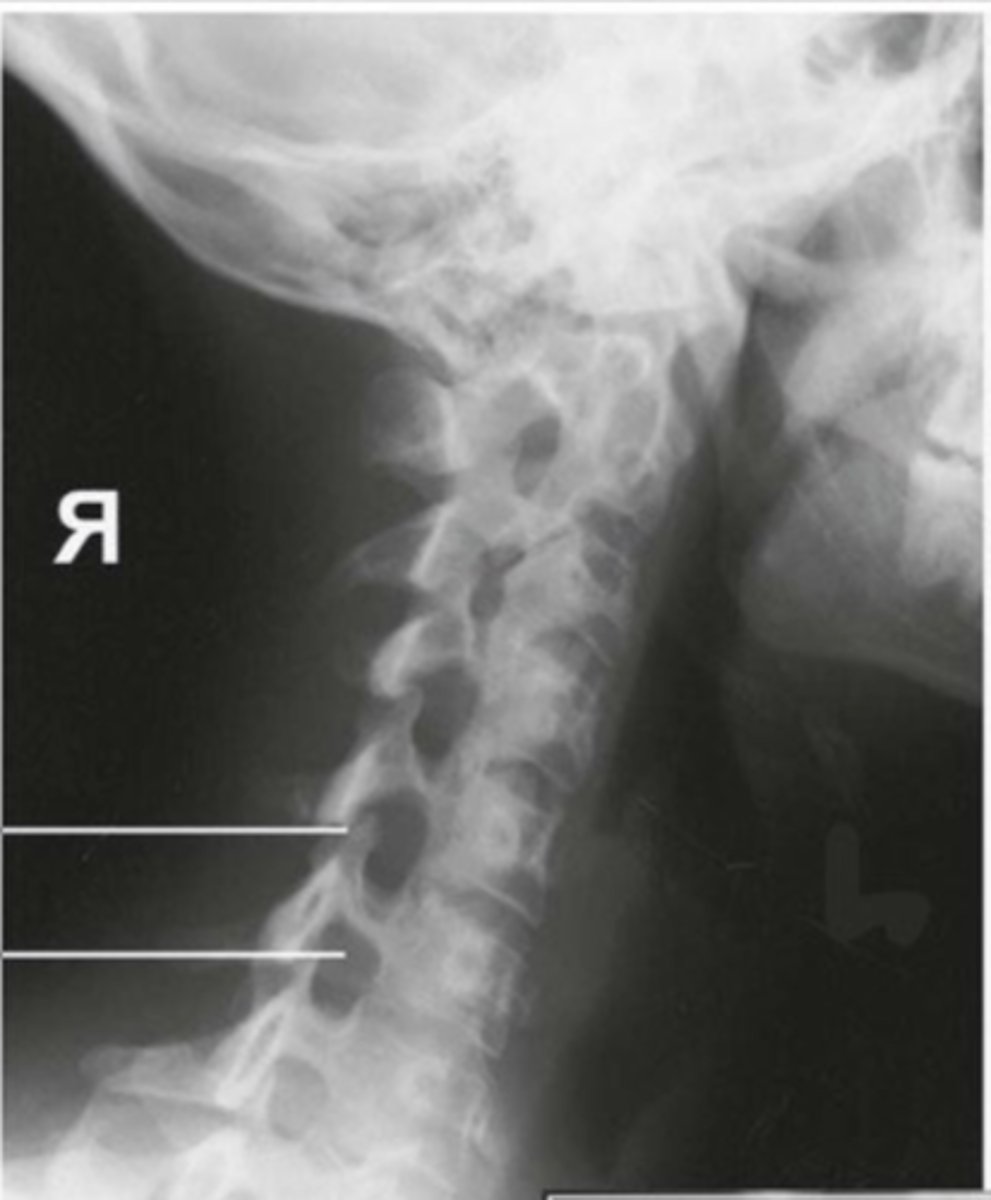
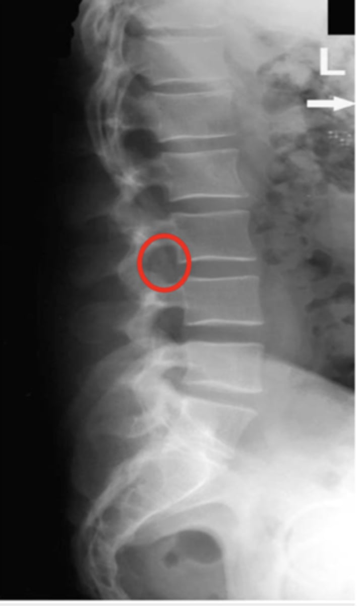
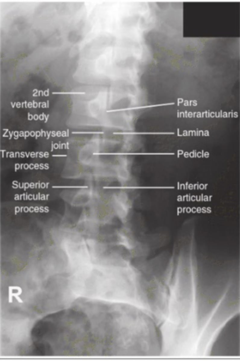
Patient is Under-rotated as the intervertebral foramina are fully not open
Identify the Positioning Error
Patient is Over-rotated as the intervertebral joints are open however the zygapophyseal (facat) joints are visualized.
Identify the Positioning Error
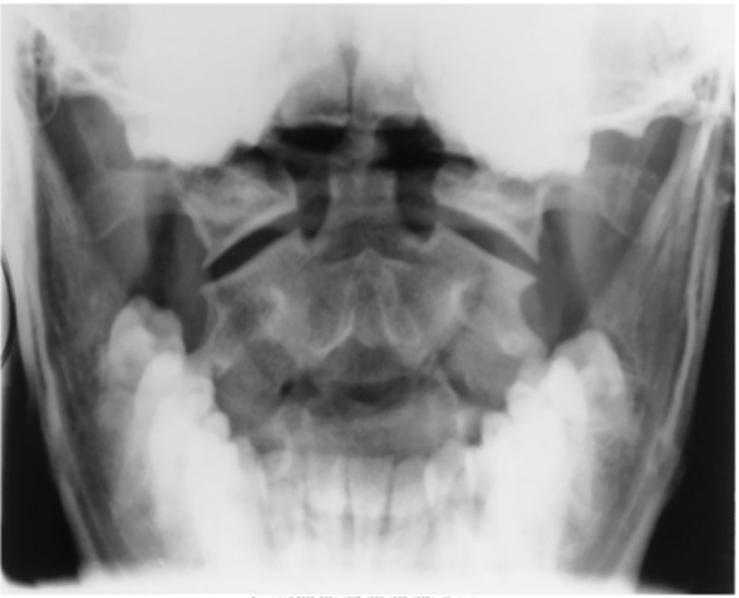
The patient's head is tilted downwards as the upper teeth are sitting below the level of the base of the skull and are superimposing the vertebrae
Identify the positioning error
The patient's head is tilted upwards as the upper teeth are sitting above the base of the skull and the upper teeth look like a smiley face
Identify the positioning error
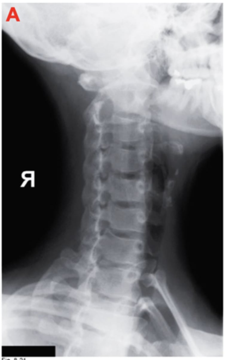
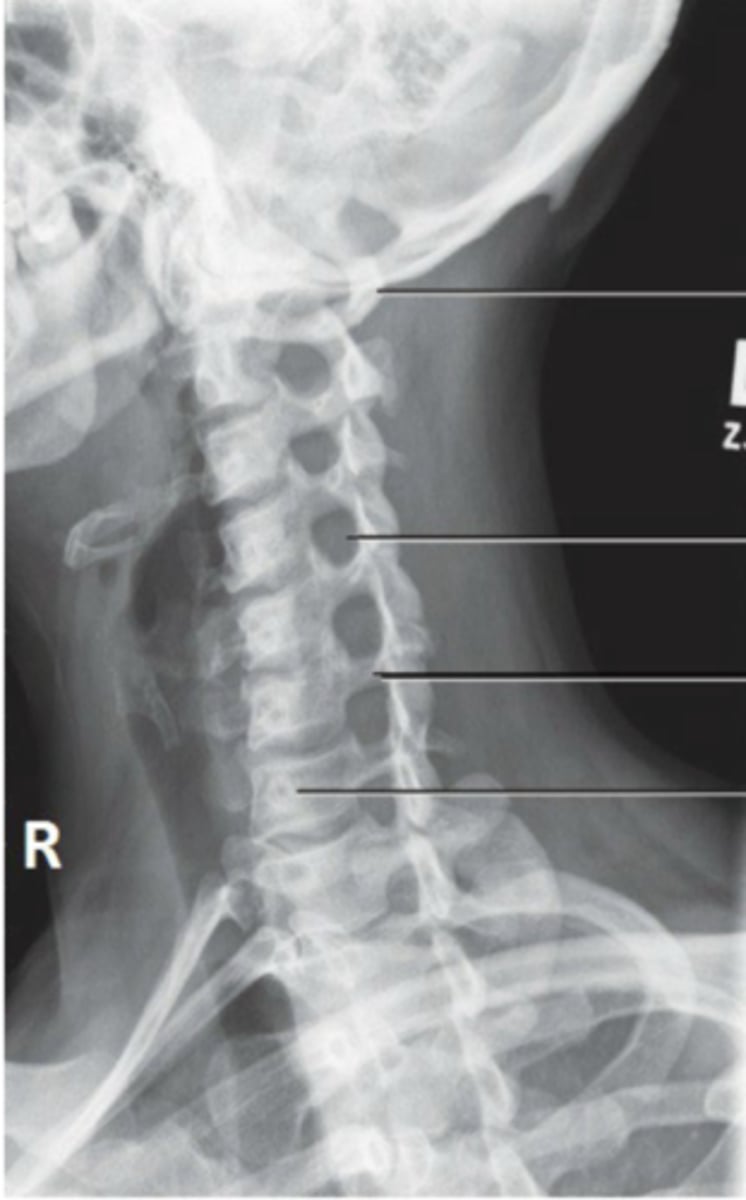
1. The patient is rotated to the left as the Spinous processes are situated on the right side of the vertebra.
2. The patient is tilted backwards as the joint spaces on the inferior cervical vertebrae are beginning to close. The spinous processes are also becoming elongated, moving inferiorly, indicating the patient is tilted backwards/towards the IR.
Identify the positioning errors
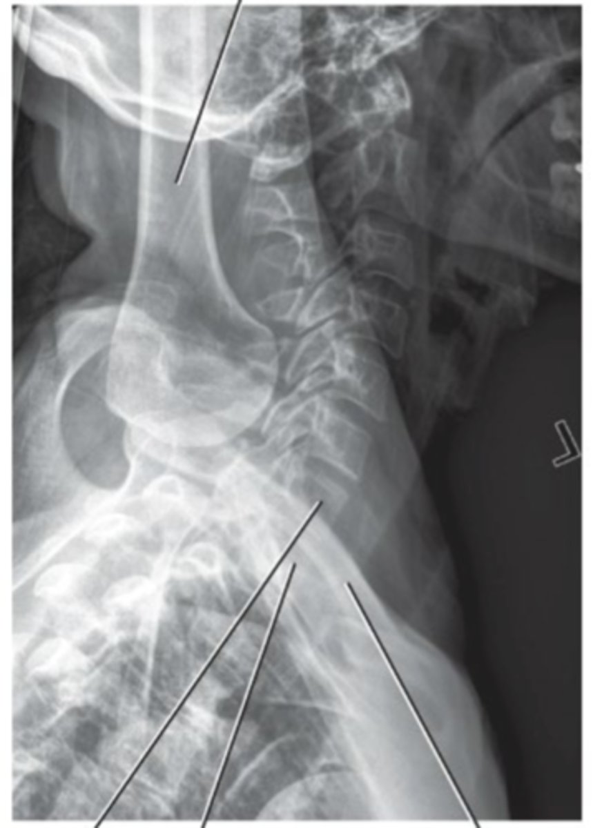
Patient is Rotated to the Right/posteriorly rotated as the articular processes are separated anterior-posteriorly. The right articular process appears larger indicating patient is rotated to the right.
Identify the positioning error
Patient is tilted as the articular processes are not superimposed from superior to inferior.
Identify the positioning error
Patient is rotated to the Left as the left clavicle is sitting further away from the midline than the right.
Inspiration was not performed as the diaphragm is sitting hight
Identify the positioning errors
Patient is rotated as the humeral heads are not superimposed, and the edges of the vertebrae are not superimposed anteriorly and posteriorly.
Identify the positioning error
mAs is too low as the image looks grainy, and there is quantum model present.
Identify the projectional errors
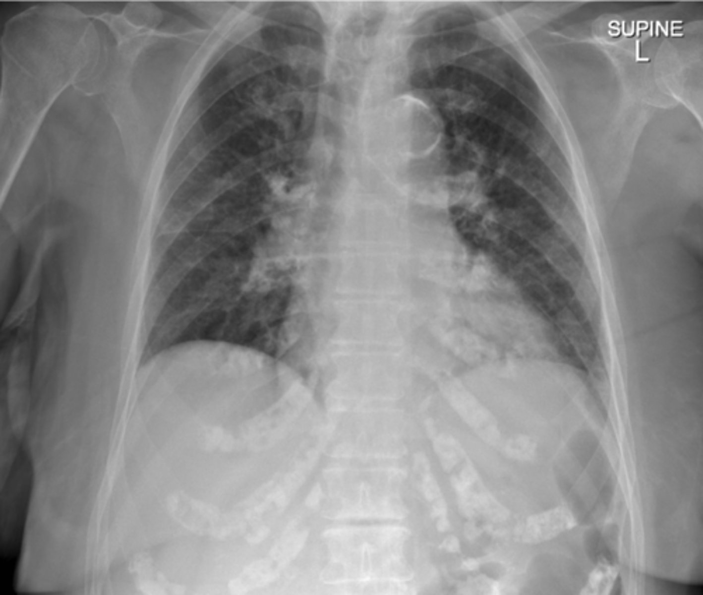
Patient is rotated and tilted as the superior/inferior and anterior/posterior borders of the vertebrae are not superimposed.
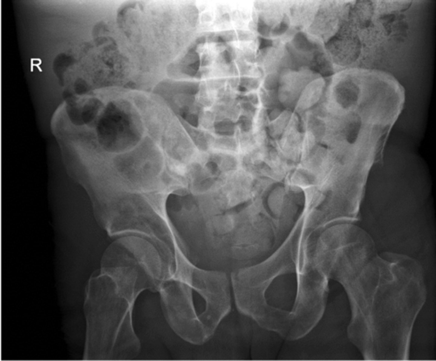
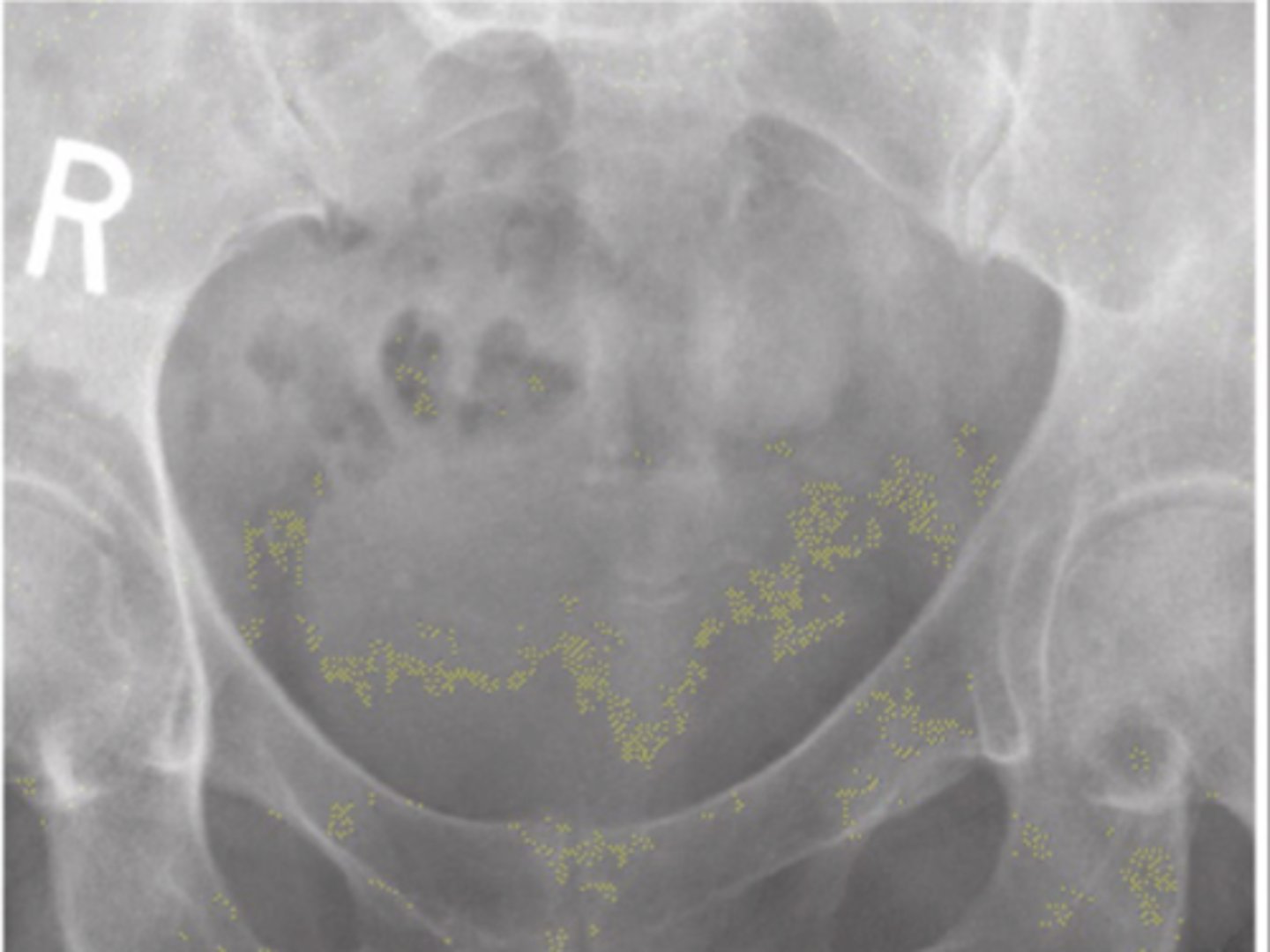
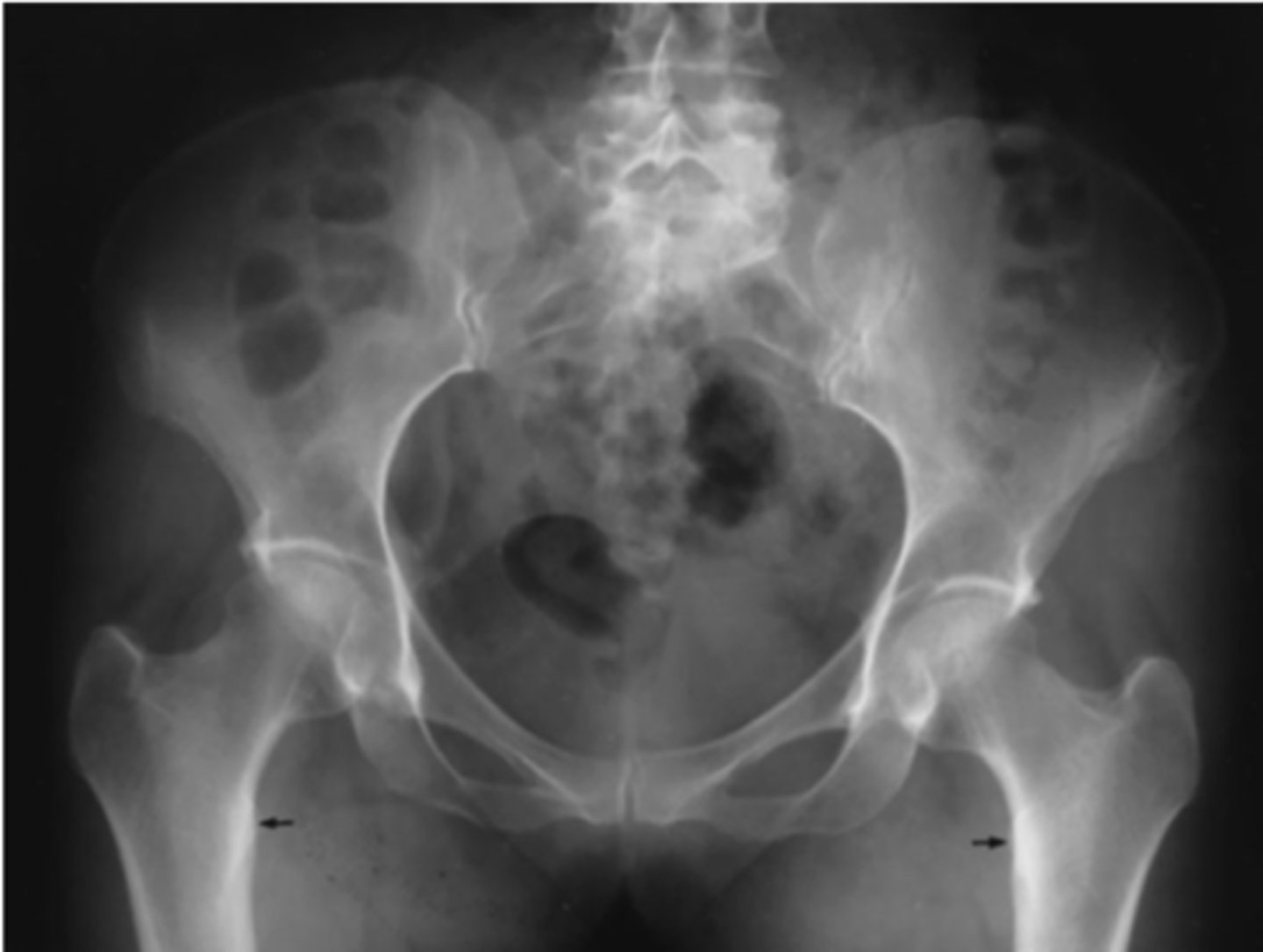
The patient is rotated to their left as the left iliac wing is more anterior than the right
Identify the positioning errors
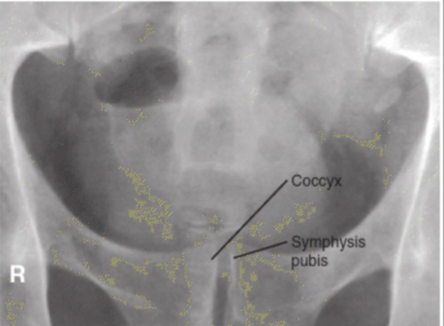
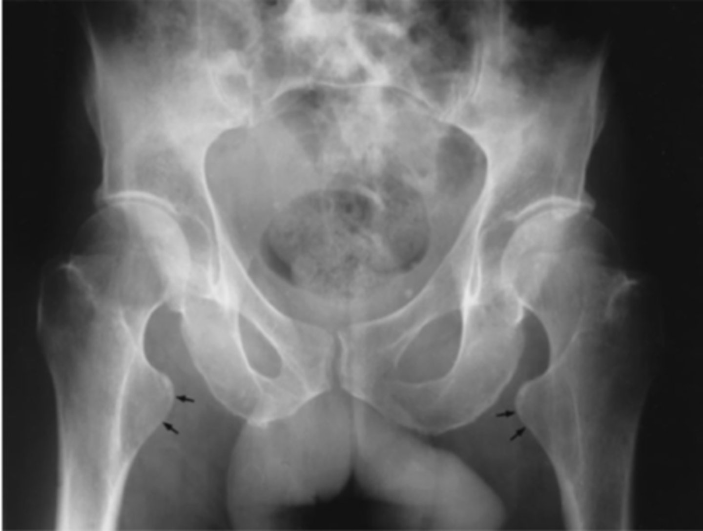
Patient is rotated to the left as the right iliac wing is foreshortened and the coccyx is not sitting at the level of the pubis synthesis
Identify the positioning error
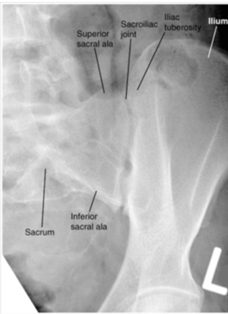
The tube is angled downwards rather than up, as the sacrum and SI joint
is foreshortened and the sacral foramina are not open
Identify the positioning error
The tube is angled too much, as the sacrum and SI joints are extremely elongated
Identify the positioning error
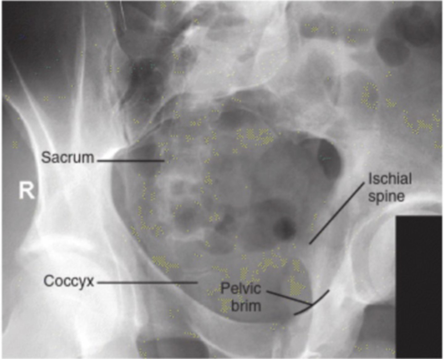
Patient is rotated posteriorly as the femoral heads and margins of the sacrum are not superimposed.
Identify the positioning error
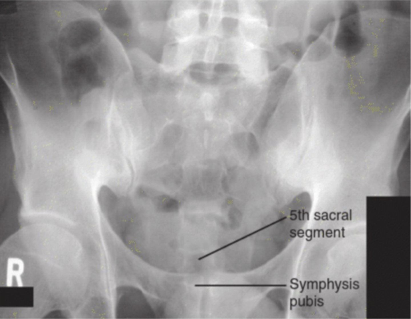
The tube is angled upwards rather than downwards for the coccygeal view, as the pubic symphysis is elongated, sacral foramina are open and the coccyx is superimposing the pubic synthesis
Identify the positioning error
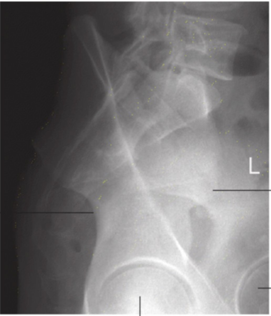
The patient is rotated to the right as the coccyx is not in line with the pubic synthesis and is sitting to the left.
Identify the positioning error
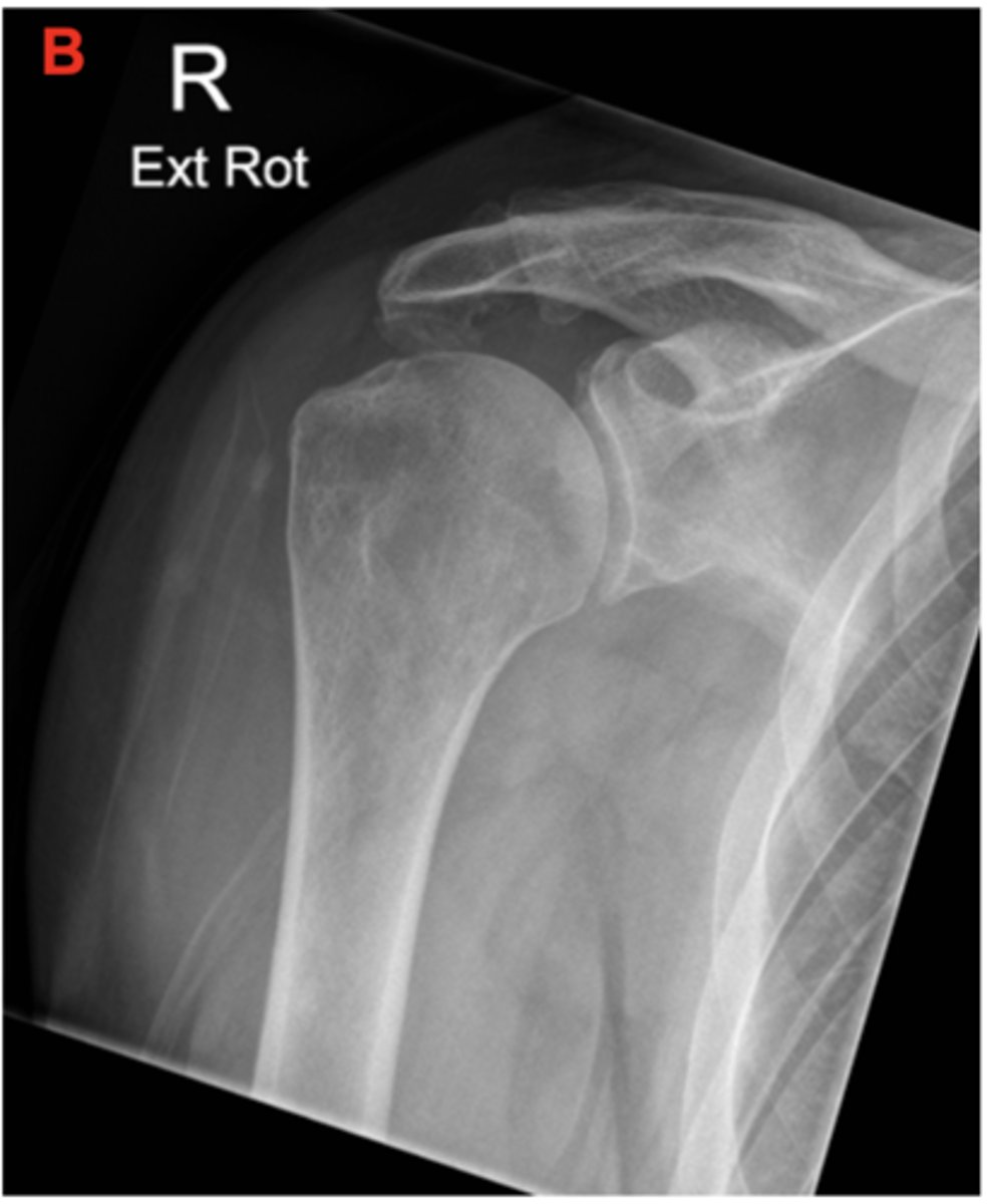
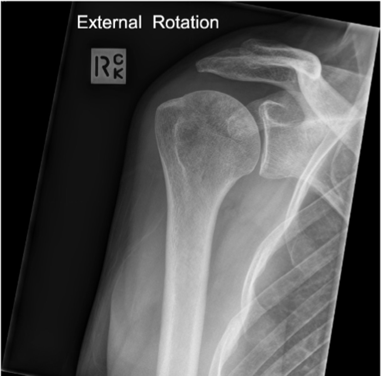
External Rotation as the greater tubercle is in profile and the lesser tubercle is not
Which Humerus rotation was present in this projection
Internal rotation as the lesser tubercle is in profile and the greater tubercle is not
Which Humerus rotation was present in this projection
Neutral roation as neither the greater or lesser tubercles are in profile
Which Humerus rotation was present in this projection
Neutral rotation/true position as the femoral neck is foreshortened and the greater and lesser trochanters are in profile.
Which femur rotation was present for this projection
Internal rotation (15-20 degrees) as the femoral neck is elongated and the lesser trochanters are superimposed with the femoral shaft.
Which femur rotation was present for this projection
External rotation as the femoral neck is extremely foreshortened and the greater trochanter is superimposed by the femoral neck
Which femur rotation was present for this projection
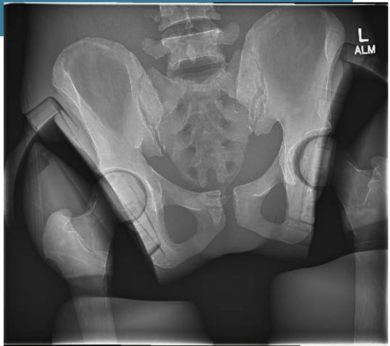
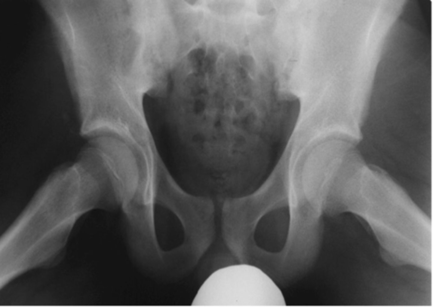
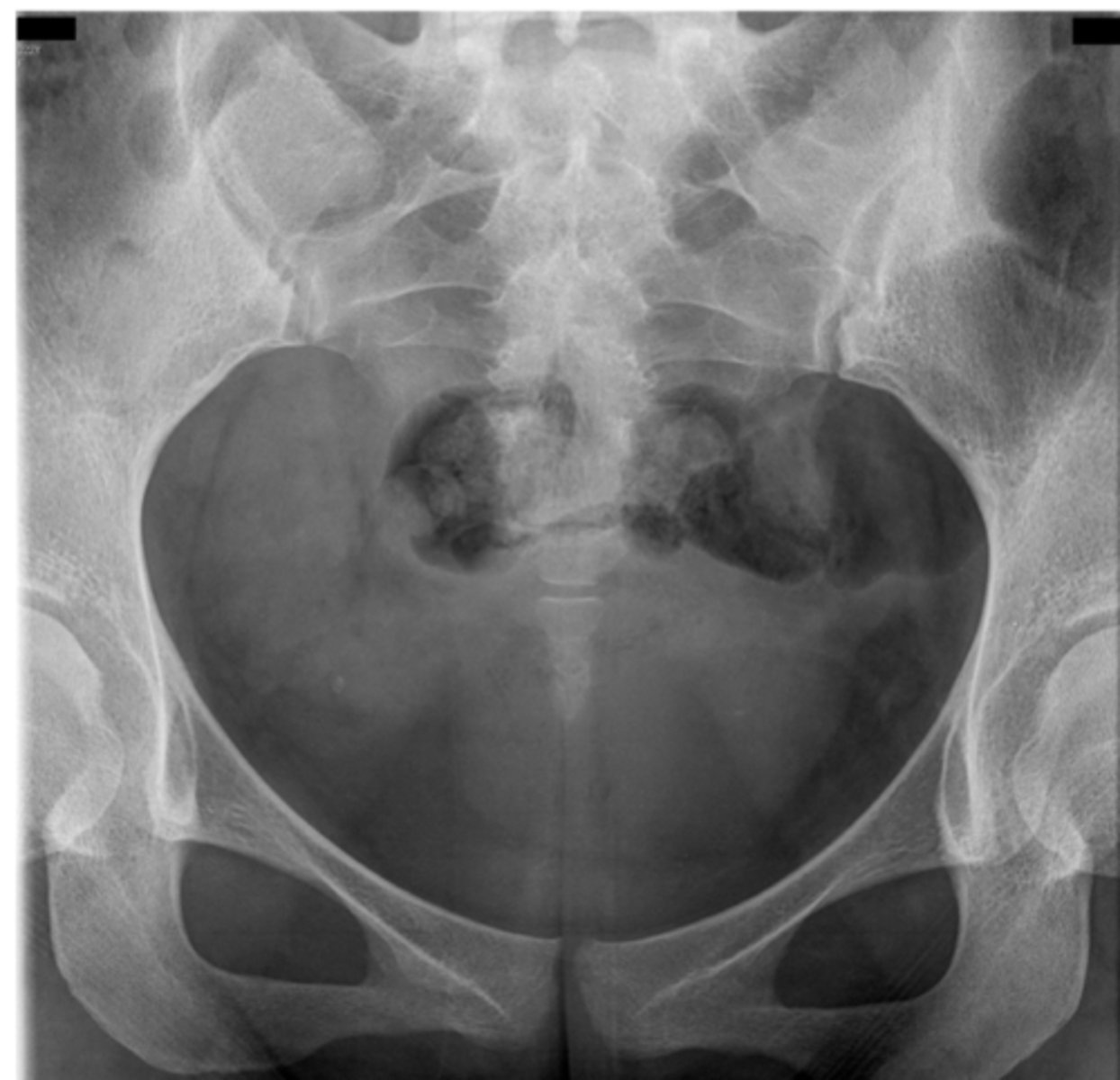
AP Pelvis
What projection is this?
AP Charnley Pelvis
What projection is this?
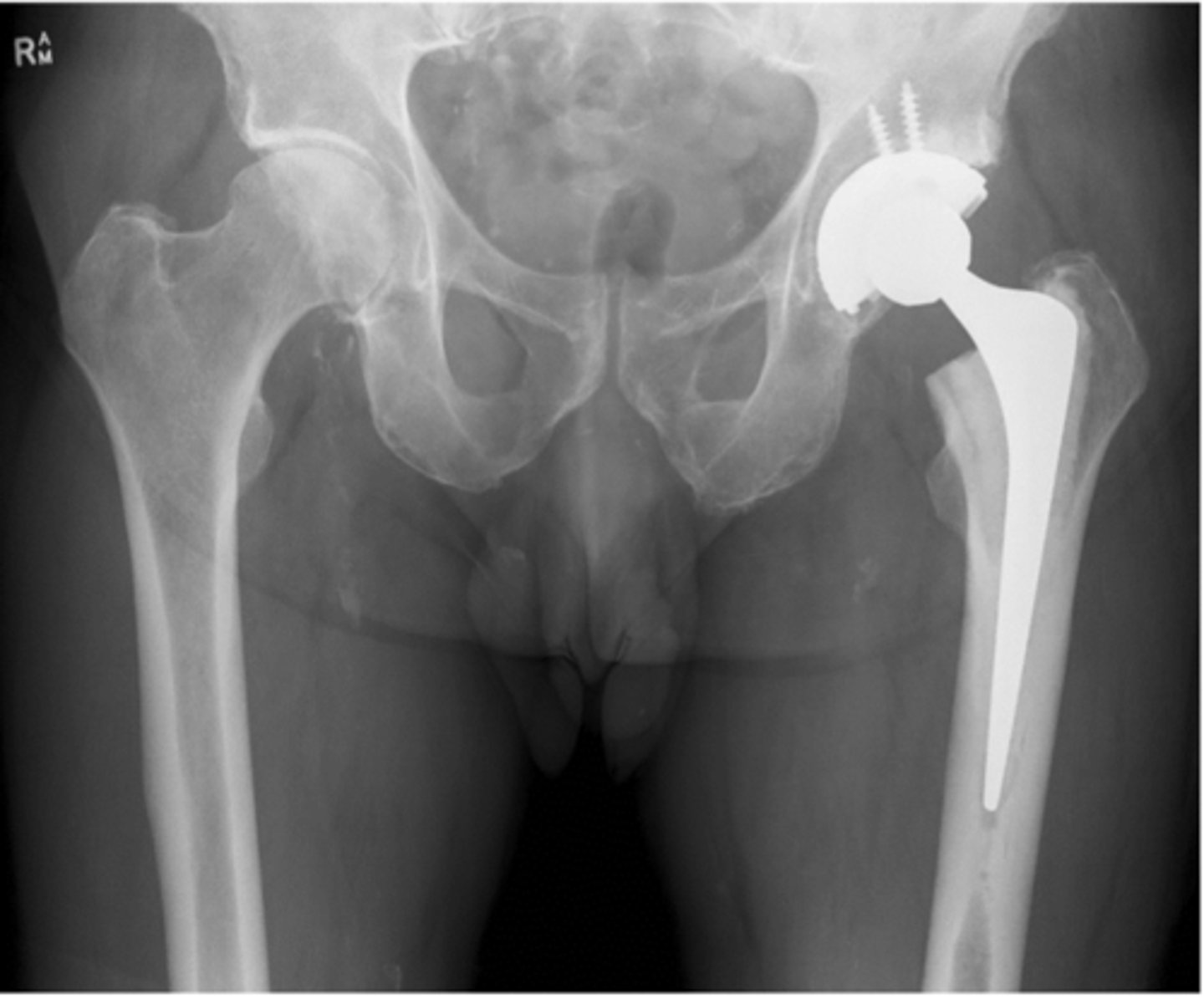
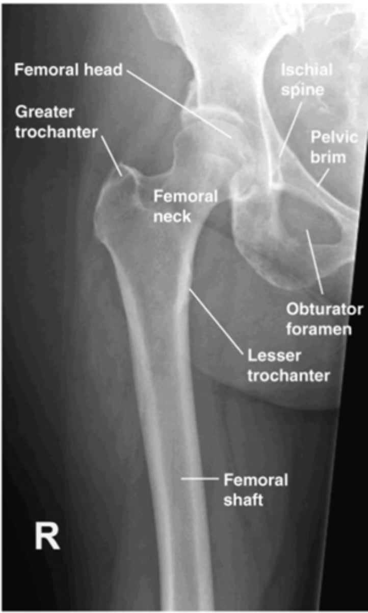
AP Hip
What projection is this?
Lateral Hip
What projection is this?
Frog leg lateral hip
What projection is this?
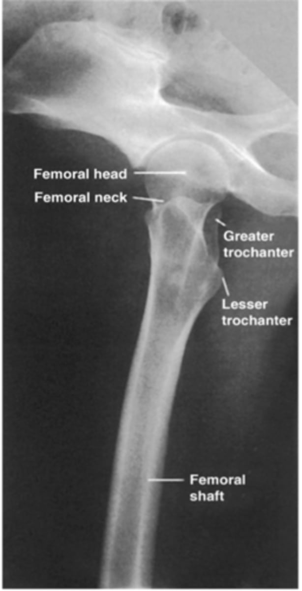
AP Proximal femur
What projection is this?
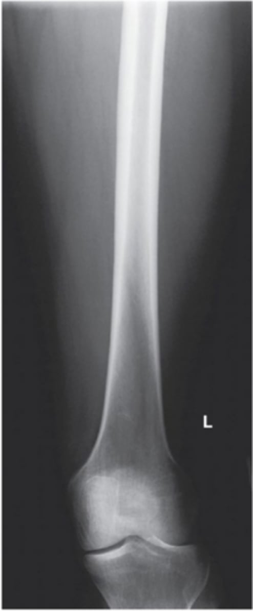
AP Distal femur
What projection is this?
Lateral Proximal femur
What projection is this?
Lateral Distal femur
What projection is this?
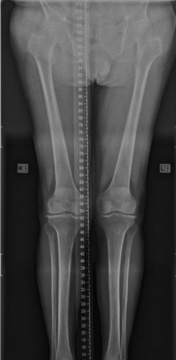
Long legs View
What projection is this?
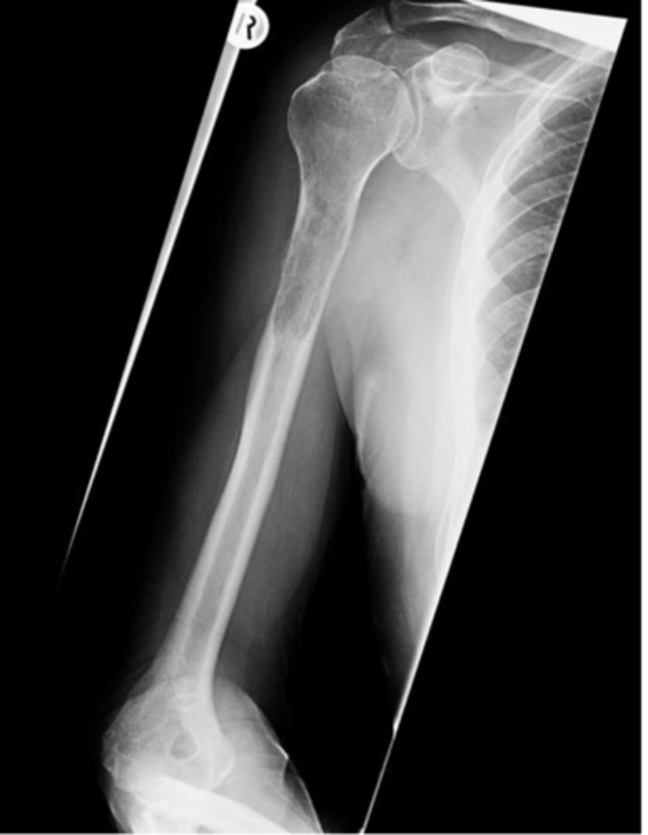
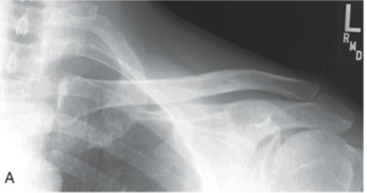
AP Humerus
What projection is this?
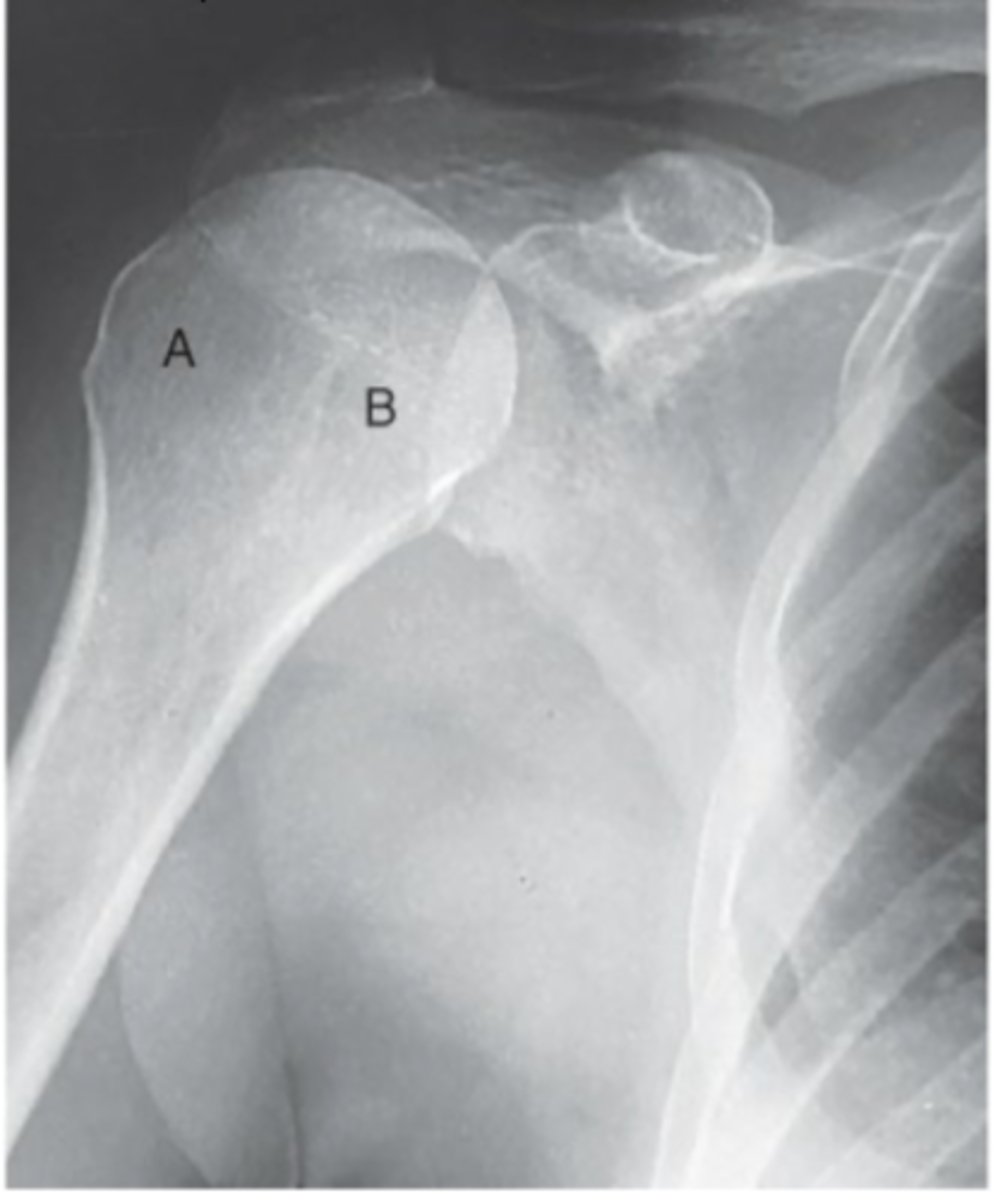
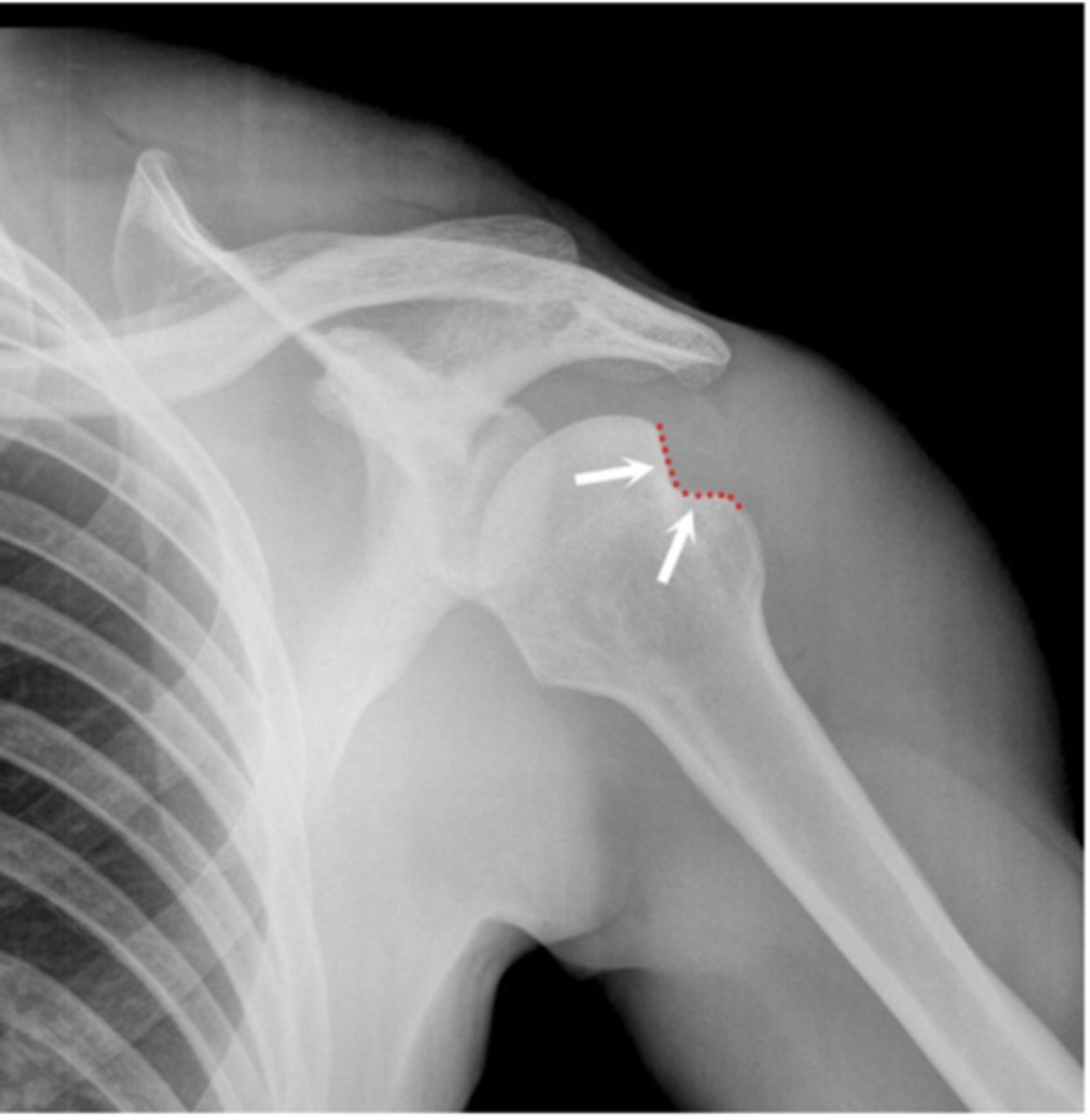
Hill Sachs defect
What deformity is seen here
Bankart Lesion
What deformity is seen here
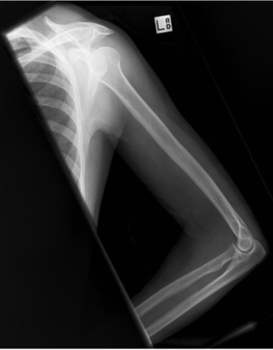
Lateral Humerus
What projection is this?
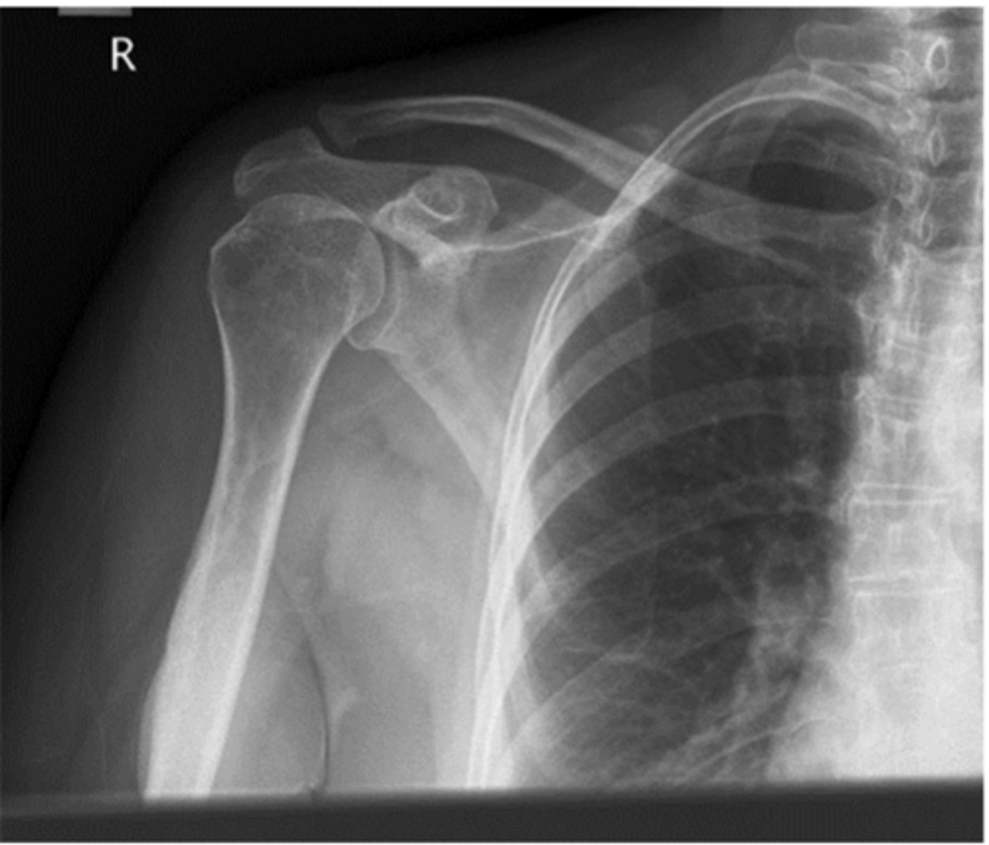
AP Shoulder
What projection is this?
GH Joint/AP Oblique
What projection is this?
Lateral Shoulder/Scap-Y
What projection is this?
Superior-Inferior (SI) Axial Shoulder
What projection is this?
Inferior-superior (IS) Axial Shoulder
What projection is this?
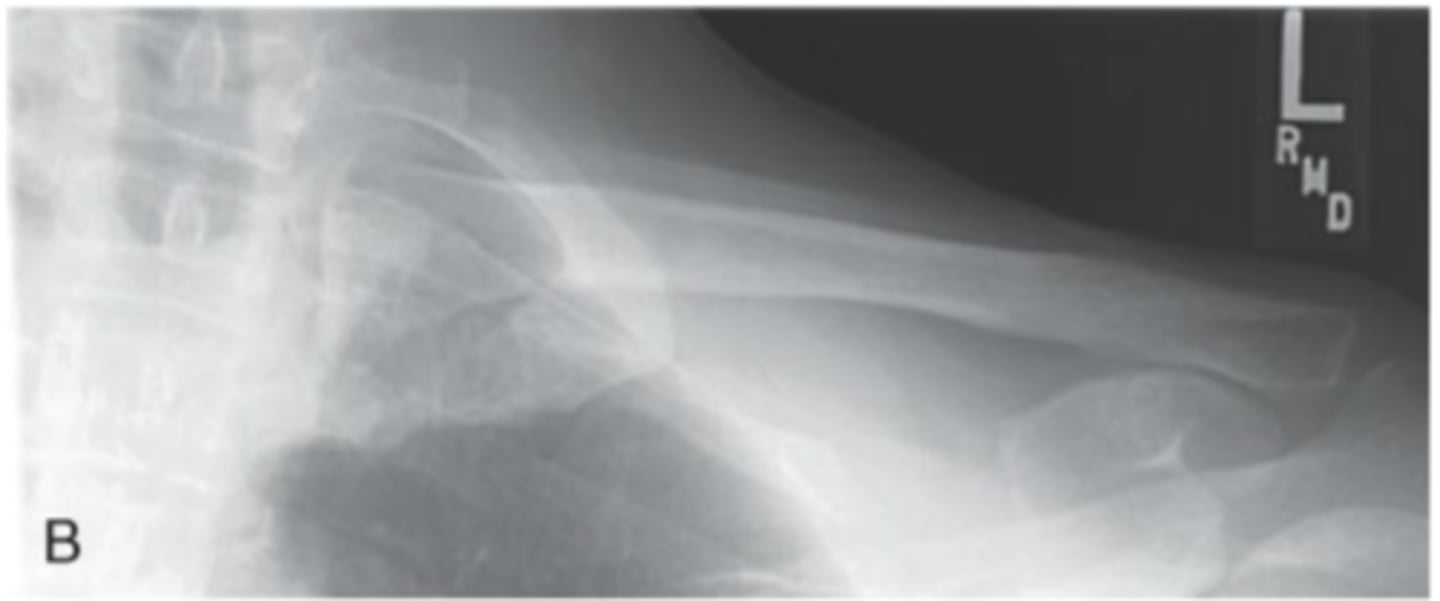
AP Clavicle
What projection is this?
AP Axial Clavicle
What projection is this?
Weight Bearing AP Acromioclavicular joint
What projection is this?
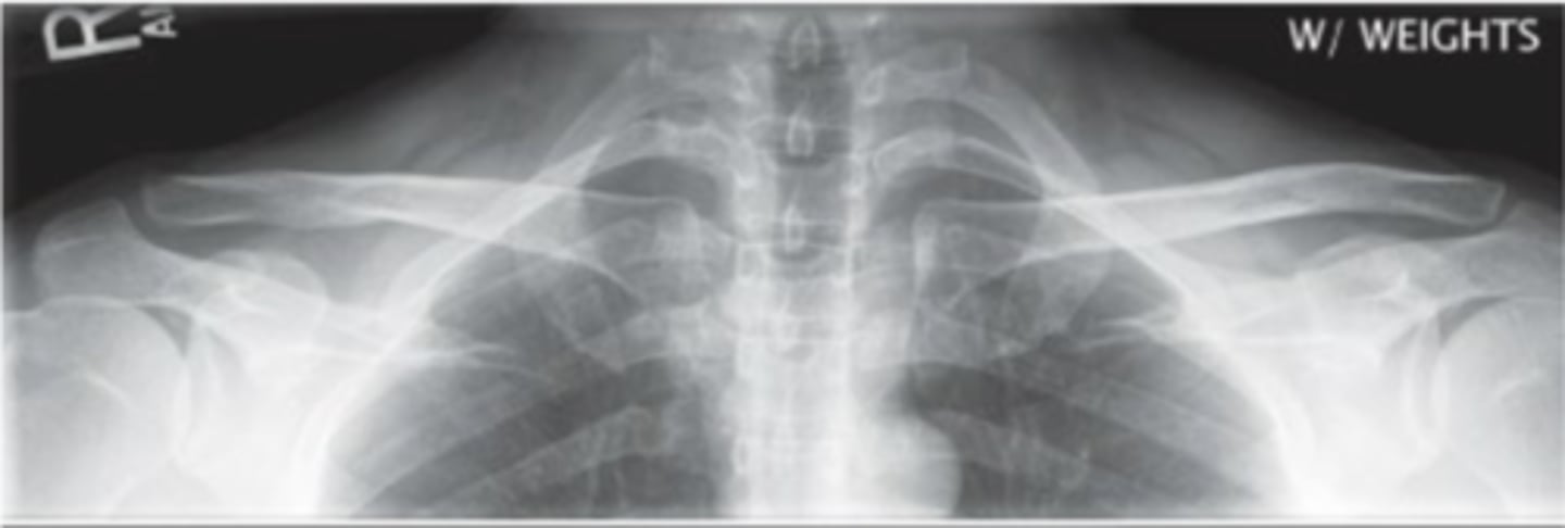
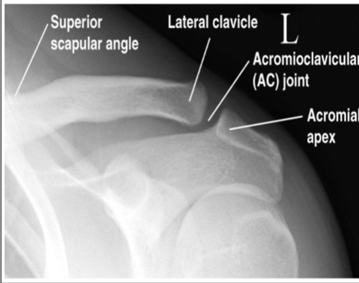
AP Acromioclavicular joint
What projection is this?
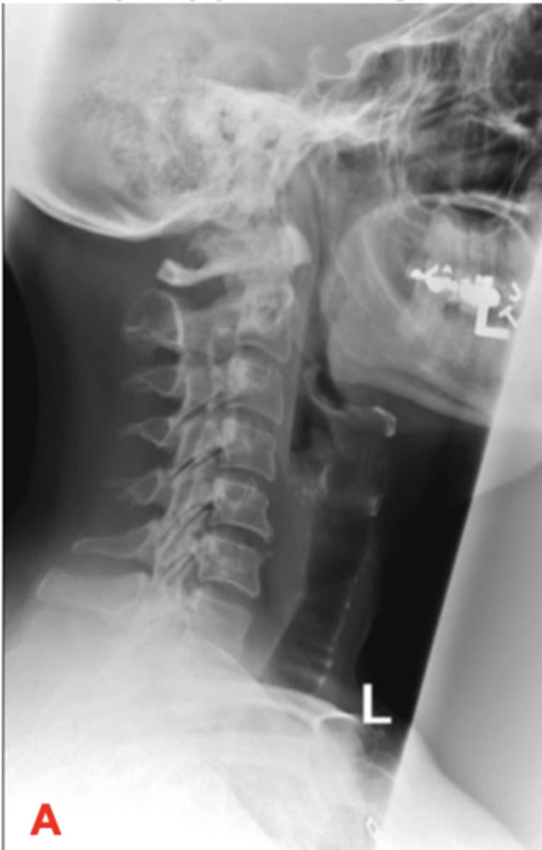
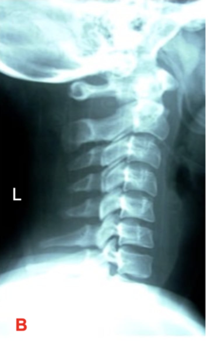
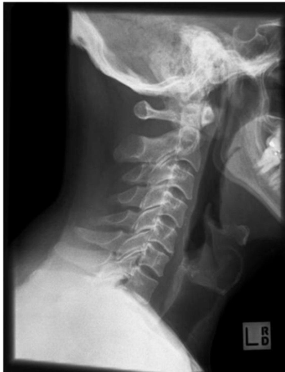
Lateral C spine
What projection is this?
Oblique C spine demonstrating the Left Intervertebral foramina
What projection is this?
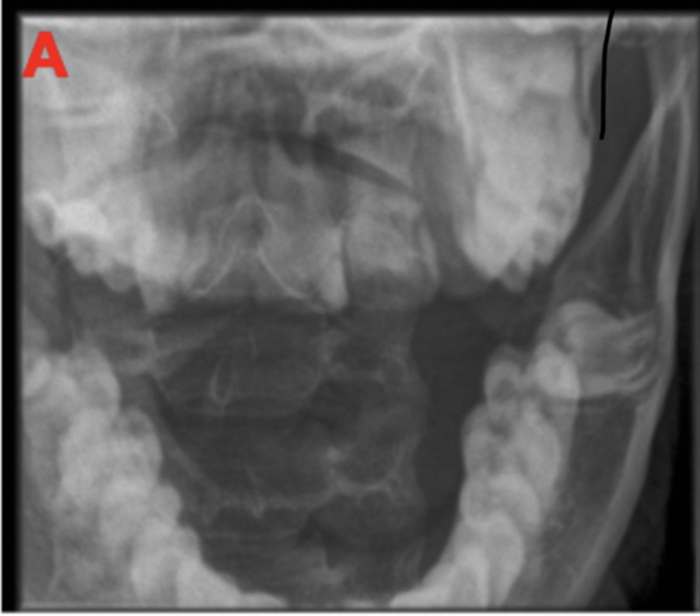
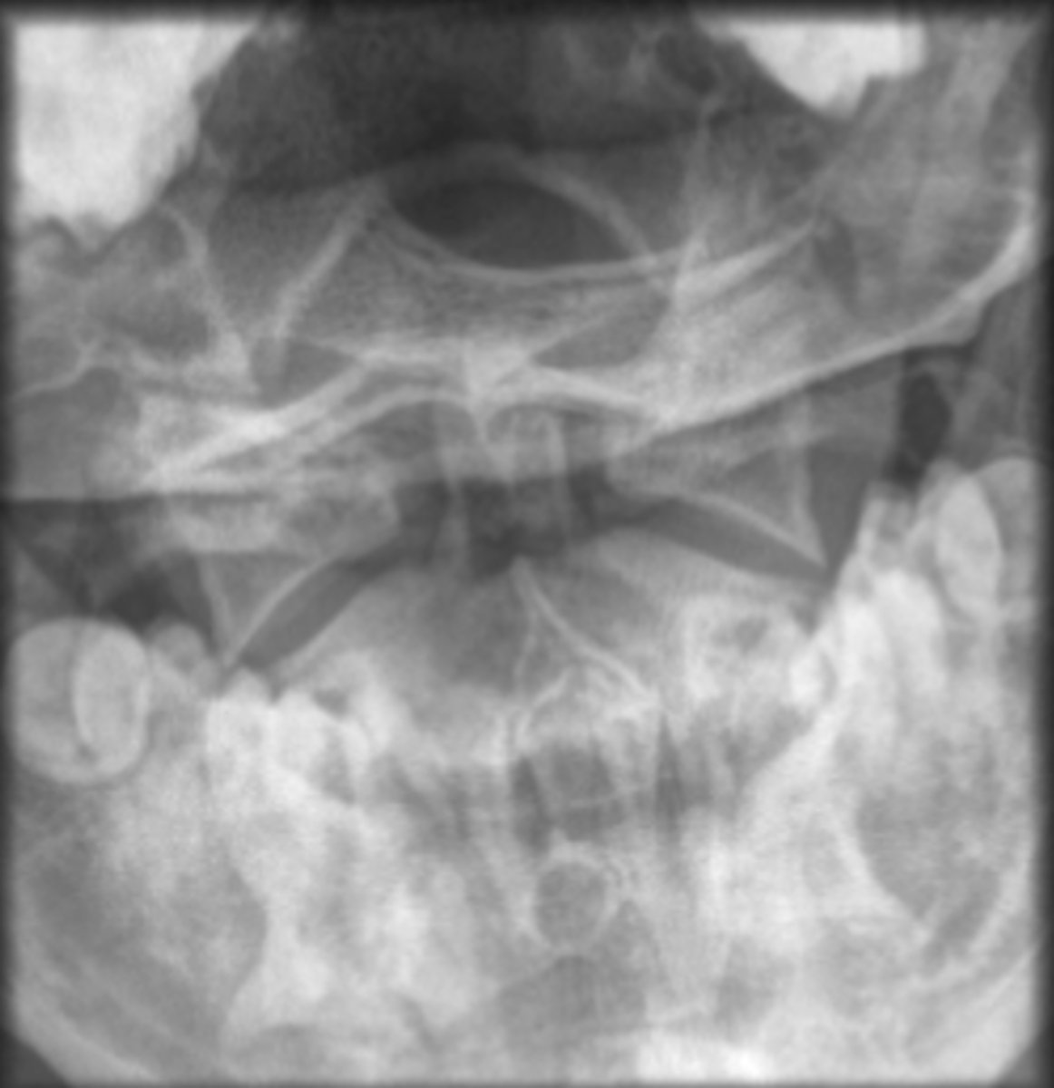
Open Mouth/C1 and 2 C spine
What projection is this?
Lateral Swimmers C Spine
What projection is this?
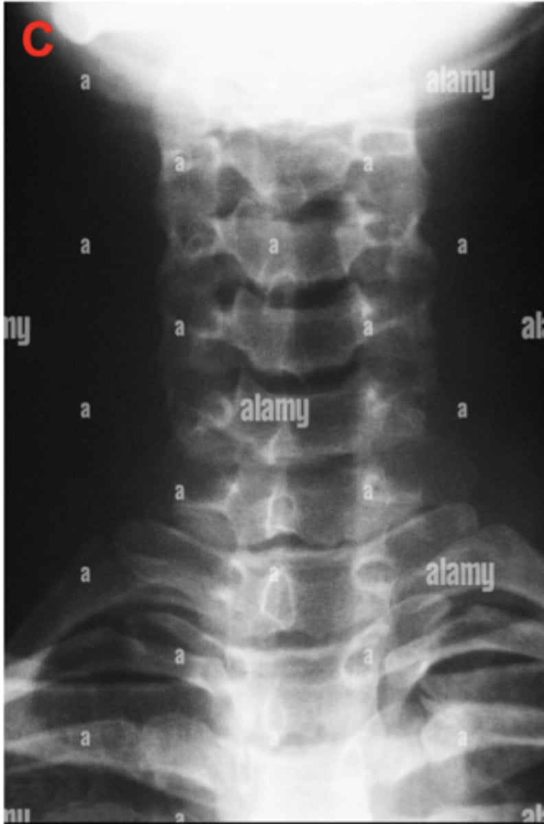
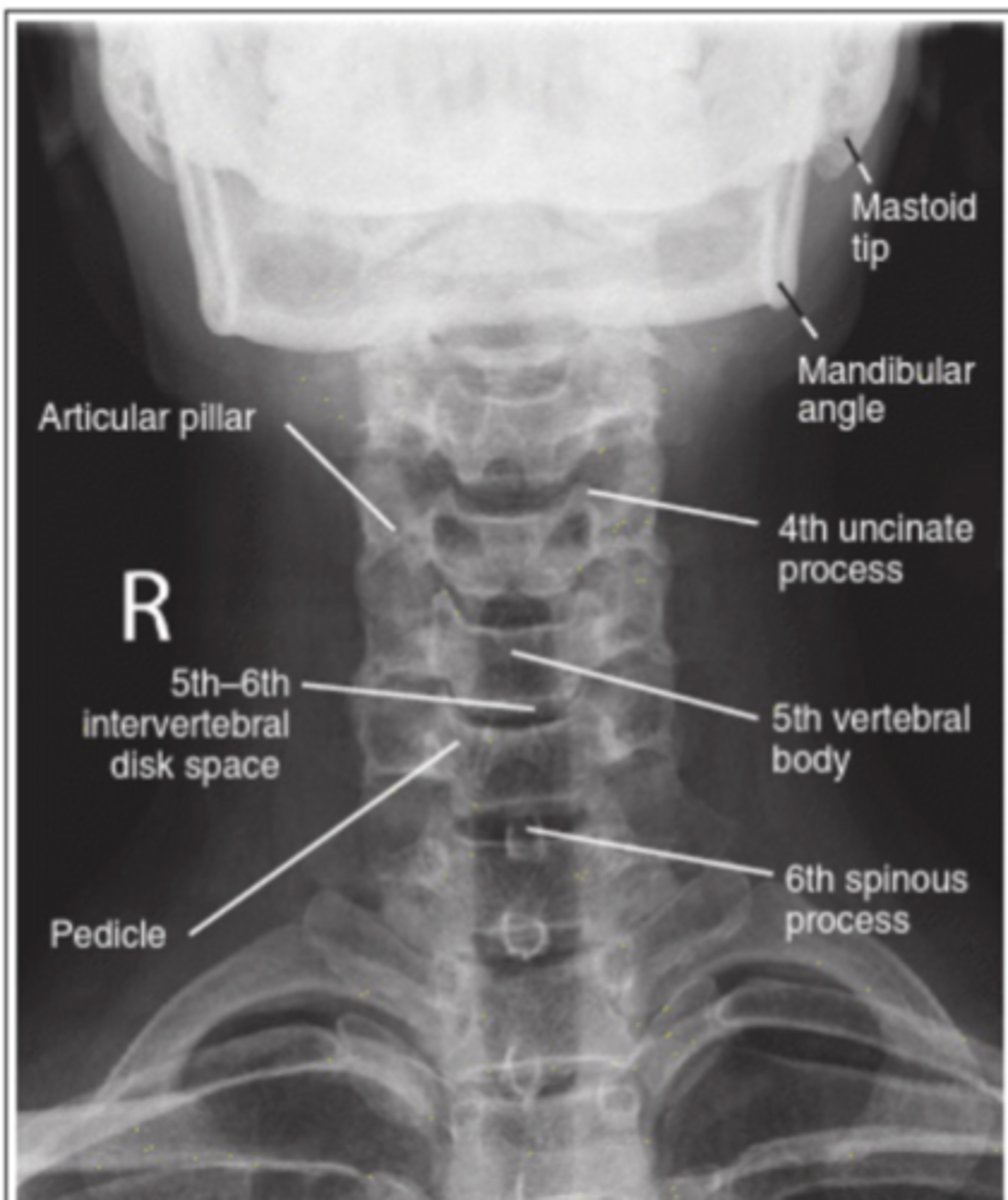
AP C spine
What projection is this?
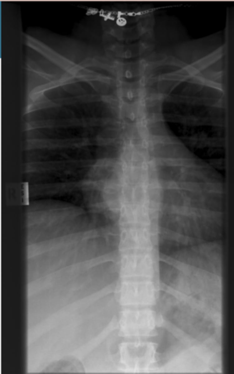
AP T-Spine
What projection is this?
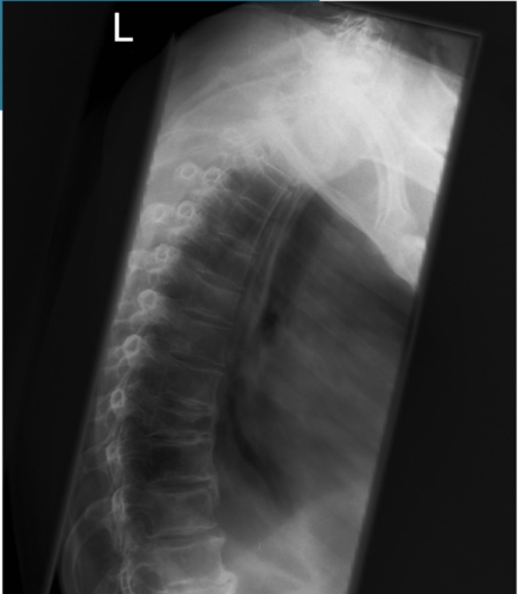
Lateral T-spine
What projection is this?
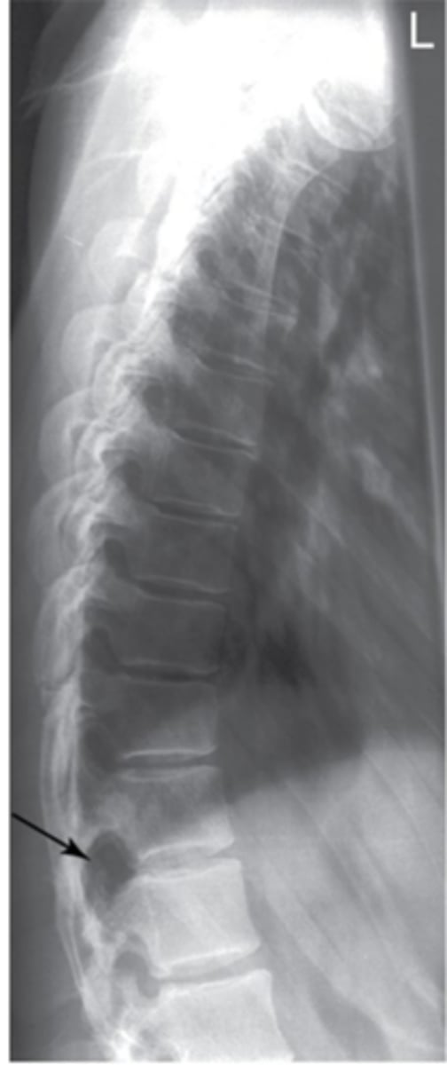
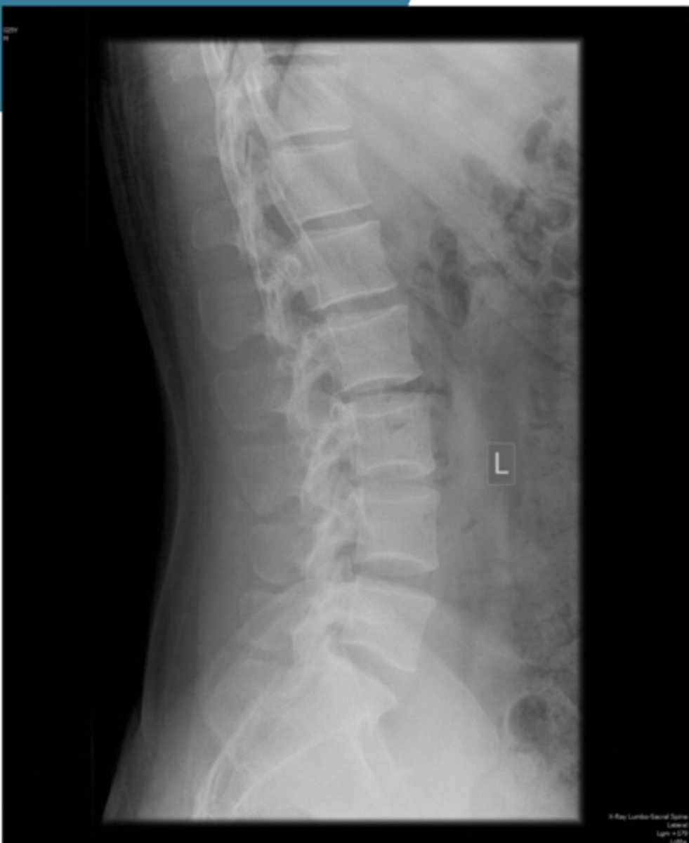
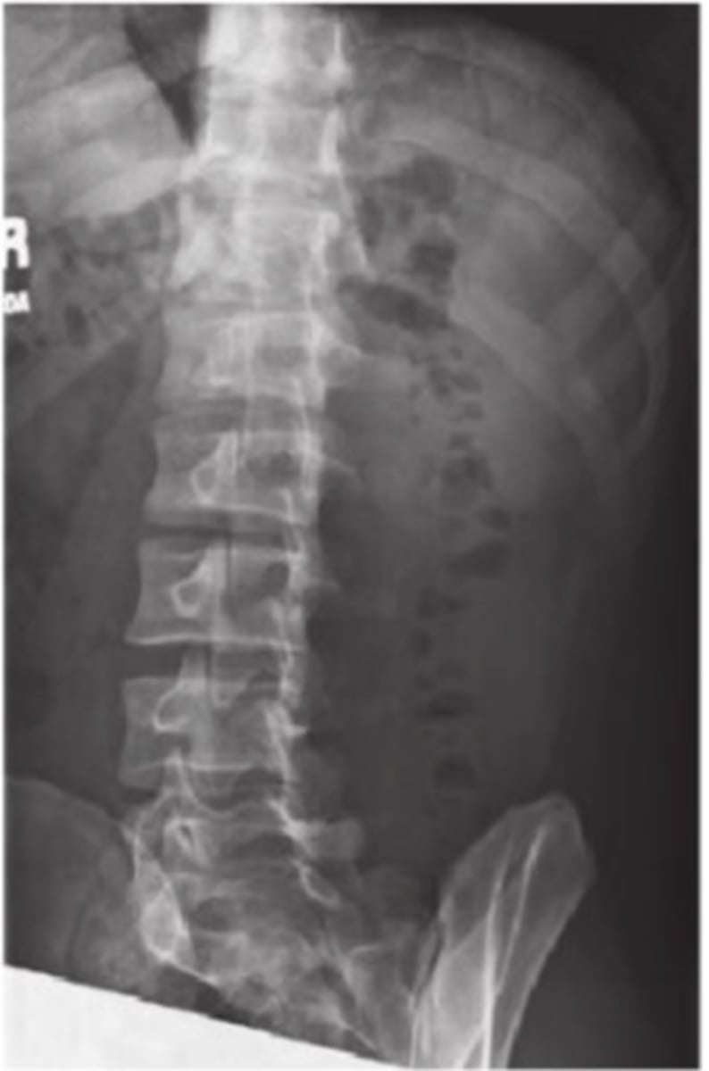
Lateral L Spine
What projection is this?
AP L-spine
What projection is this?
Oblique L-spine
What projection is this?
Oblique L-spine
What projection is this?
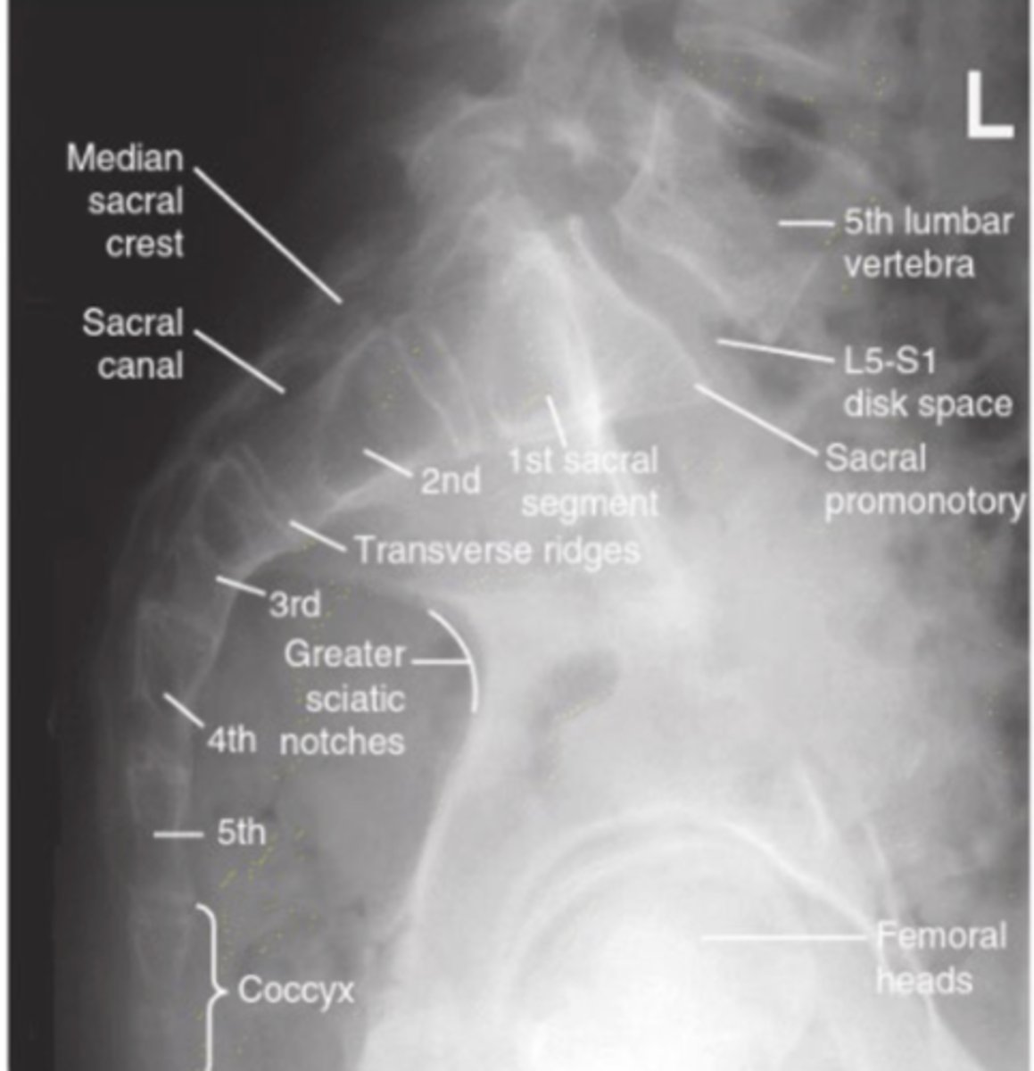
AP Sacrum
What projection is this?
Ap Coccyx
What projection is this?
Lateral Sacrum and Coccyx
What projection is this?
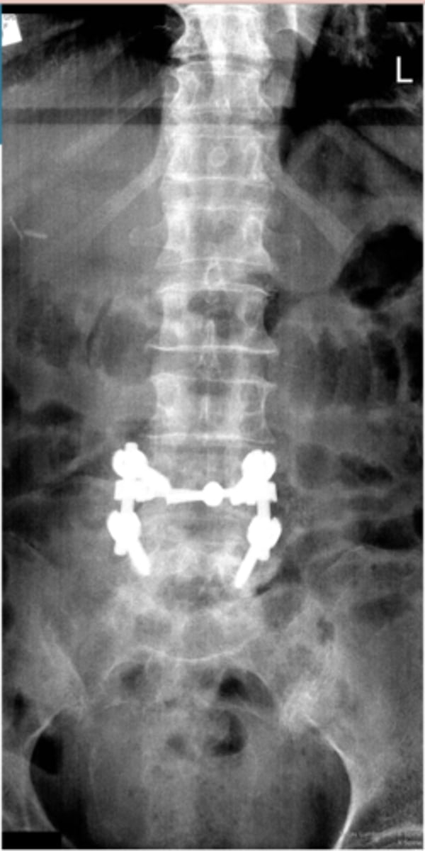
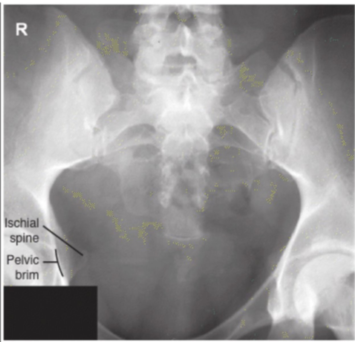
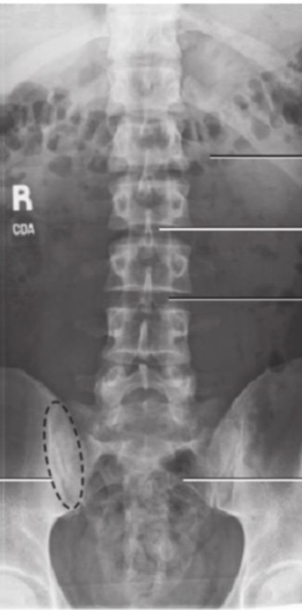
Sacroiliac joints
What projection is this?