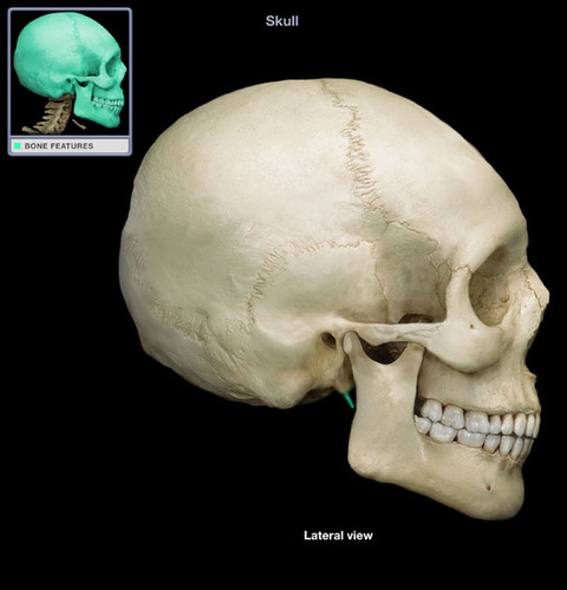
233 Unit 3 #1 Axial Skeleton -- Skull + Hyoid
1/68
There's no tags or description
Looks like no tags are added yet.
Name | Mastery | Learn | Test | Matching | Spaced |
|---|
No study sessions yet.
69 Terms
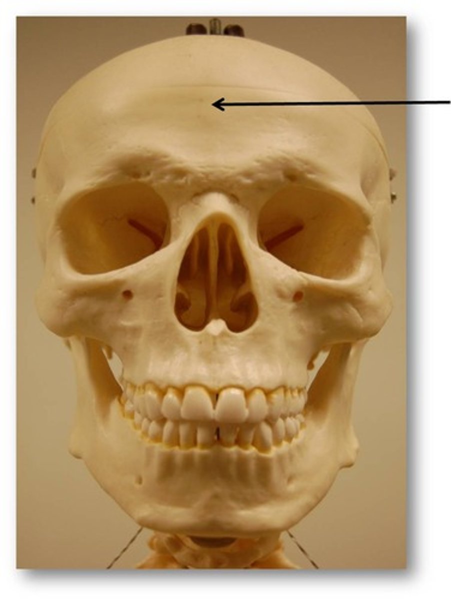
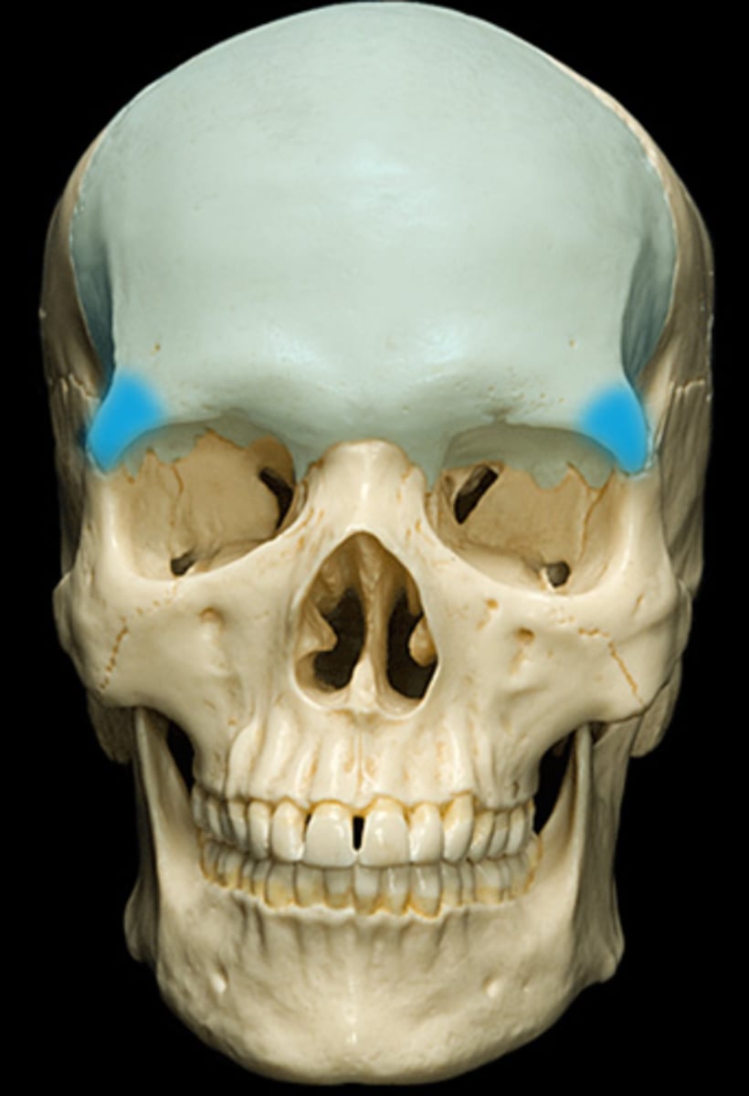
frontal bone
bone that forms the forehead
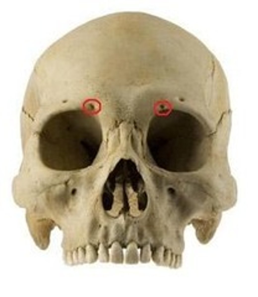
supraorbital foramen (notch)
opening above each orbit allowing blood vessels and nerves to pass
zygomatic process of frontal bone
extension of frontal bone that articulates with zygomatic bone at lateral border of orbit
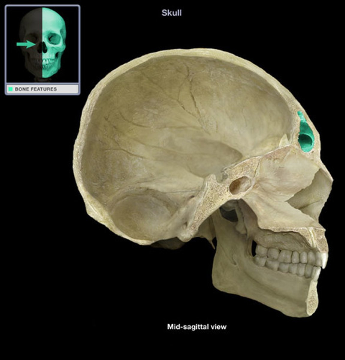
frontal sinus
cavity within the frontal bone
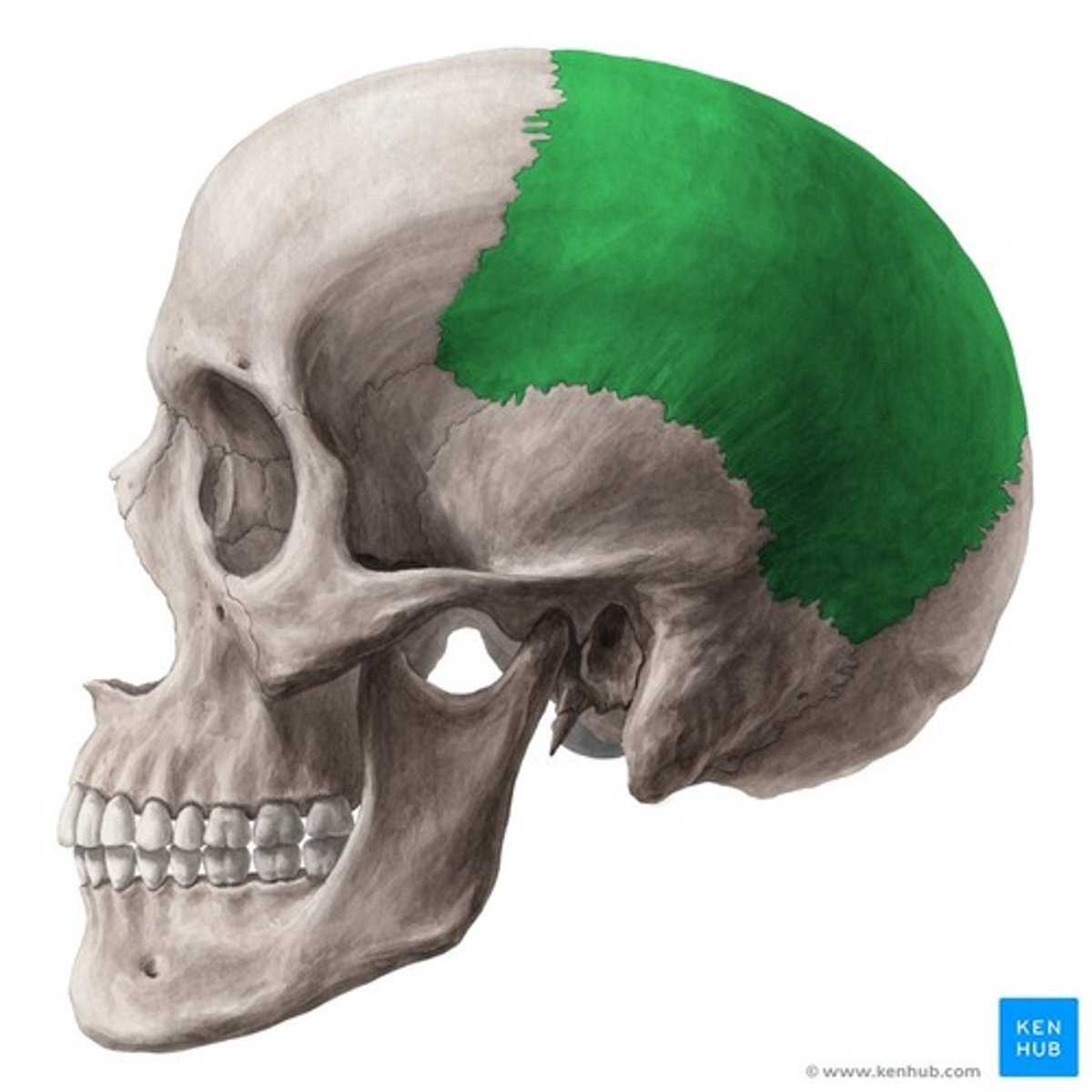
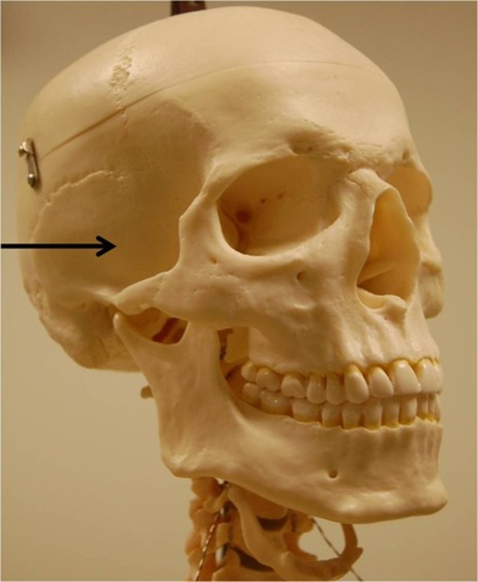
parietal bone
forms sides of the skull
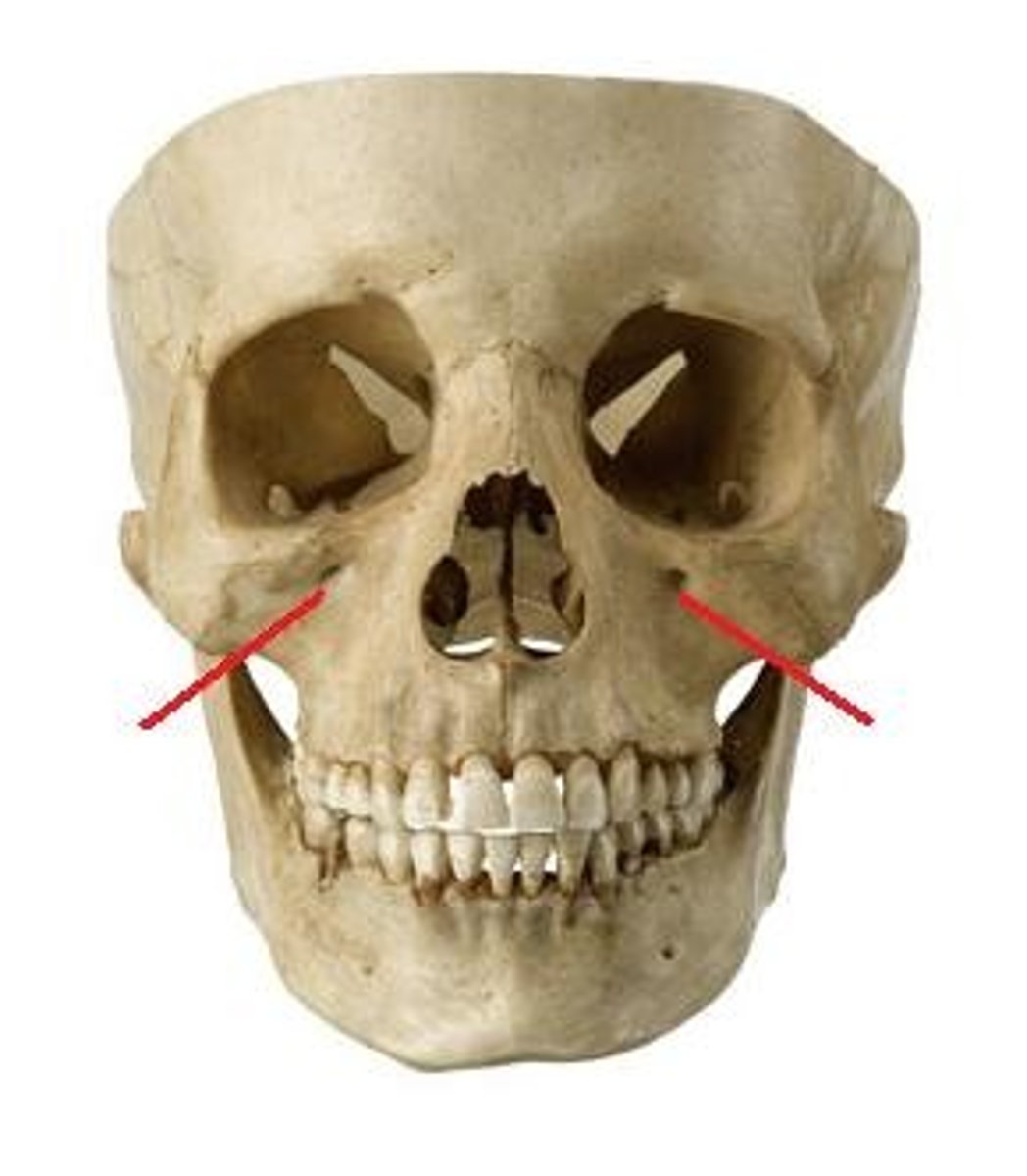
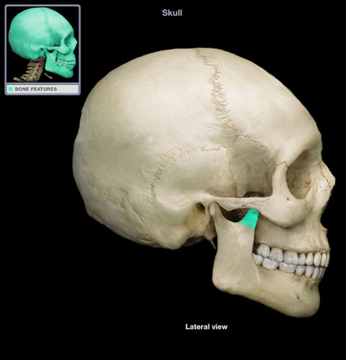
Zygomatic bone
cheek bone

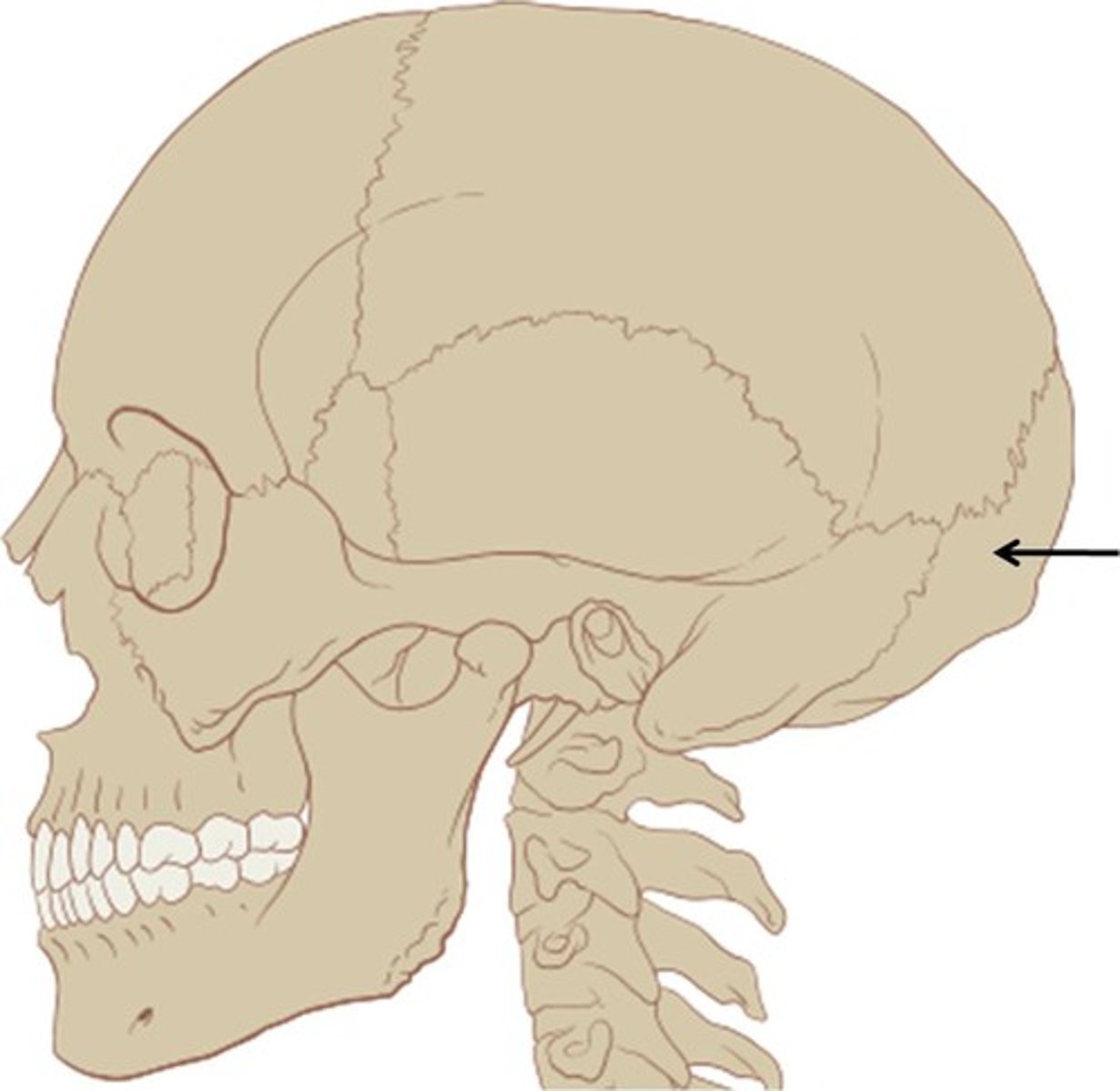
occipital bone
bone at the back of head
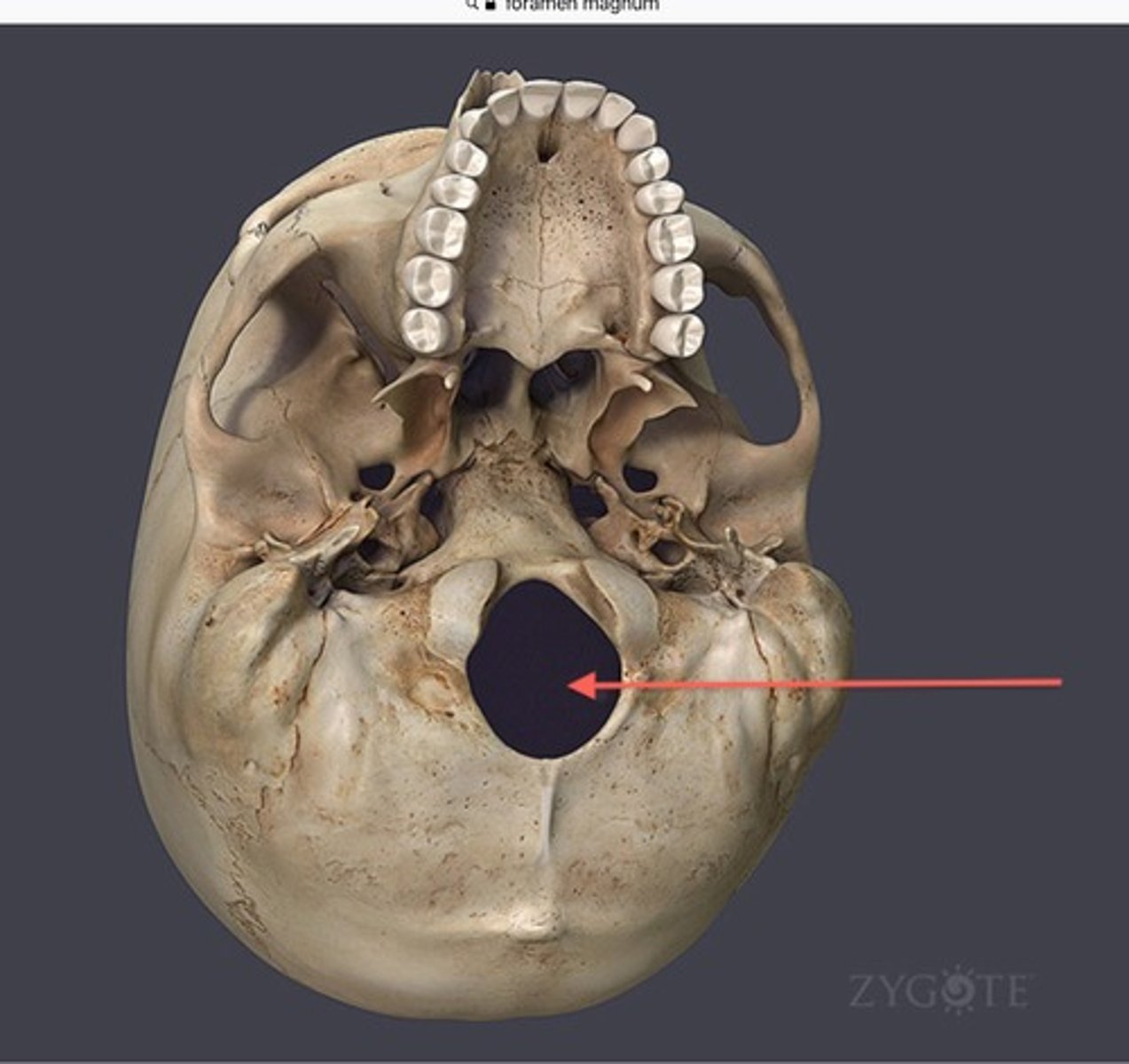
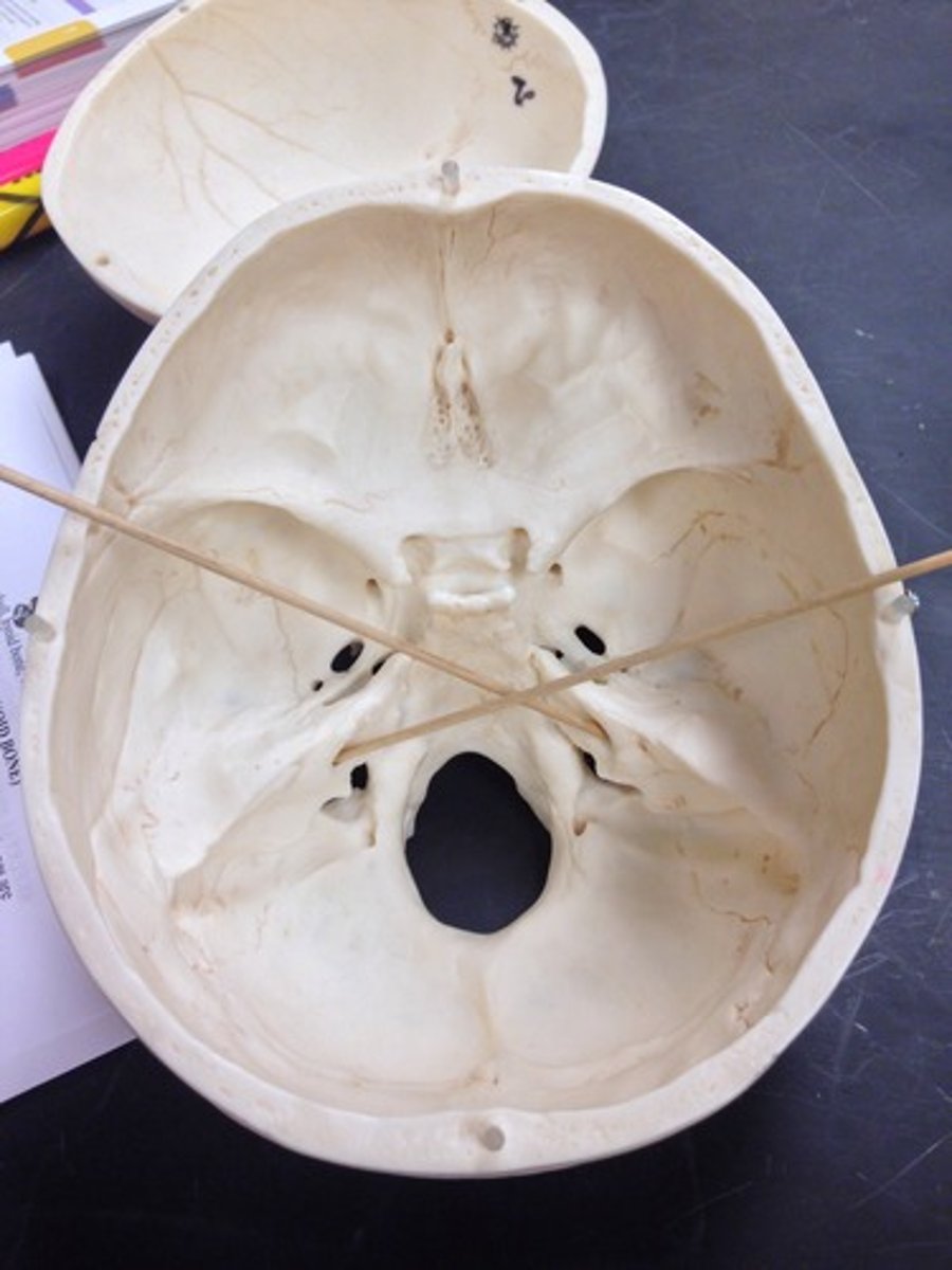
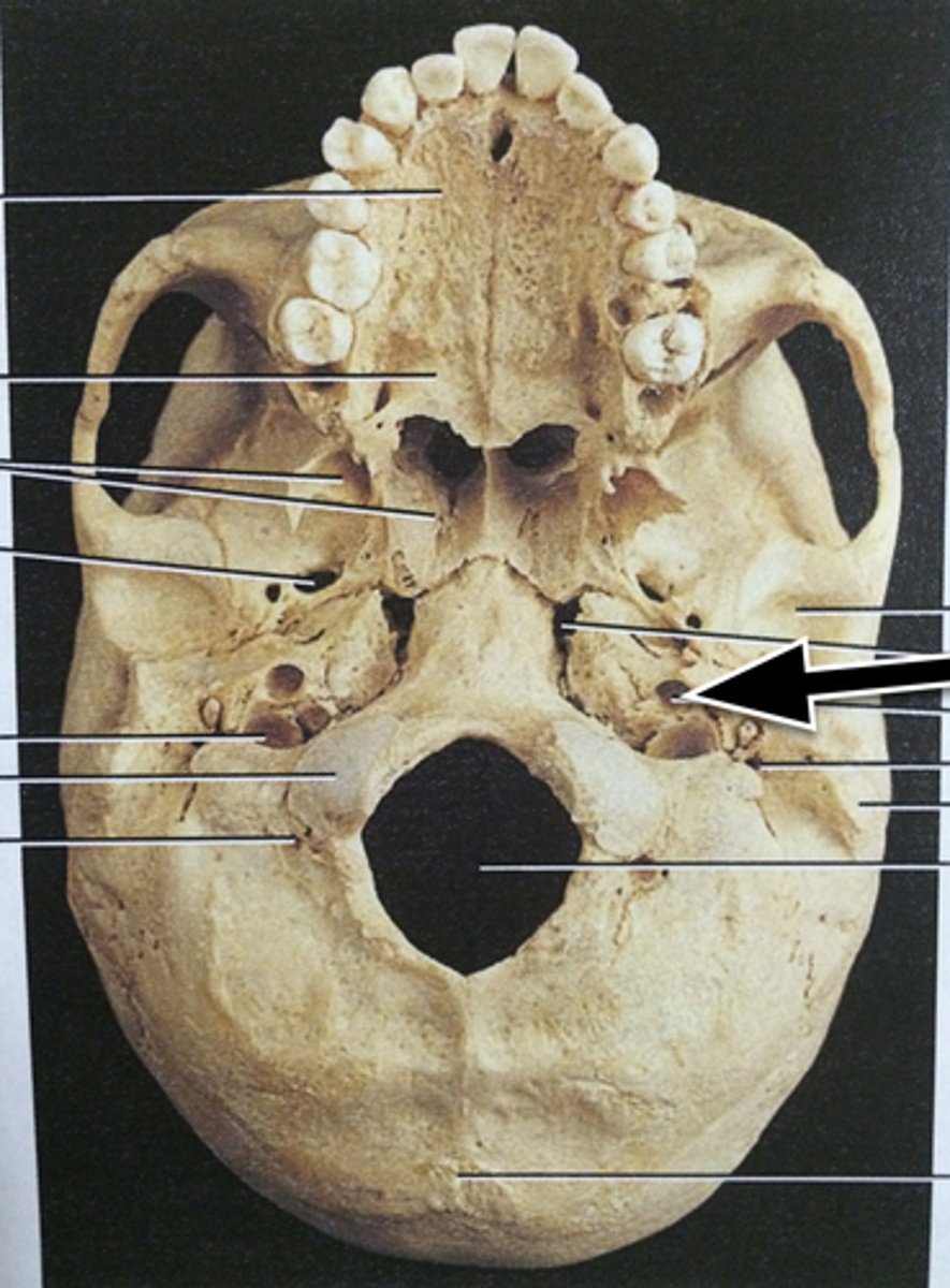
foramem magnum
large hole in the base of the skull, spinal cord enters here
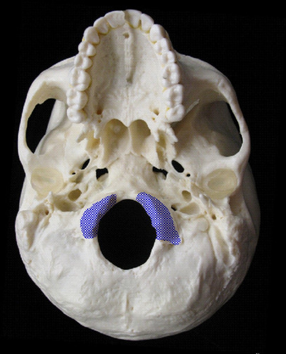
occipital condyles
articulate with first cervical vertebra (atlas)
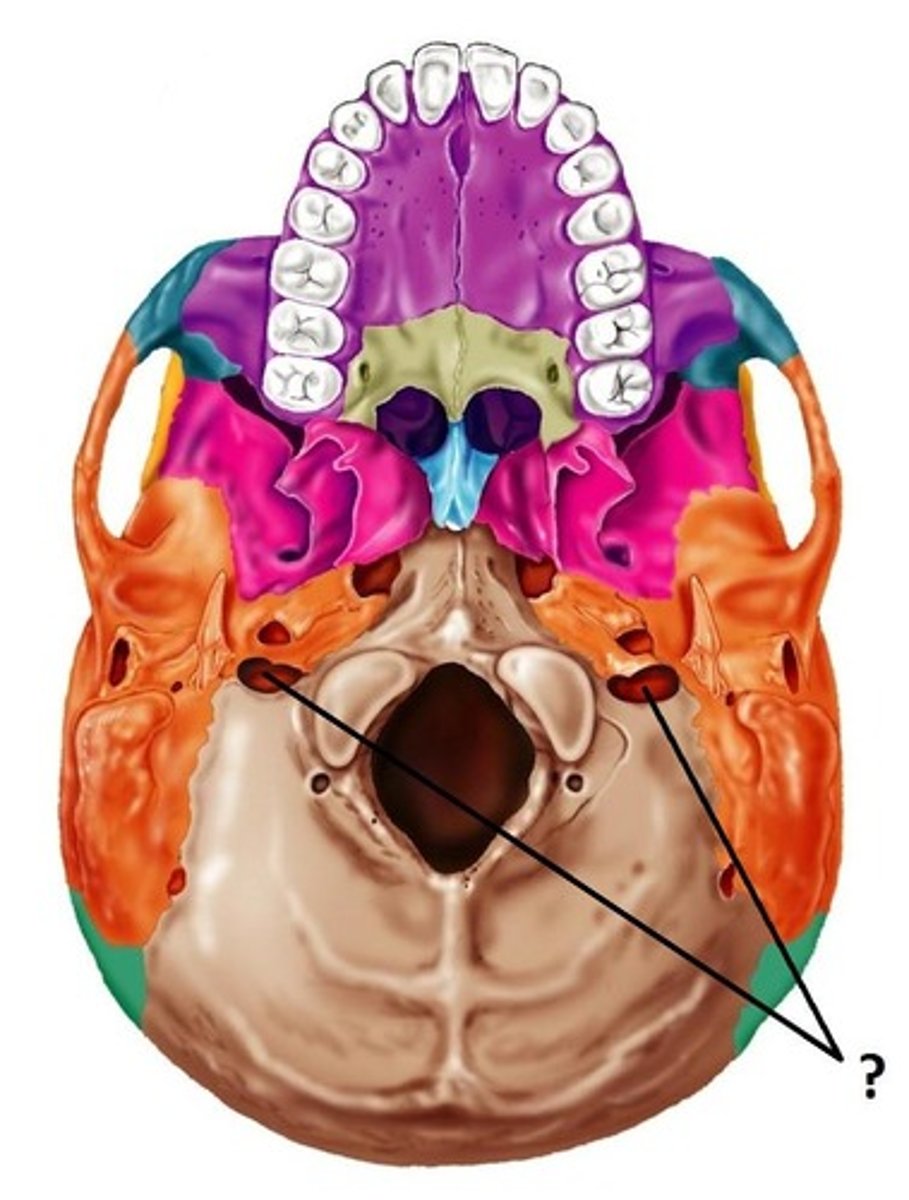
jugular foramen
Name this foramen.
temporal bone
bone that contains the external auditory meatus and articulates with the mandible
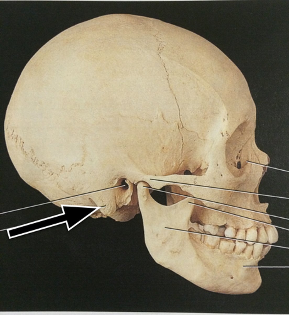
mastoid process
round projection on the temporal bone behind the ear
styloid process
sharp process extending downward from the temporal bone on each side of the skull
external auditory meatus (canal)
outer ear canal
internal auditory meatus (canal)
canal located between the jugular foramen and carotid canal from superior view
zygomatic process of temporal bone
extension from the temporal bone that forms the posterior portion of the zygomatic arch
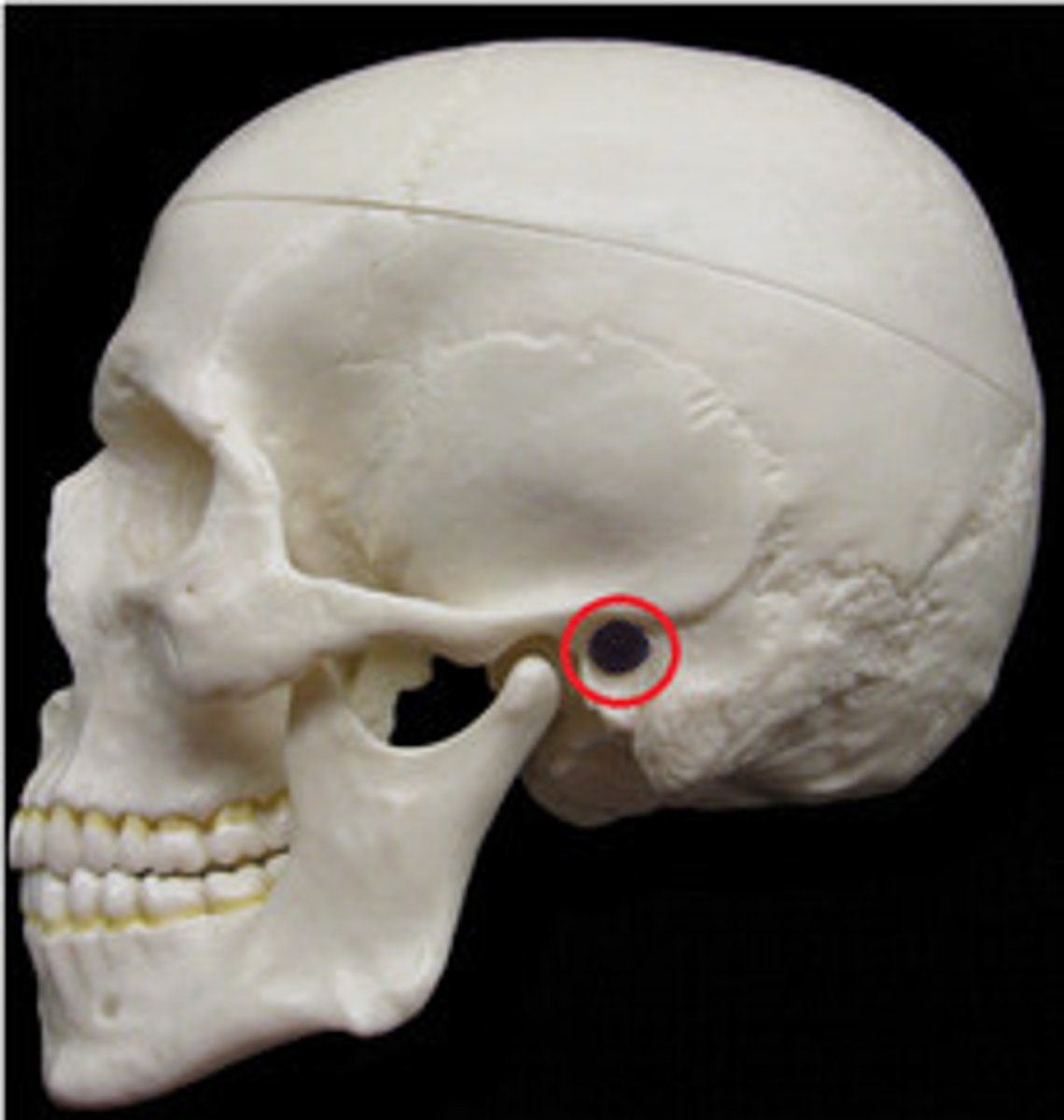
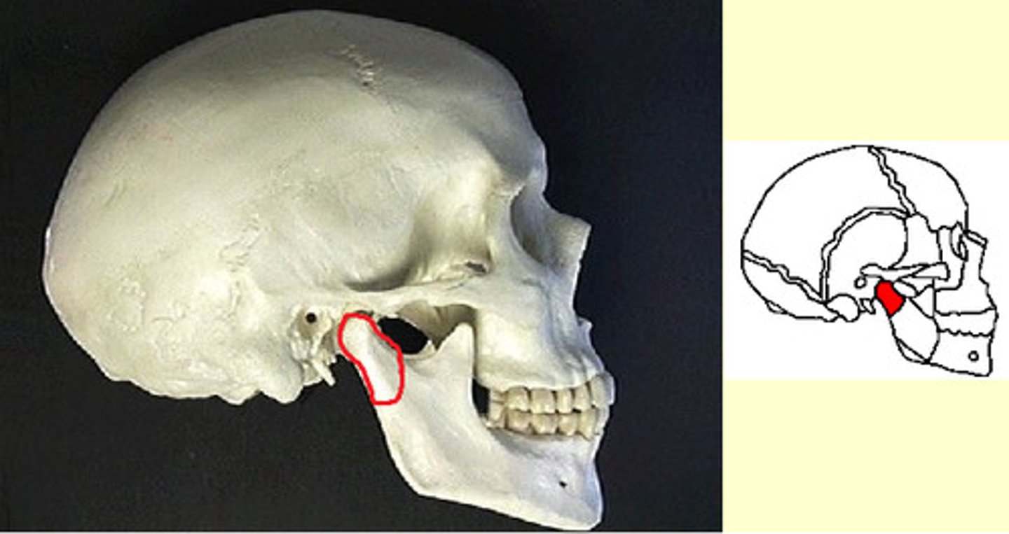
mandibular fossa
the depression in the temporal bone into which the condyle of the mandible fits
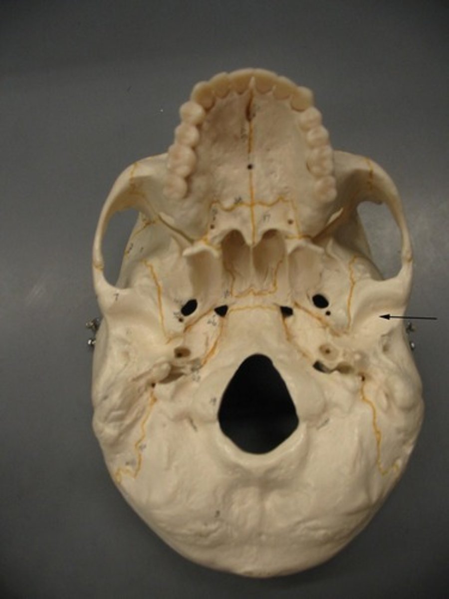
carotid foramen (canal)
opening for carotid artery
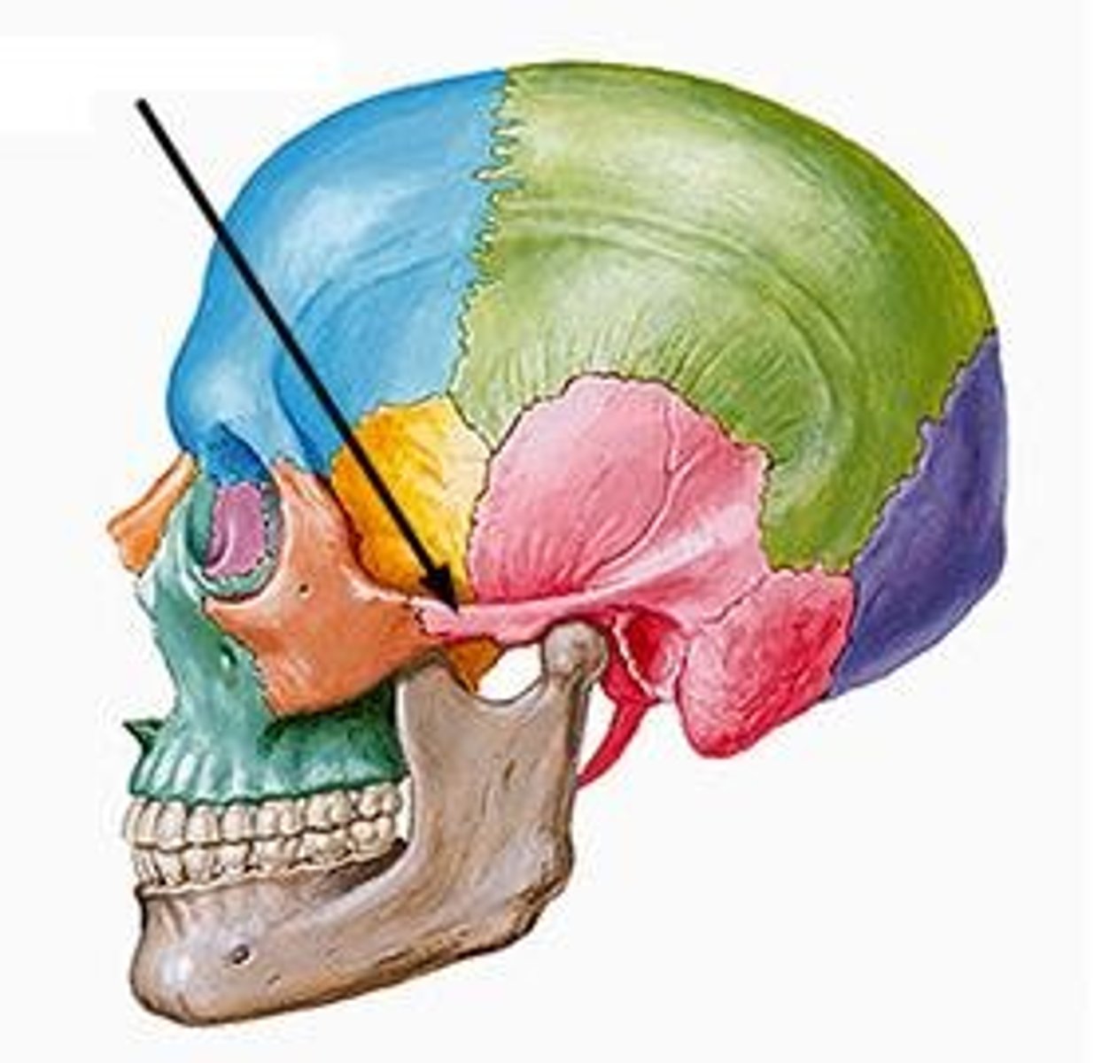
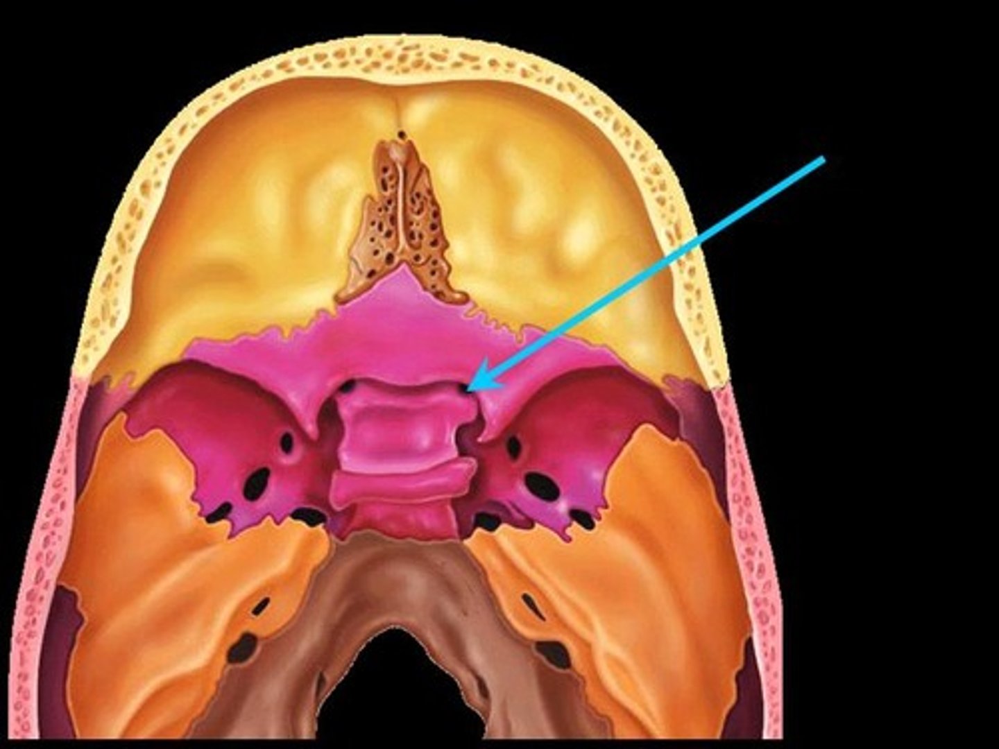
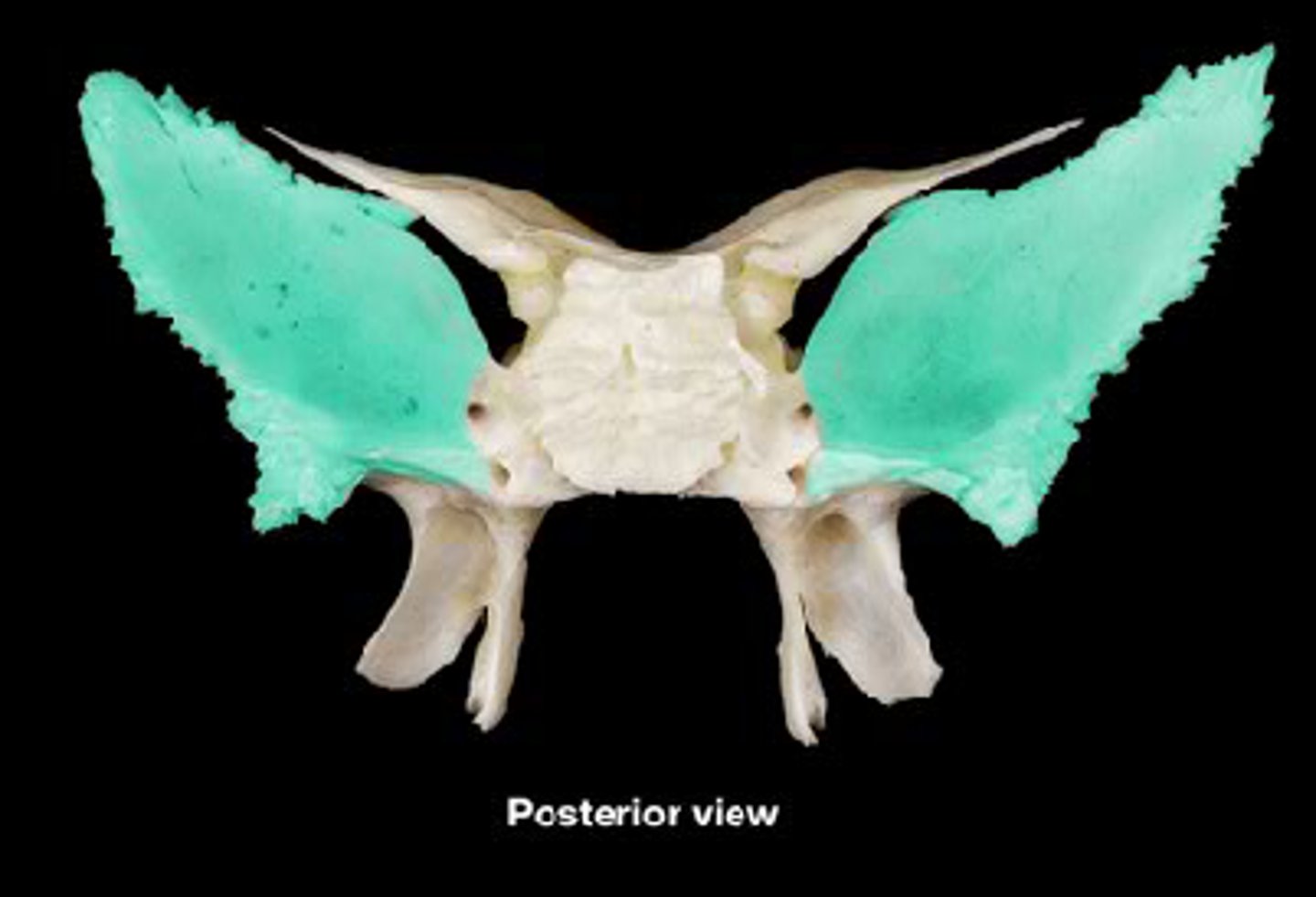
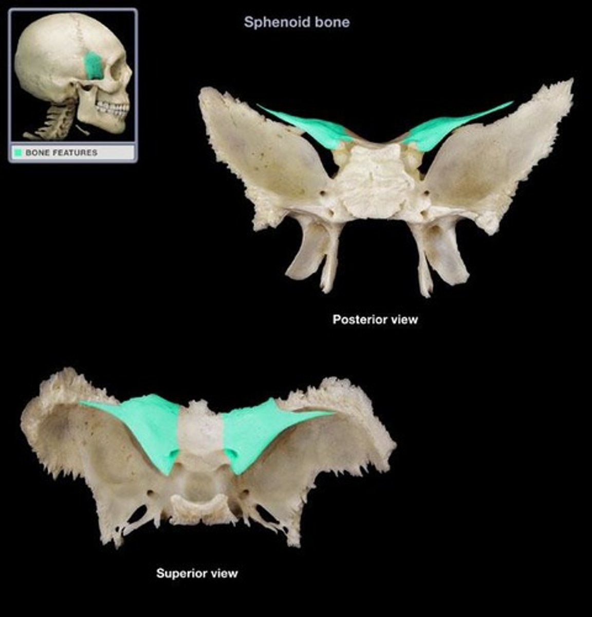
sphenoid bone
Bone that joins all of the bones of the cranium together--shaped like a butterfly or bat
greater wing of sphenoid bone
posterior and inferior to lesser wing on the interior of the skull, also visible in the posterior orbit and lateral side of the skull
lesser wing of sphenoid bone
anterior and superior to greater wing on the interior of the skull
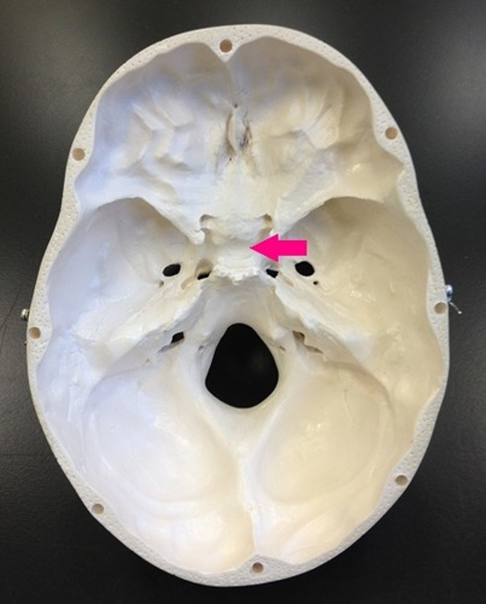
sella turcica
depression in the sphenoid bone where the pituitary gland is located (hypophyseal fossa)
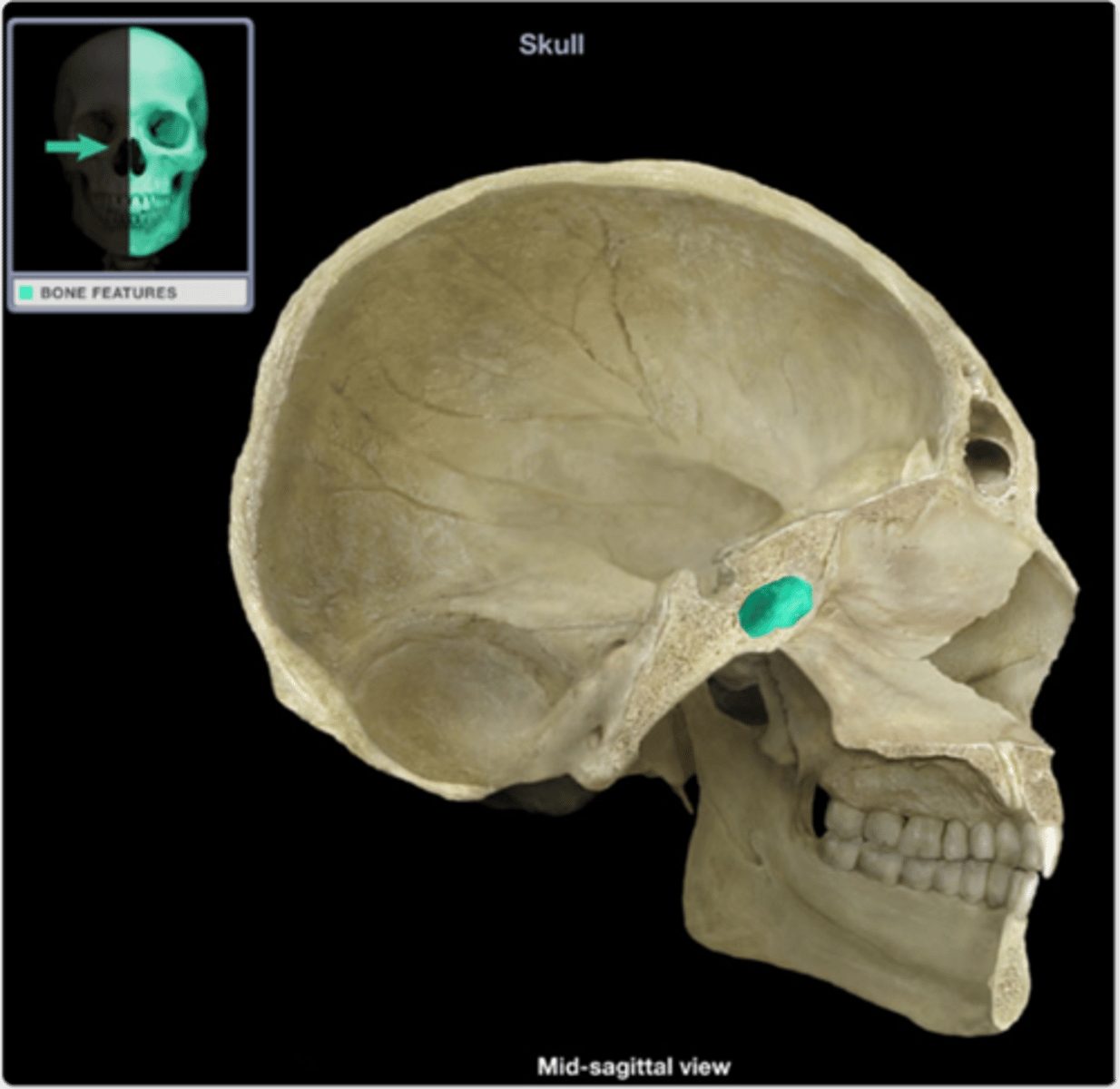
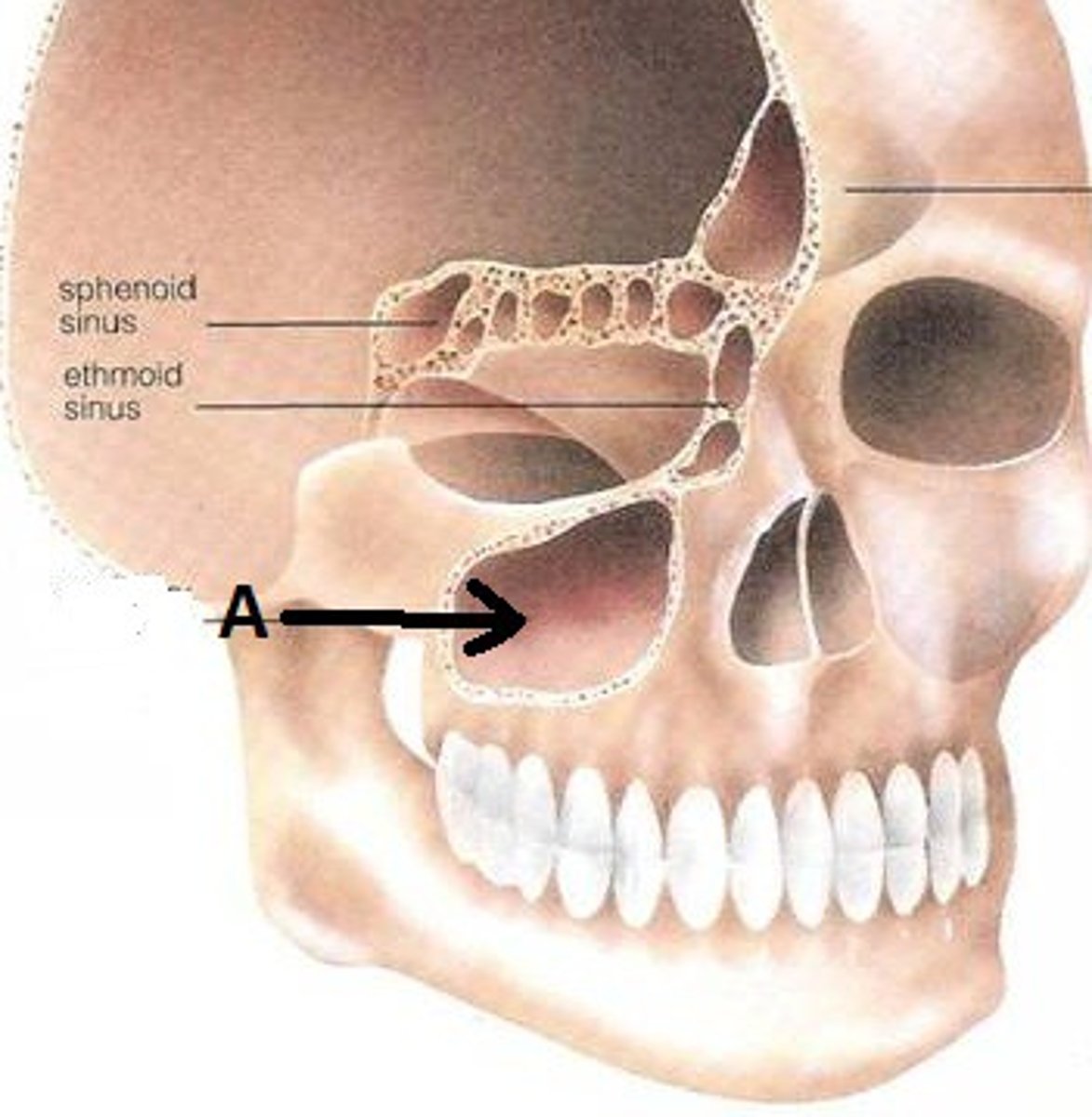
sphenoid sinus
sinus that sits just UNDER the sella turcica. It is posterior to the nose, and slightly superior
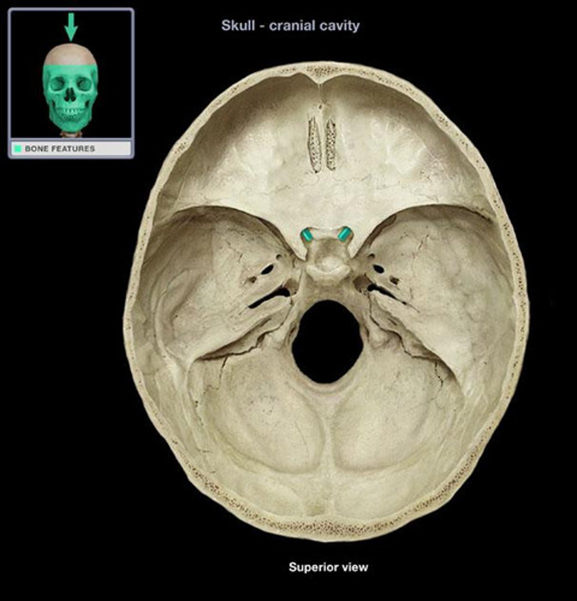
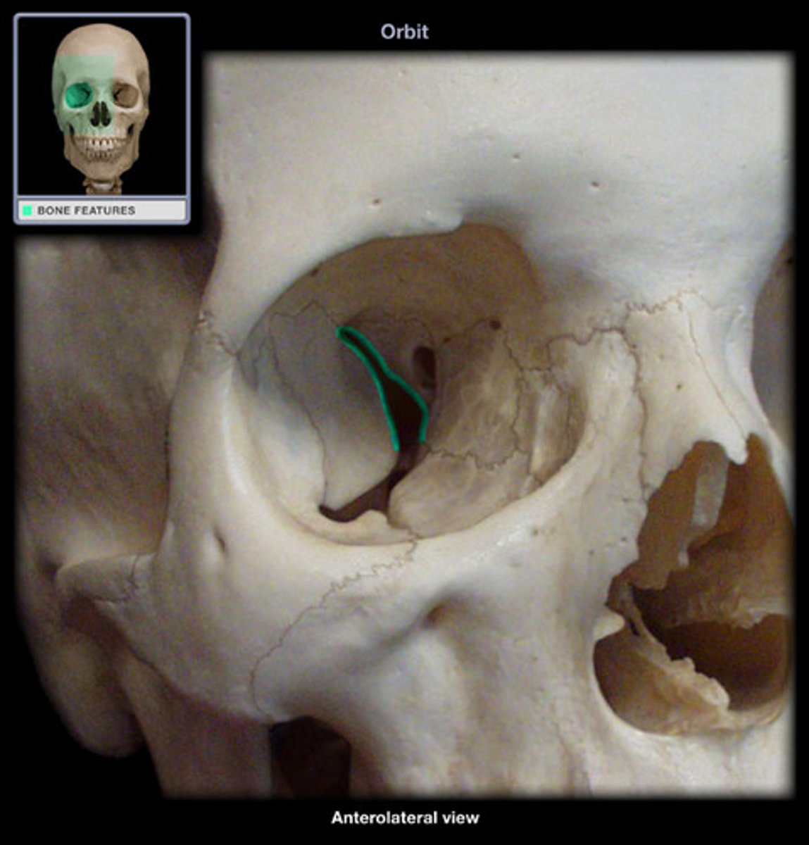
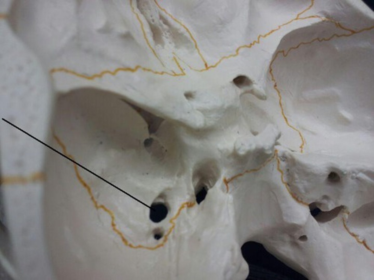
optic foramen (canal)
opening located just anterior to sella turcica to allow optic nerves to pass from eyes to brain
superior orbital fissure
opening of the sphenoid bone at the back of the eye socket
foramen rotundum
tucked under the lesser wing of the sphenoid, anterior to the foramen ovale
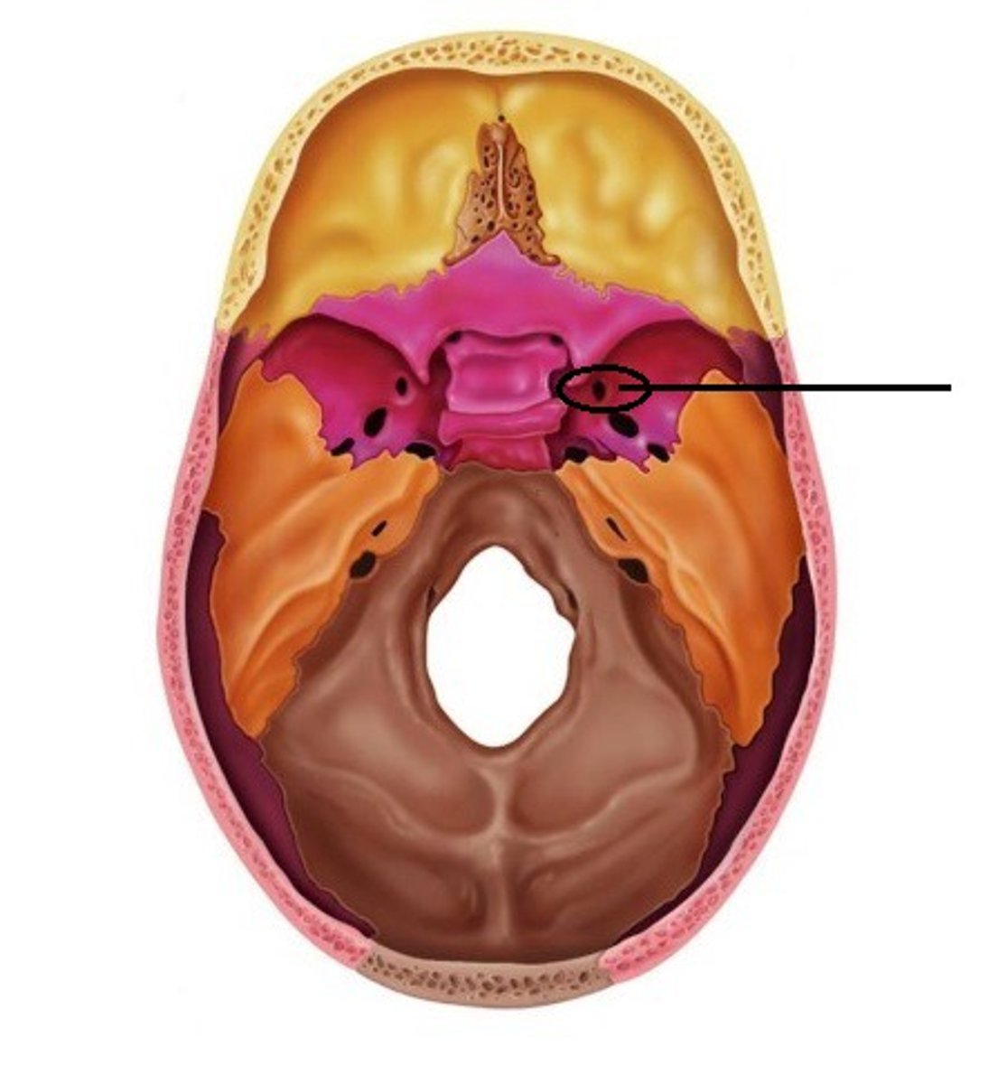
foramen ovale
posterior to foramen rotundum; it sits between the media lacerate foramen and lateral foramen spinosum.
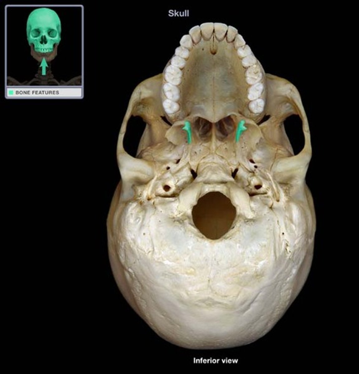
pterygoid process
Process of the sphenoid bone, consisting of two wing-shaped plates that sit at the back of the palate
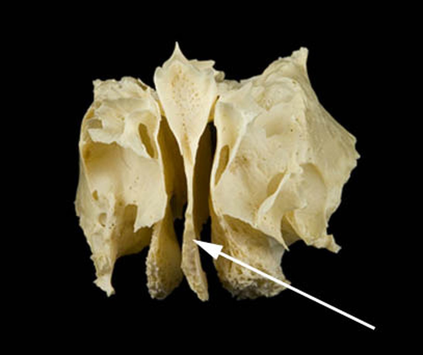
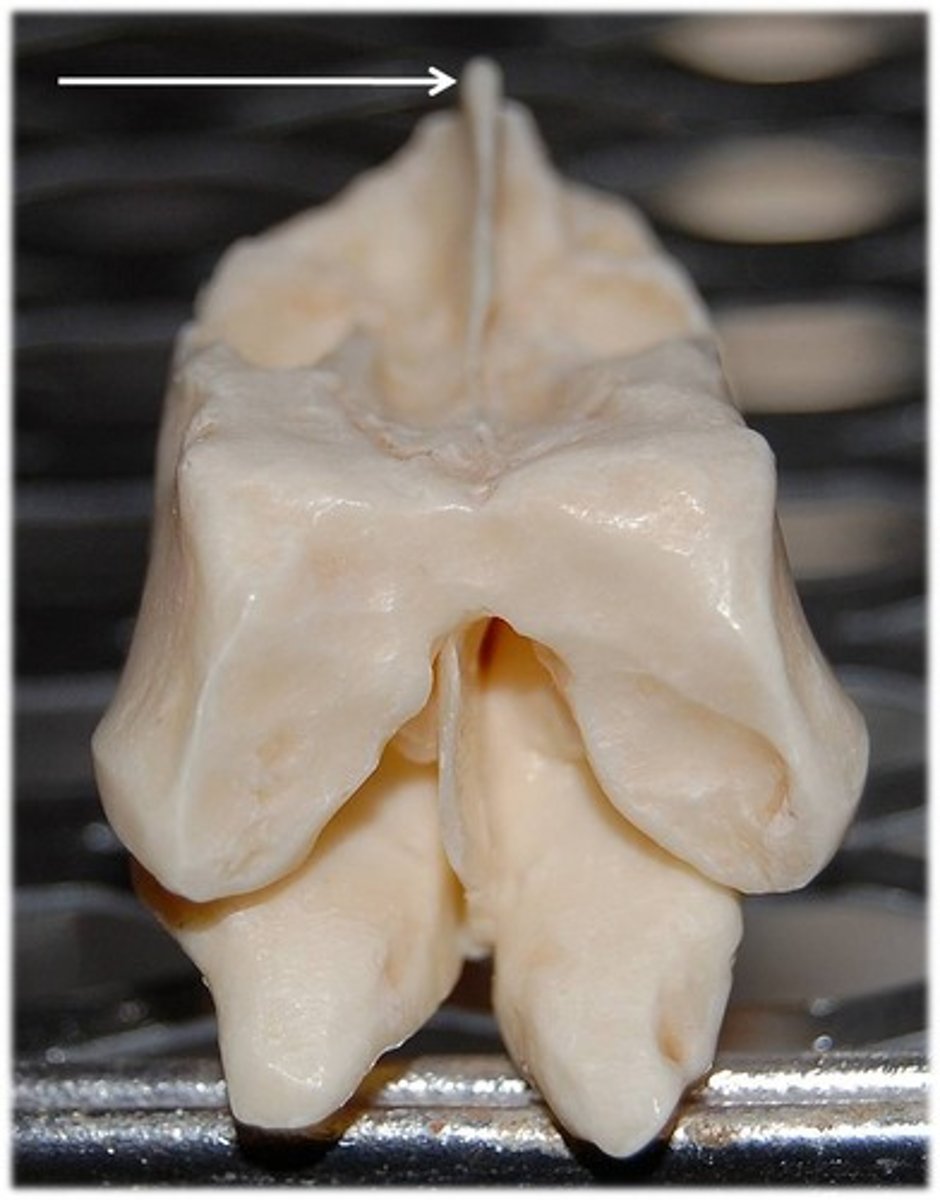
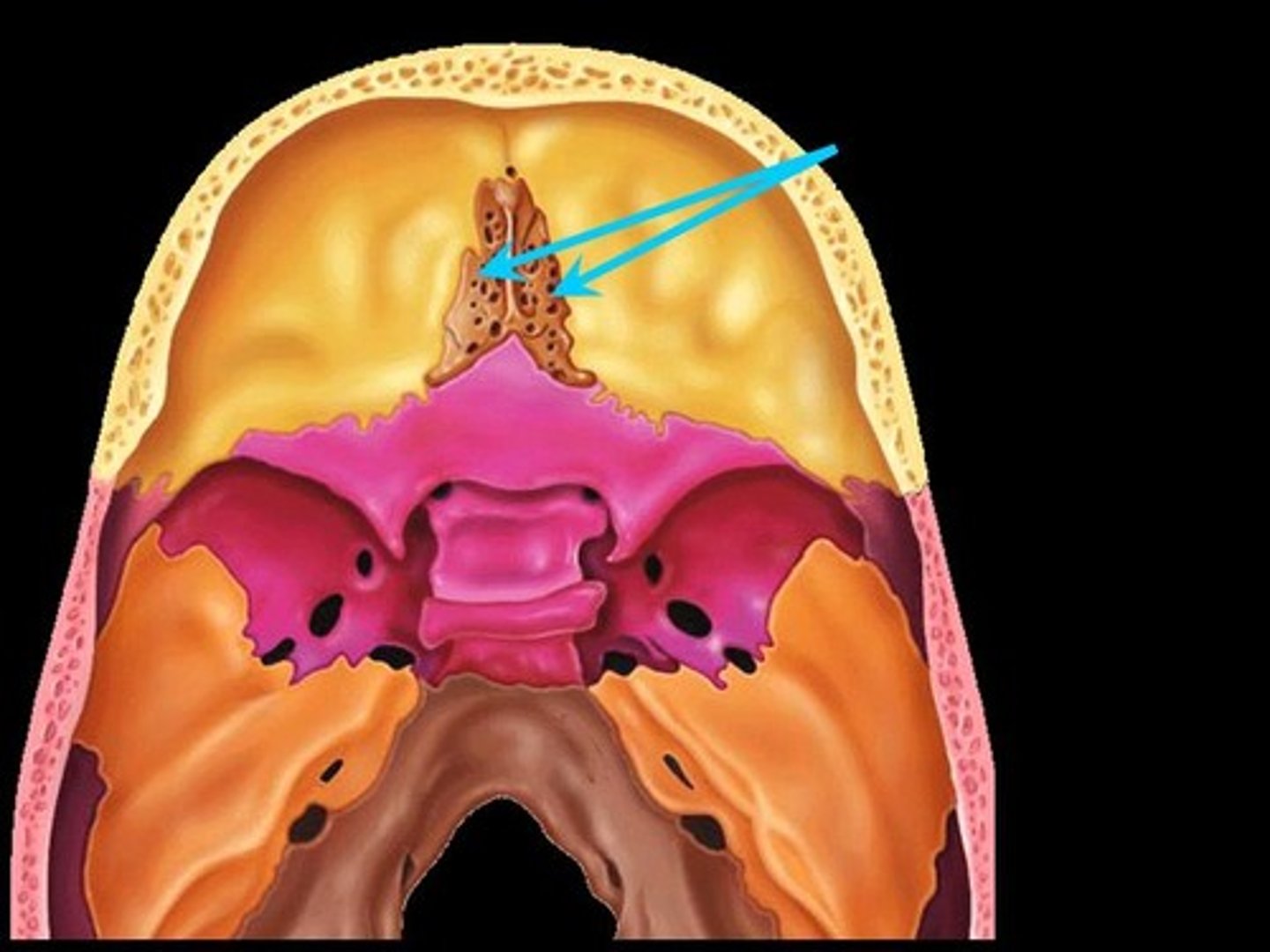
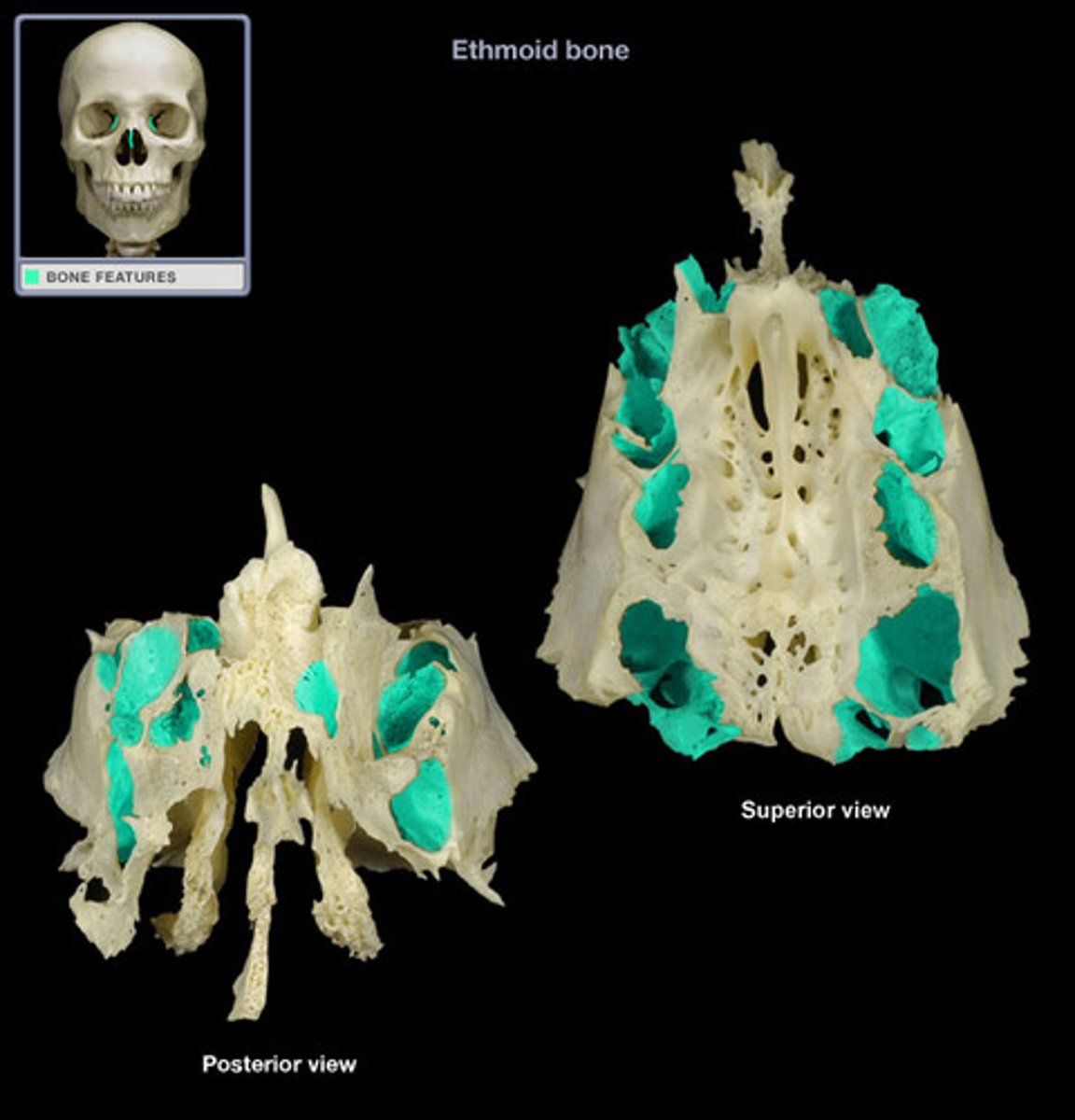
ethmoid bone
Light spongy bone between the eye sockets; forms part of the nasal cavities.
crista galli
ethmoid bone - "rooster's comb" sticks up into cranial cavity
cribiform plate
superior surface of the ethmoid; perforated by foramina that olfactory nerves travel through, which provide sense of smell
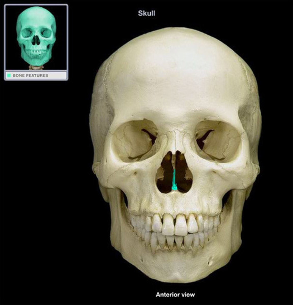
perpendicular plate
ethmoid bone, forms superior part of nasal septum
superior nasal conchae
top-most scroll-like projections of ethmoid bone--NOT VISIBLE from anterior view of skull
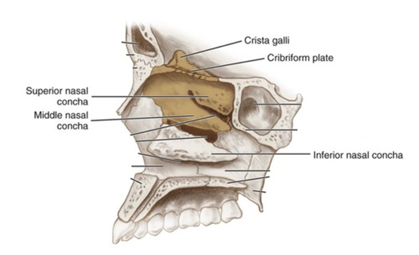
middle nasal concha
middle scroll-like projections of ethmoid bone--THESE ARE VISIBLE from anterior view of skull
inferior nasal conchae
The lowermost scroll-shaped bones on the sidewalls of the nasal cavity (these are NOT part of the Ethmoid bone)
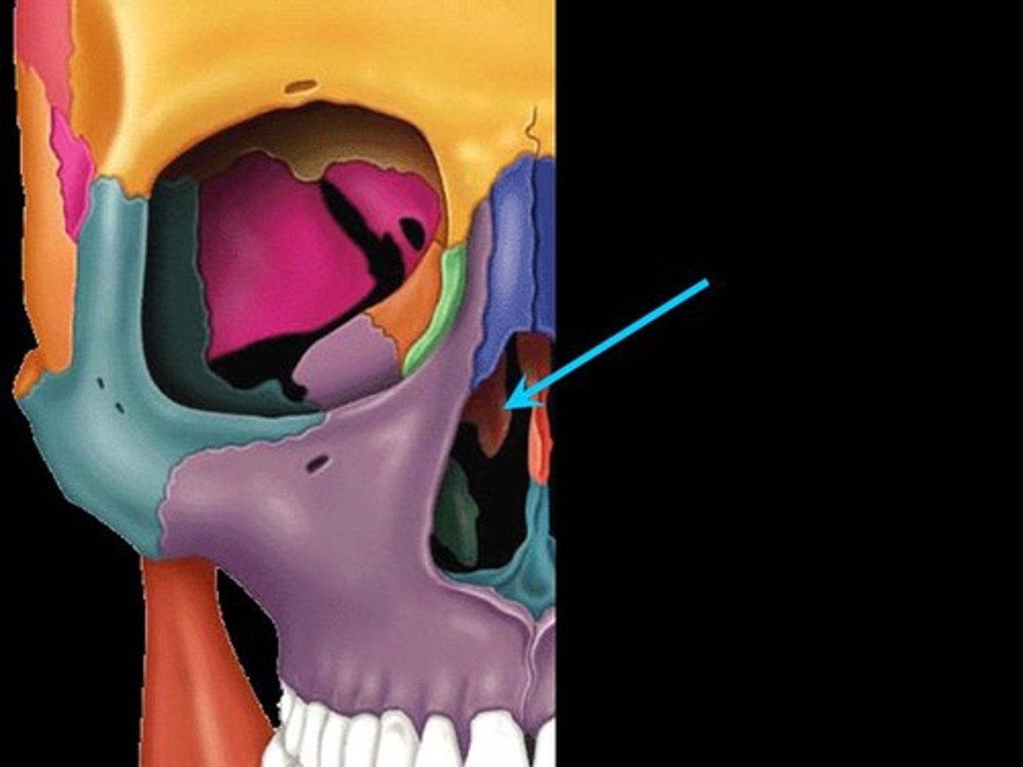
ethmoid sinus
Nasal Sinus located between the nose and the orbits.
Vomer
forms the inferior portion of the nasal septum
nasal bone
bridge of nose
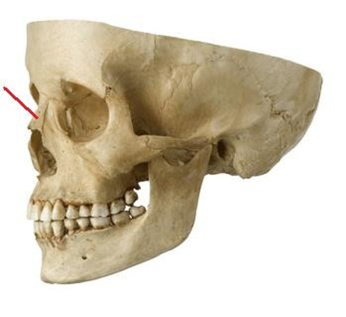
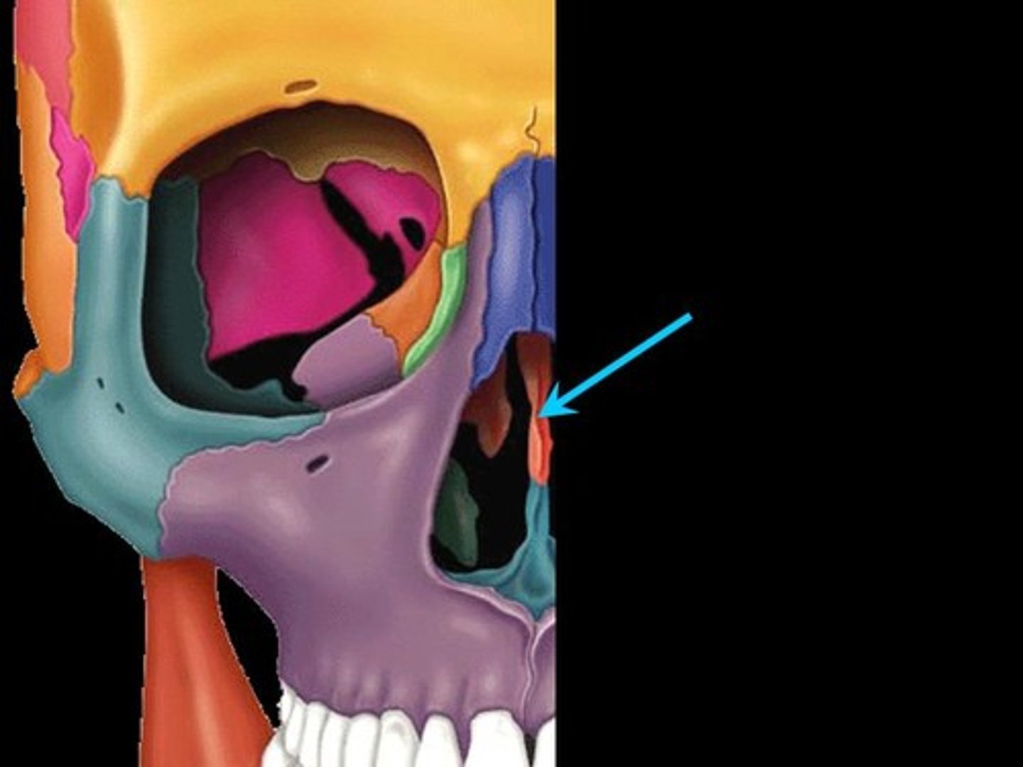
lacrimal bone
small rectangular bone making up part of the front inner walls of each eye socket
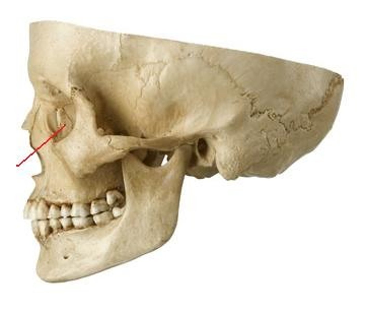
lacrimal canal
consists of a duct at the inner corner of each eye
Maxilla
upper jaw--contains all your upper teeth
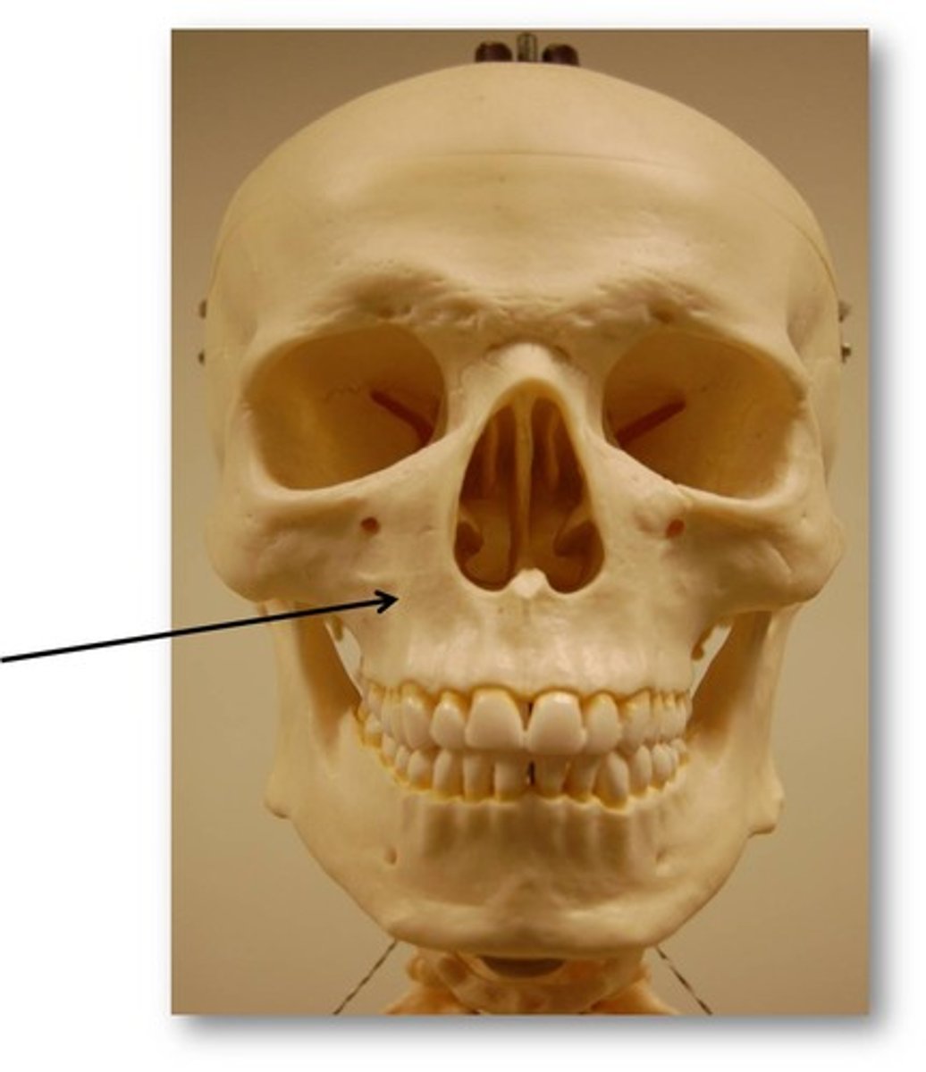
alveolar process
the grooves that show where the teeth sit within the maxilla

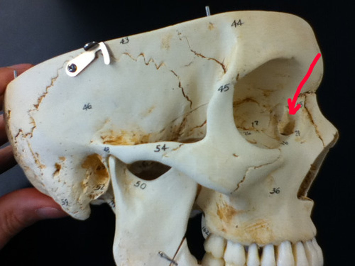
infraorbital foramen
opening under the orbit--carries nerves and blood vessels the the nasal region
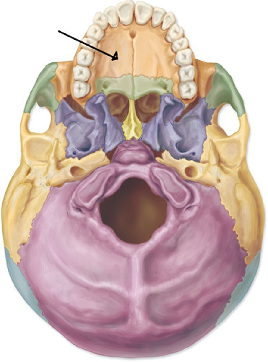
palatine process
of the MAXILLLA BONE - forms the anterior portion of the hard palate (roof) of the mouth
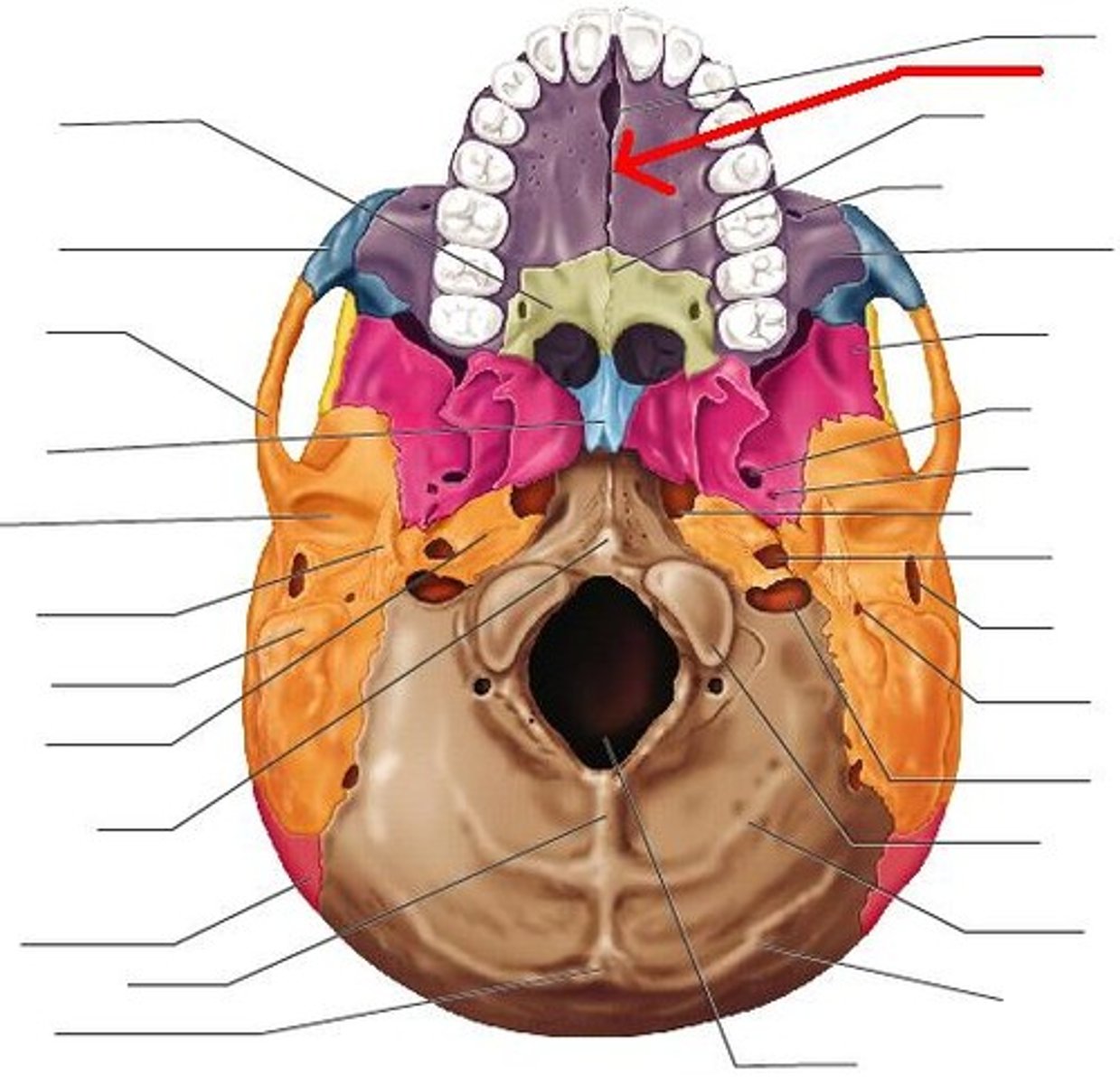
median palatine suture
median suture between bones of the palate
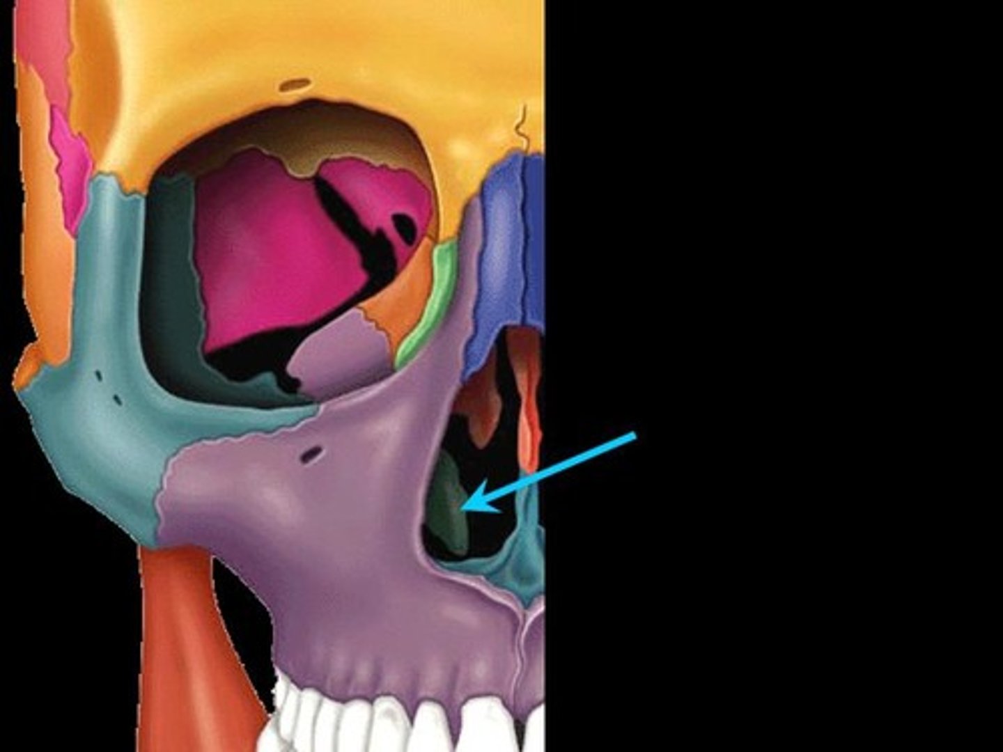
maxillary sinus
sinus on either side of the nasal cavity below the eyes
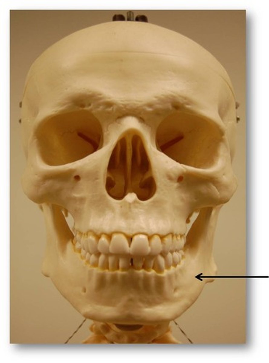
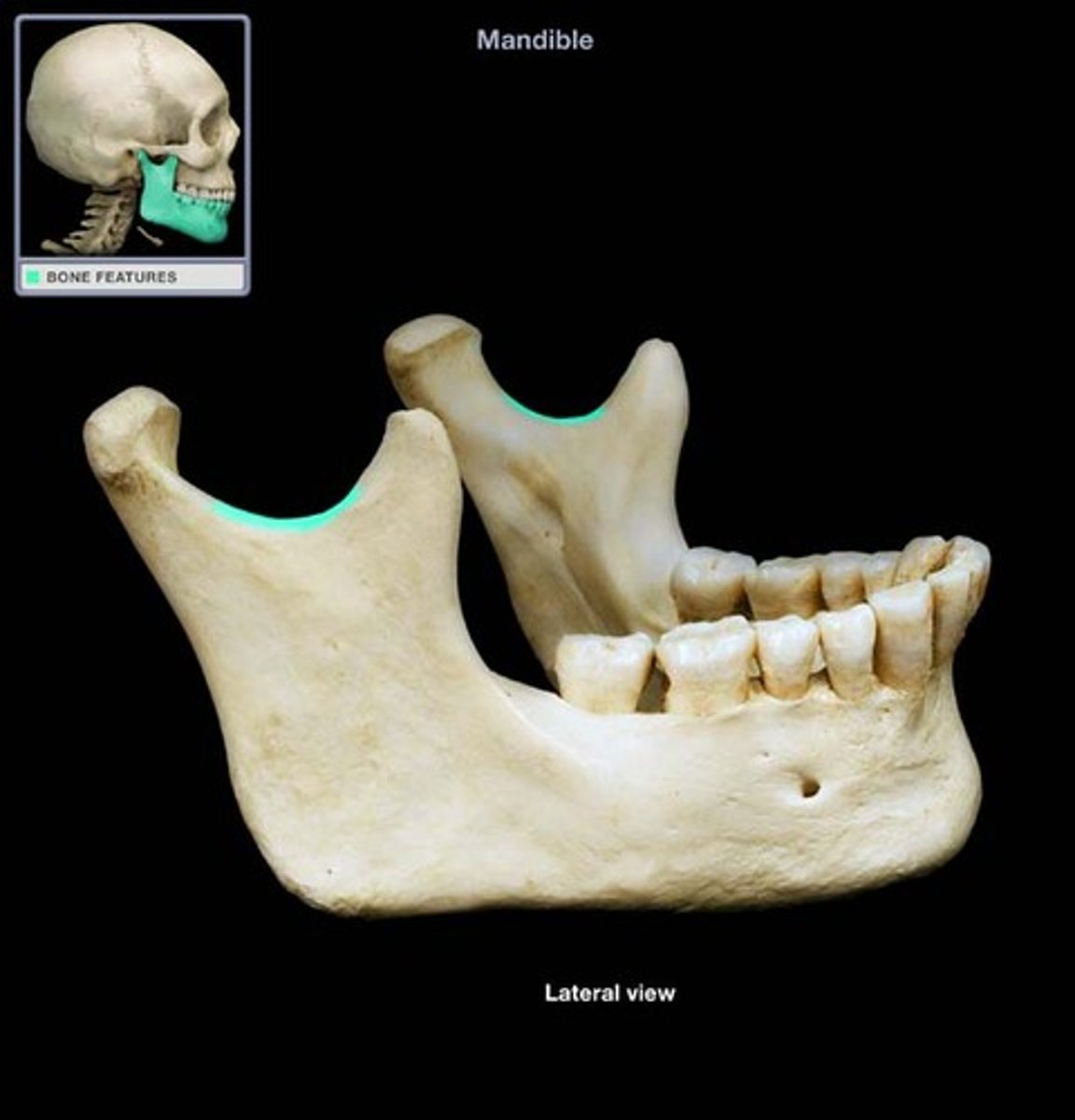
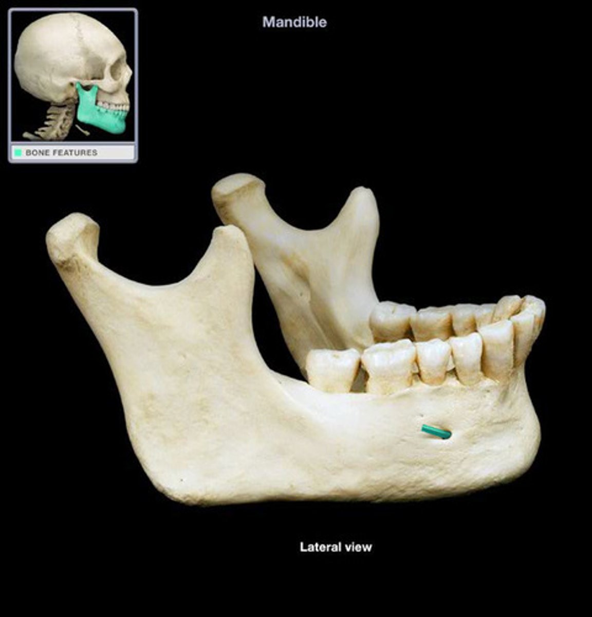
Mandible
lower jaw
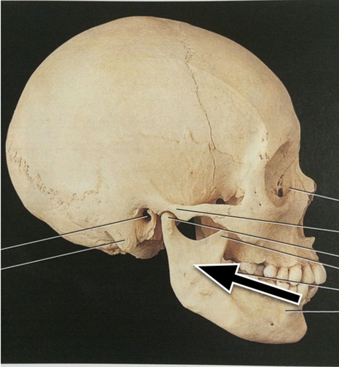
body of mandible
horizontal portion of the mandible

angle of mandible
The lower posterior of the ramus
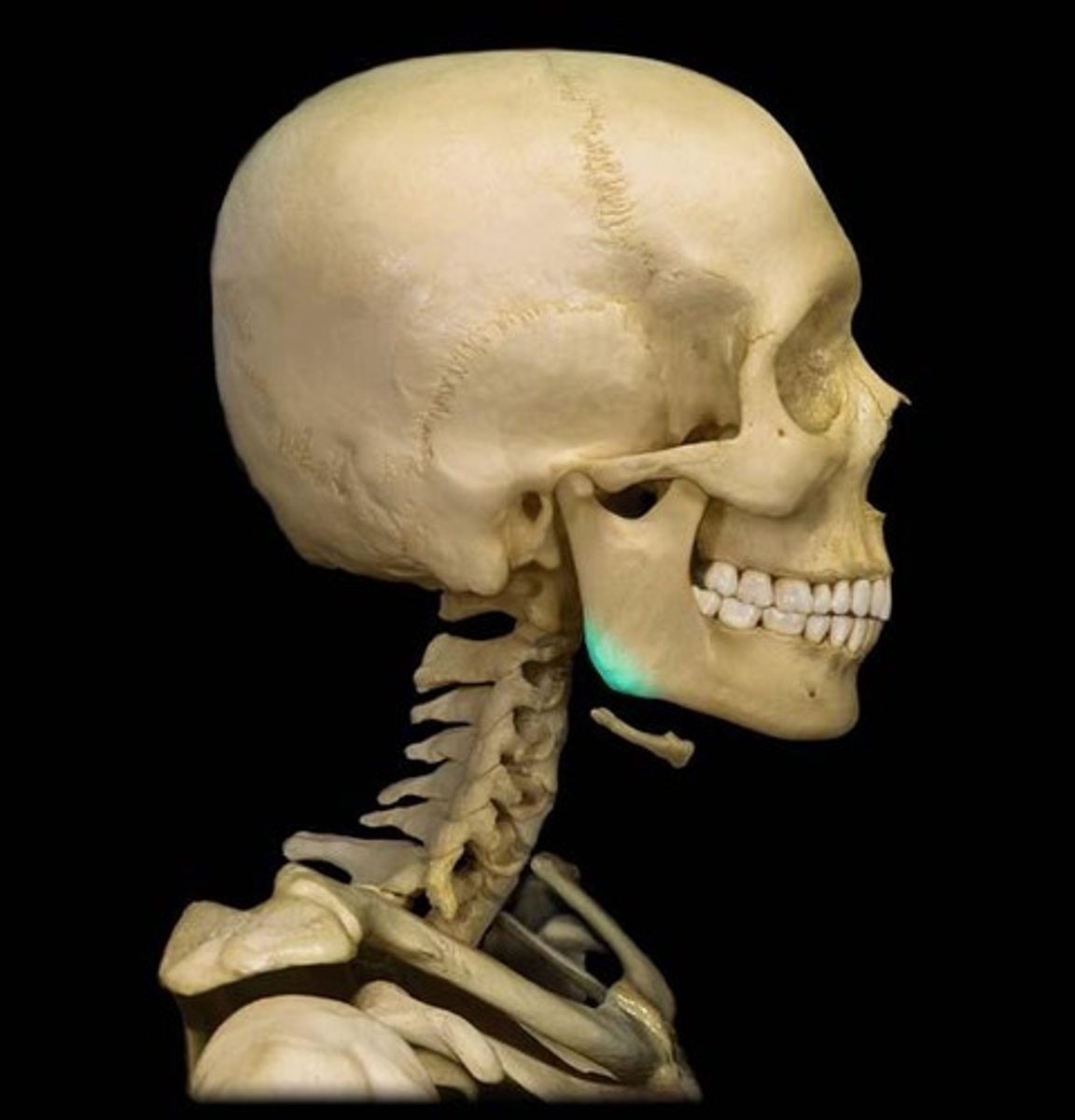
ramus of mandible
vertical part of mandible
condylar process of mandible
articulates with temporal bone
mandibular notch
coronoid process of mandible
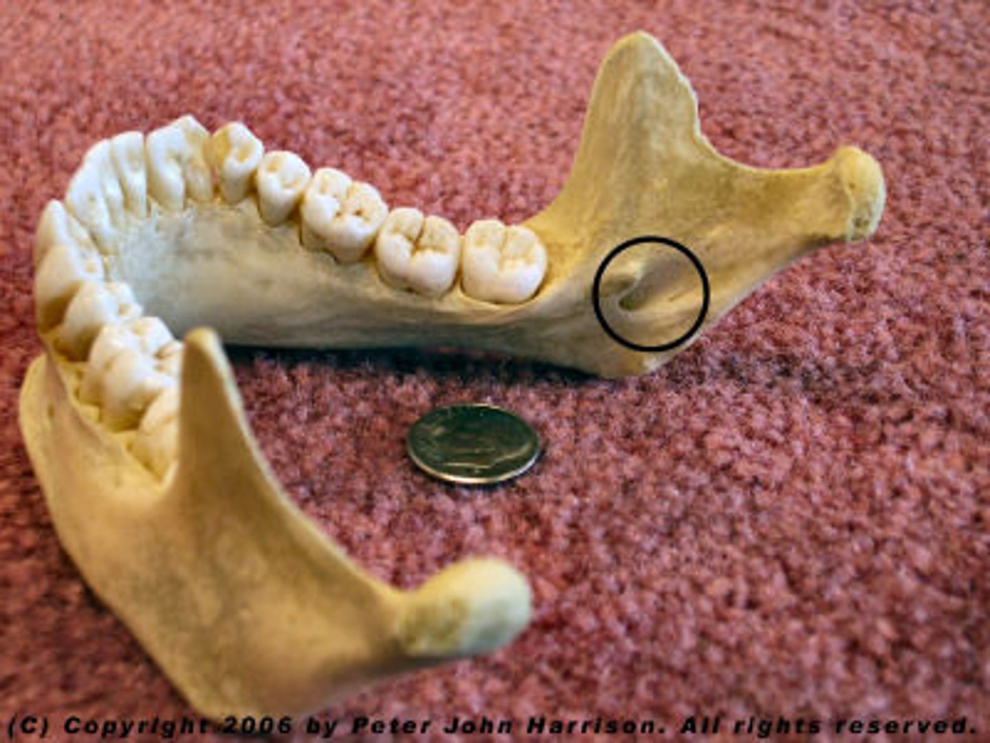
mandibular foramen
on internal surface of ramus of jaw
alveolar process of mandible
bumpy, ridge-like projections where the teeth are held
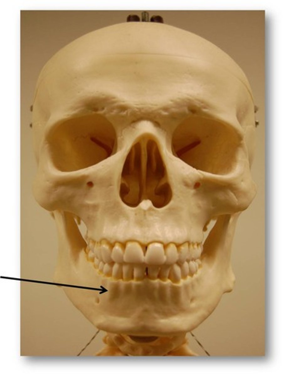
mental foramen
foramen on anterior part of jaw
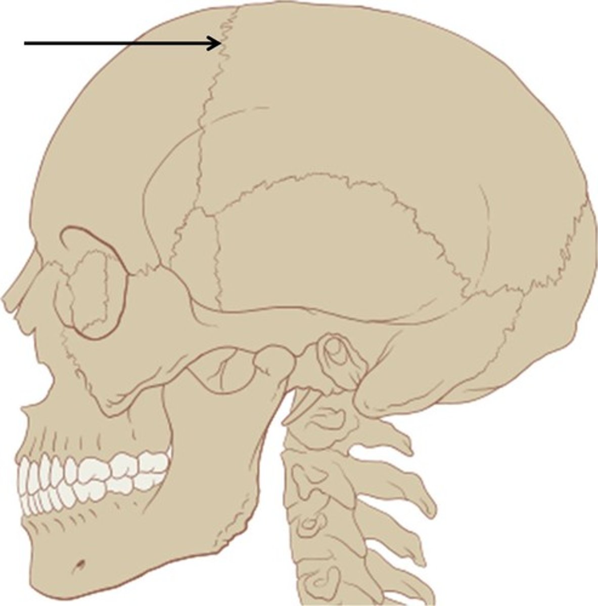
coronal suture
between frontal and parietal bones
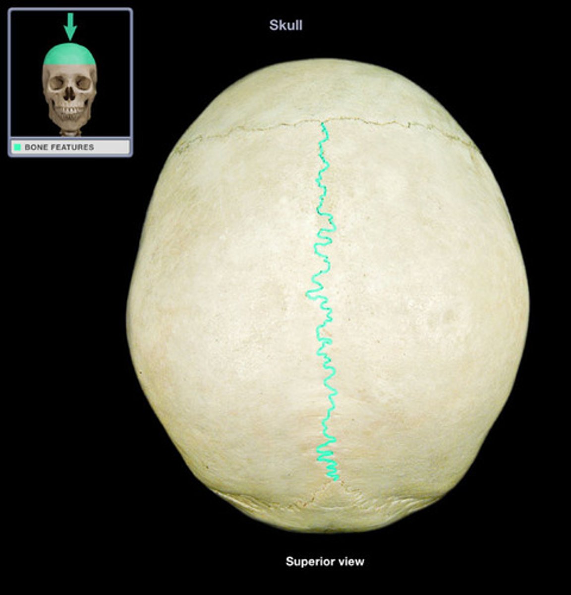
sagittal suture
between parietal bones
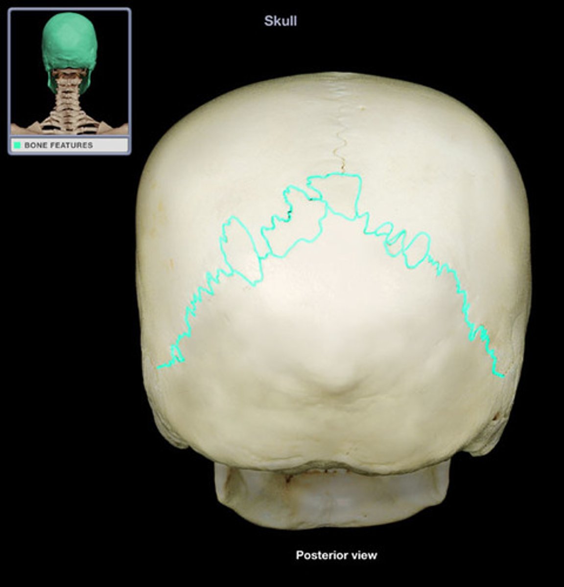
Lambdoidal Suture
suture that separates parietal bones from occipital bone
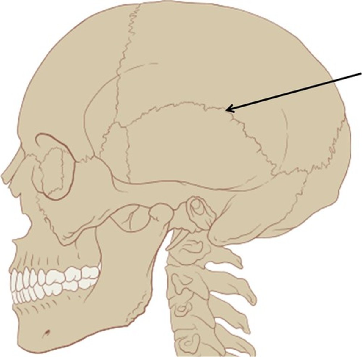
squamous suture
Between parietal and temporal bones
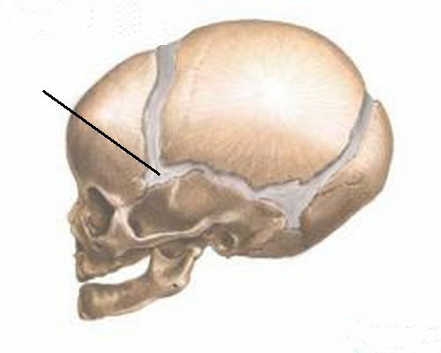
anterior fontanel
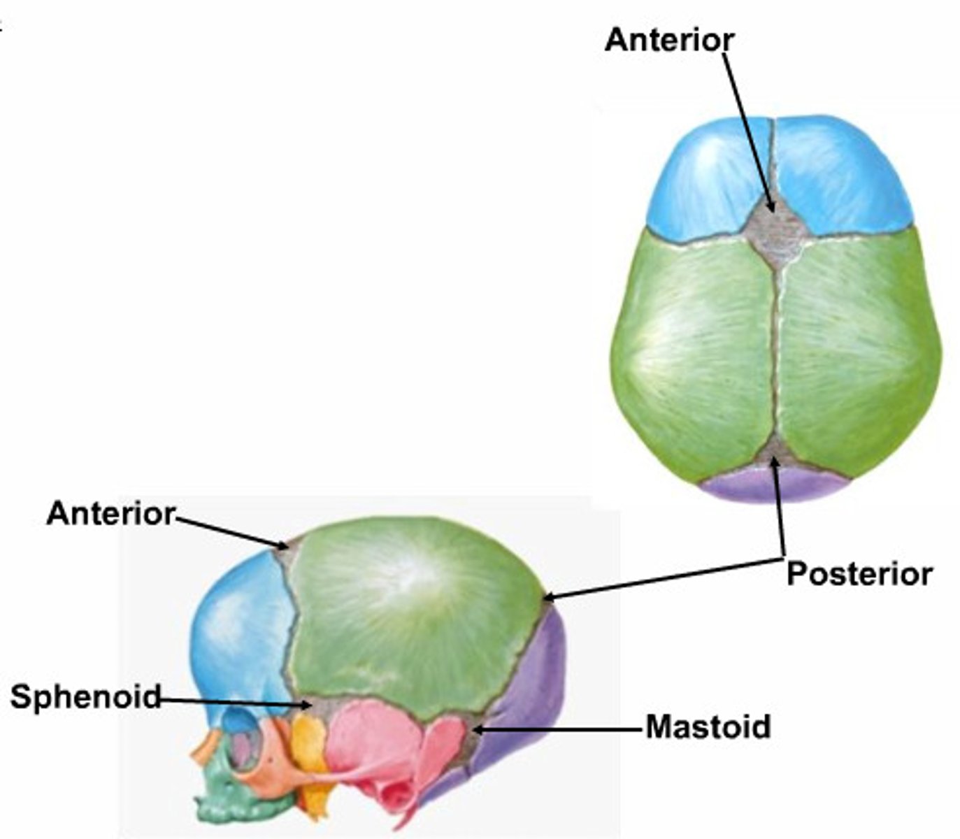
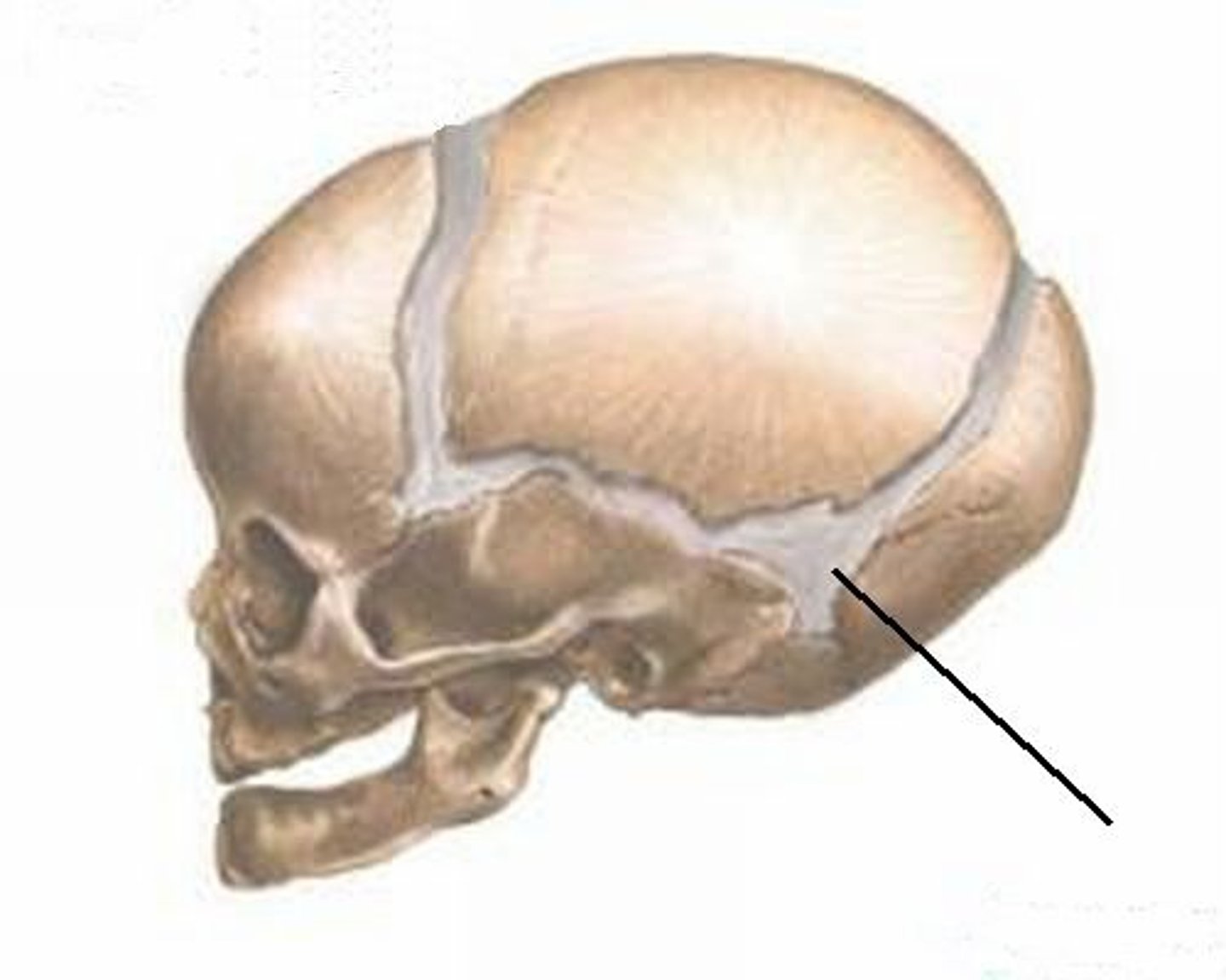
posterior fontanel
back soft spot on fetal skull between parietal and occipital bone
anterolateral (sphenoid) fontanel
at the junction between the squamous sutures and the coronal suture
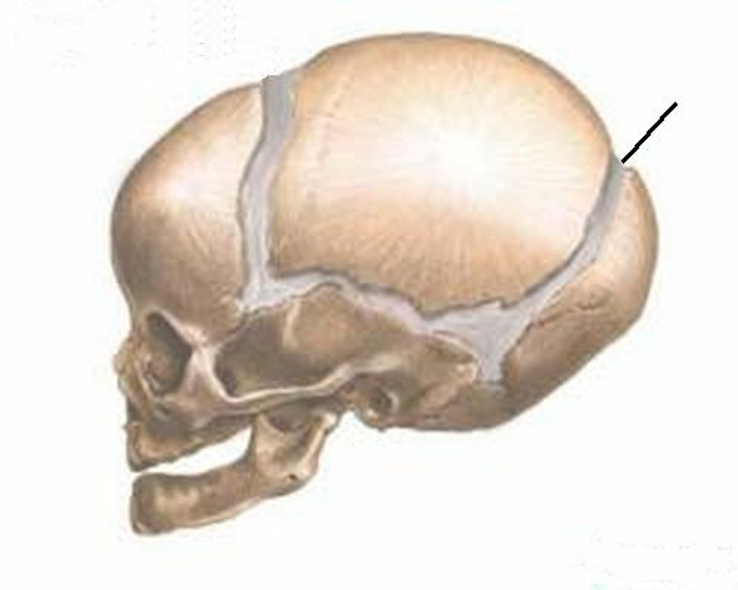
posterolateral (mastoid) fontanel
located between the parietal, occipital, and temporal bones
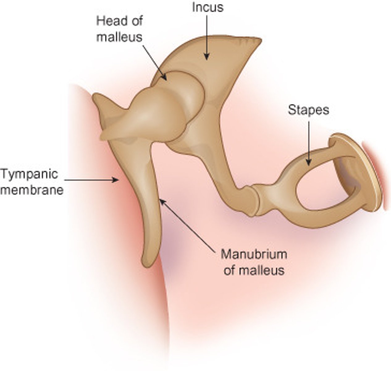
ear ossicles
malleus, incus, stapes
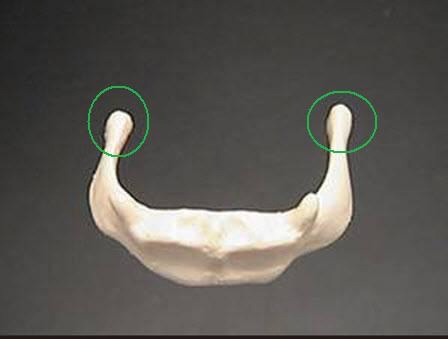
hyoid bone
a U-shaped bone in the neck that supports the tongue.
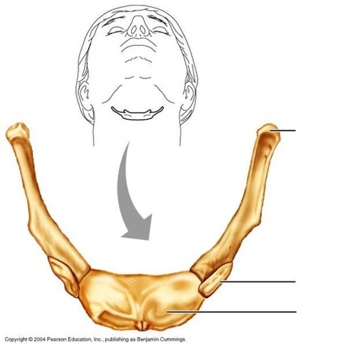
body of hyoid
horizontal surface of hyoid bone

lesser horn of hyoid bone
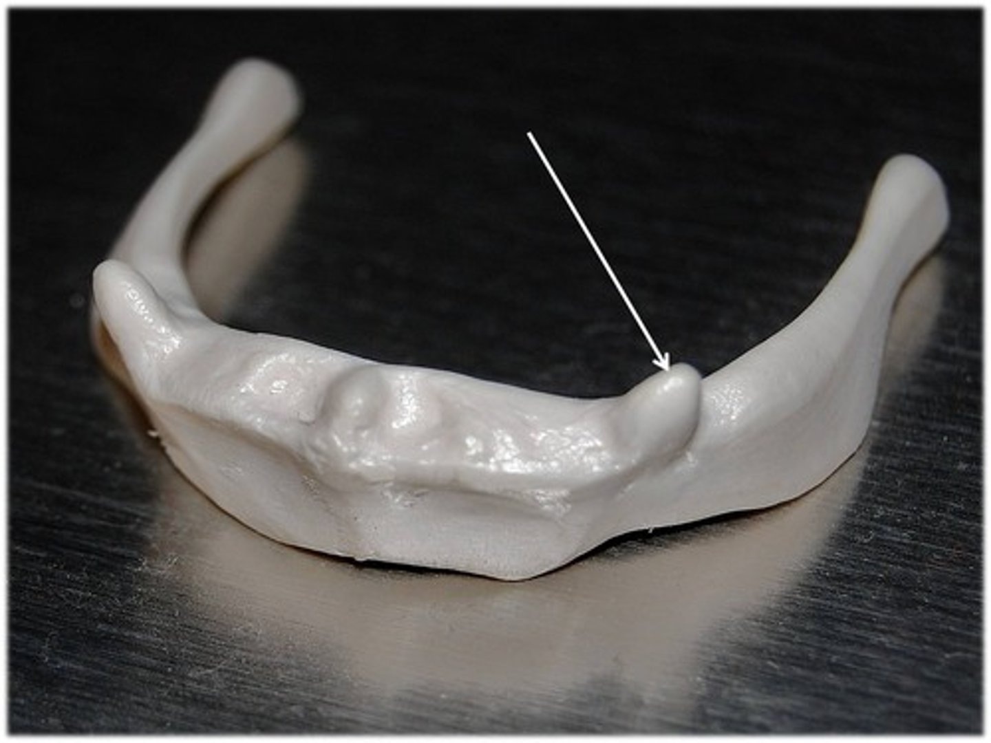
greater horn of hyoid bone
the larger, posterior projection from the body of the hyoid bone