SS - PALATE & PHARYNX
1/31
There's no tags or description
Looks like no tags are added yet.
Name | Mastery | Learn | Test | Matching | Spaced | Call with Kai |
|---|
No analytics yet
Send a link to your students to track their progress
32 Terms
PHARYNX - DEFINITION
DEFINITION
Pharynx is a muscular tube of connective tissue/mucosal → ‘musculo-tendinous tube’
It is 12cm = divided into 3 parts
Cone-shaped
Oesophagus = food pipe
Pharynx = function of carrying food AND air
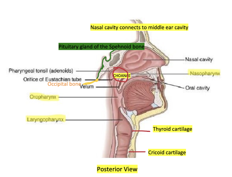
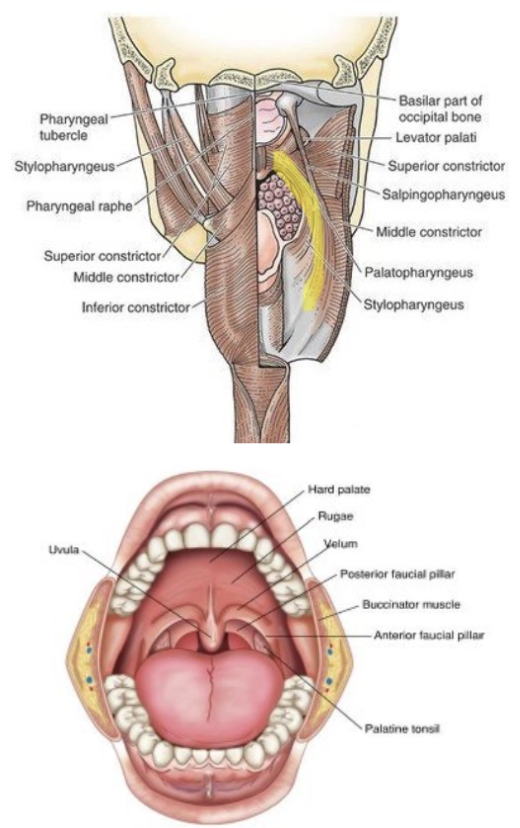
Where sphenoid bone & occipital bone meet, its union is called the basisphenoid or Basi oxyford. At this point, there is an elevation = pharyngeal tubercle
EXTENDS FROM
Nasal cavities (pharyngeal tubercle of occipital bone)
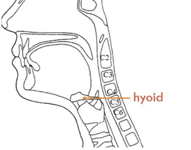
Vocal folds (cricoid cartilage at the level of C6)
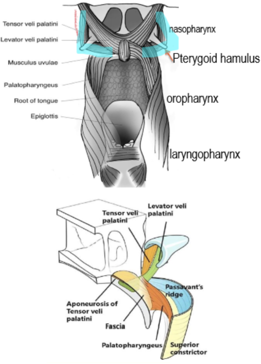
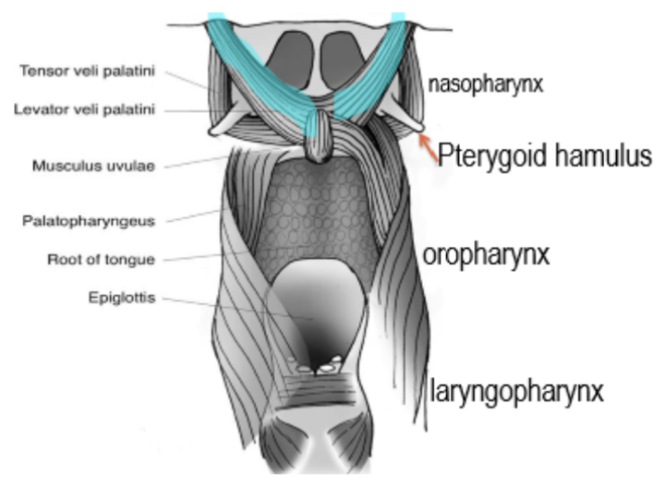
PHARYNX SPLIT INTO 3 PARTS
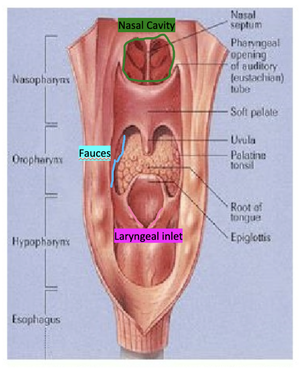
Nasopharynx: the choanae connects the nasal cavity to nasopharynx
Oropharynx: the fauces connect the oral cavity to oropharynx
Laryngopharynx: the aditus laryngitis (laryngeal inlet) connects the larynx to laryngopharynx

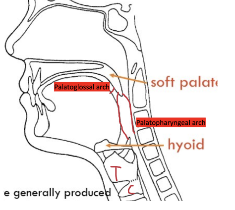
TERMINOLOGY
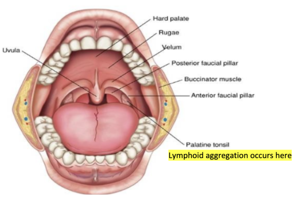
Fauces - region between the anterior faucial pillar (palatoglossal arch) & posterior faucial pillar (palatopharyngeal arch)
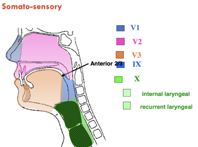
Palatoglossal arch connect posterior ⅓ of tongue to anterior ⅔ of tongue
Palatine tonsil is within the fauces
Velum = soft palate

STRUCTURE OF PHARYNX 1 - INNER MUCOUS MEMBRANE
Innermost layer will always secrete something.
It is a mucous membrane which has palatine glands and pharyngeal glands to pour out secretions inside.

STRUCTURE OF PHARYNX 2 - PHARYNGEAL APONEUROSIS
This is the middle CT layer
Dense CT sleeve
Attached to base of the skull, hyoid & thyroid cartilage
Gives attachment to many pharyngeal muscles
Strengthened posteriorly by fibrous band = median raphe
Next to mucosa (1st layer) you have submucosa (2nd layer) which is made up of connective tissue. Major component of CT = collagen. This layer has more condensation of collagen & expansile collagenous tissue.
Aponeurosis is important because it forms the structure of the attachment to muscles.
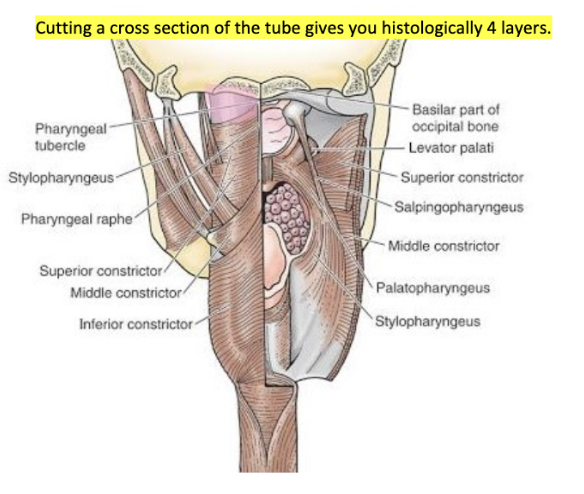
STRUCTURE OF PHARYNX 3 - MUSCLE LAYER
External circular muscles
3rd layer is what you can see. Two types…
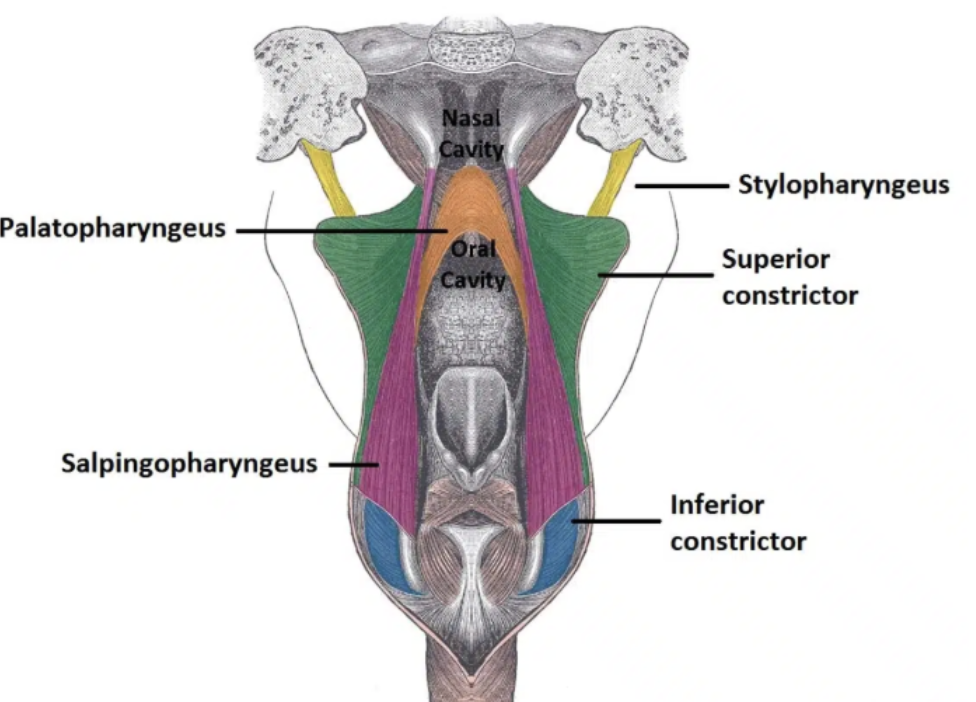
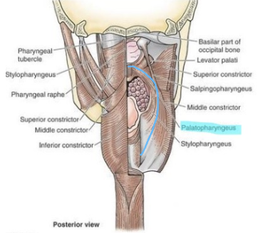
External Longitudinal muscles = the external ones that are getting attached from the bones above. They act to shorten and widen the pharynx, and elevate the larynx during swallowing
Stylopharyngeus
—> Originates from the styloid process of the temporal bone and inserts onto the pharyngeal wall.
—> Innervated by glossopharyngeal nerve (IX)
Palatopharyngeus
—> Originates from the hard palate of the oral cavity and inserts onto the pharyngeal wall.
—> Innervated by the vagus nerve (X)
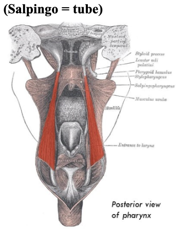
Salpingopharyngeus
—> Originates from the Eustachian tube and inserts onto the pharyngeal wall.
—> Innervated by the vagus nerve (X)
—> In addition to contributing to swallowing, it also opens the Eustachian tube to equalise the pressure in the middle ear.
2) Circular muscles = muscles that try to get attached from CT, and then get attached back to median raphe proper. Constrictors of the pharynx…
Superior
Median
Inferior
FUNCTIONS OF PHARYNX
Act as a passageway for food: from oral cavity to oropharynx.
Act as a passageway for air: from nasal or oral cavity through pharynx into larynx and trachea.
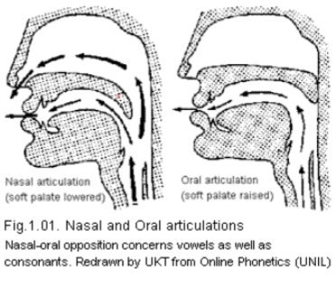
valve needed to prevent inhalation of food (soft palate)
muscles perform in such a way that food does not regurgitate back
Equalise air pressure between middle ear and atmosphere via opening of auditory tube
To mitigate against high altitude pressures = yawn or swallow or suck a popsicle to put pharyngeal muscles into contraction.
This is because there's a tube that connects the nasopharynx to the middle ear cavity.
This tube is called an auditory tube or eustachian tube, pressure in both should be equalised, other ways if the pressure in the middle ear cavity is higher, you get an uncomfortable feeling. Tympanic membrane**
Basically, nasopharynx is guarded by muscles, and those muscles try to neutralise the pressure acting in the middle ear cavity.
Function in swallowing: moves bolus toward esophagus
Function in speech
alteration of vocal tract shape → change in resonance
constriction of vocal tract → change air flow → turbulence.
Protective function
palatine tonsil = lymphoid aggregation. This is suitable for an immune defence mechanism = protective in nature.
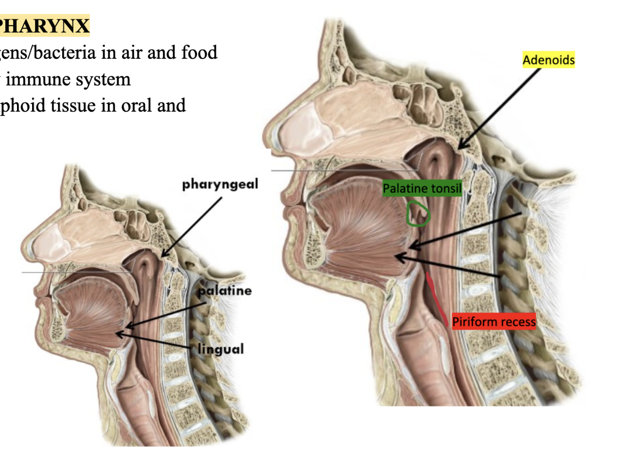
PROTECTIVE FUNCTION OF PHARYNX
Pharynx is exposed to allergens/bacteria in air and food
Tonsils part of early immune system
aggregations of lymphoid tissue in oral and pharyngeal cavities.
contains 3 sets which form a ring around the oral cavity.
Palatine
between palatoglossal and palatopharyngeal arches
largest in children, shrink after puberty
Lingual
root of tongue
Pharyngeal (adenoids)
posterior nasopharynx
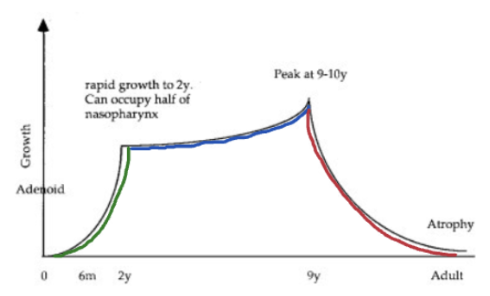
PHARYNGEAL TONSIL/ADENOID GROWTH
Size and growth may affect acoustic properties of vocal tract
Peaks at 2yrs
Then static peak
Then declines
In adults (usually above 15yrs) you don't see adenoids anymore
Hypertrophy can obstruct nasopharynx
Mask short soft palate
FUNCTIONS OF PHARYNX IN ARTICULATION
Elevate pharynx as a whole (so elevate larynx)
Change length of resonating column of air above larynx
Decreased length (via relatively elevated larynx) associated with higher pitch
increased pharynx length with lower pitch
Changing lung volume affects pharynx
increased lung volume exerts a greater downward pull on the larynx via the trachea – lengthens pharynx and lowers pitch.
Fine control of pitch occurs in the larynx via laryngeal muscle
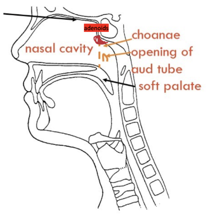
NASOPHARYNX
Nasal cavity communicates with nasopharynx through choanae
Static in size & shape as it has bone on top & bottom (limits mobility)
Resonating chamber
Respiration
Equalise air pressure
Auditory tube links nasopharynx & middle ear cavity
Lymphoid aggregation in roof of nasopharynx = adenoids. Infection that comes to nasopharynx will be trapped by adenoids. This will make them enlarged and block nasal cavity, affecting speech & breathing only through the mouth. Children will always have their mouths open.
OROPHARYNX
Oral cavity communicates with oropharynx through fauces
Dynamic
Mobile soft palate & tongue
Soft palate will be easily trying to control oropharyngeal isthmus so food doesn't regurgitate in nasal cavity, acts like a valve going up and down
Respiration & digestion
Resonating chamber
Voiced fricatives are produced with larger pharynx

LARYNGOPHARYNX
Larynx communicates with laryngopharynx through laryngeal inlet
Inferior - aditus laryngitis (entrance to larynx) at C6
Sits behind the larynx & seated just behind laryngeal inlet
Modifies laryngeal tone
Helps with digestion
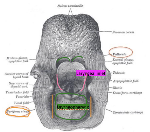
VALLECULAE & EPIGLOTTIS
Openings around the epiglottis reduce the risk of food entering the lungs
Valleculae spaces = space between tongue & epiglottis ‘formed by the attachment of the tongue base to the epiglottis via the glossoepiglottic folds’ (like ‘valley’)
Epiglottis = anterior part of laryngeal inlet which has elastic cartilage which folds back & tried to close when food enters the oral cavity, otherwise food will pass into larynx & trachea
PIRIFORM RECESS
Pyriform sinus recess = On either side of laryngeal inlet in laryngopharynx, there are 2 extensions = 'piriform recess' ‘between aryepiglottic fold and thyroid cartilage’
– directs bolus laterally and away rom larynx towards oesophagus
Piriform recess = where fish bones/chicken bones may get stuck, or some people try to smuggle drugs in this sinus 'smugglers pouch'
CT DEFINITION
Aponeurosis: thin sheet of connective tissue that provides attachment for muscles
Raphe: union of two lateral halves
PHARYNGEAL MUSCLE ATTACHMENTS:
Pharyngeal aponeurosis
Between mucous and muscular layers of pharynx
Attached to occipital bone
Continuous laterally with palatine aponeurosis
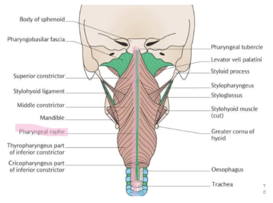
Pharyngeal raphe
Extends from the pharyngeal tubercle on the occiput to the posterior oesophagus
Narrow band of connective tissues at posterior joining of the pharyngeal constrictor muscles
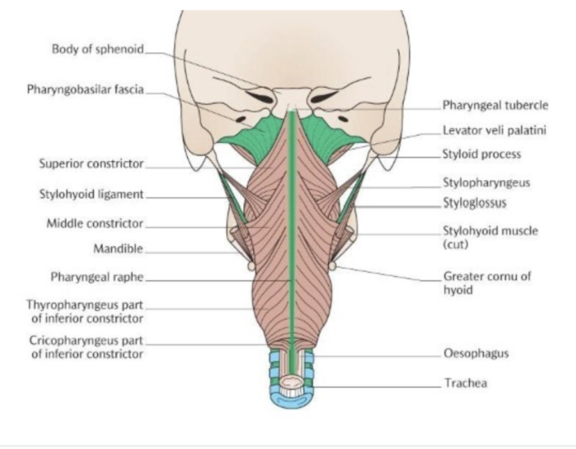
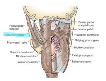
MUSCLES OF PHARYNX - EXTERNAL LAYER:
External layer consisting of 3 constrictors
Circular muscles with fixed points
Superior constrictor
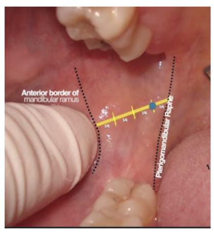
pharyngeal tubercle, pharyngobasilar fascia & pterygomandibular raphe to pharyngeal raphe
Middle constrictor
Hyoid to pharyngeal raphe
Inferior constrictor
i. Thyropharyngeus – thyroid cartilage to pharyngeal raphe
ii.Cricopharyngeus – cricoid cartilage to opening of oesophagus
Posterior attachment to pharyngeal raphe

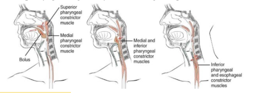
PHARYNX & SWALLOWING
Sequential constriction of pharynx helps to push the bolus down into the oesophagus
This is considered an involuntary phase in swallowing (second stage)
Cricopharyngeus is generally contracted acting as a sphincter
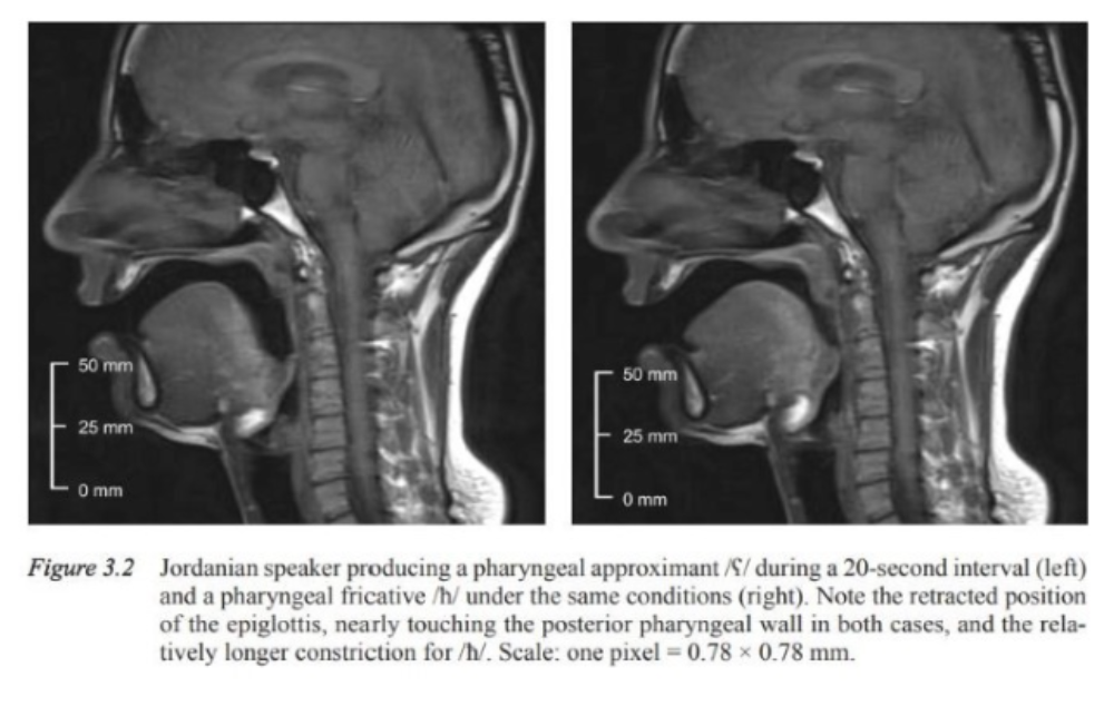
PHARYNX & ARTICULATION
Pharyngealization is a secondary articulation ie. simultaneous articulation at two different places by which the pharynx or epiglottis is constricted during the articulation of the sound.
Does not appear in English - appears in arabic language
LONGITUDINAL MUSCLE - STYLOPHARYNGEUS
ACTION | ATTACHMENT |
|
|
LONGITUDINAL MUSCLE - PALATOPHARYNGEUS
ACTION | ATTACHMENT |
|
|
LONGITUDINAL MUSCLE - SALPINGOPHARYNGEUS
ACTION | ATTACHMENT |
|
|
PALATE
Consists of hard palate and soft palate (velum)
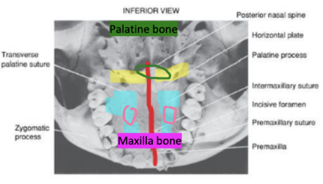
Hard palate:
Separates nasal and oral cavities.
Formed by the palatine process of maxilla and palatine bones.
Two bones meet at the intermaxillary suture (or median palatine suture)
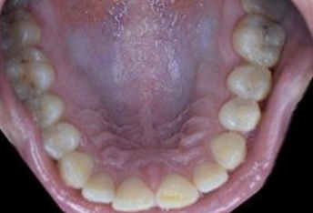
FEATURES OF HARD PALATE
Palatal vault (arch)
height of vault affects acoustics
variable
Rugae
ridges of mucous membrane
facilitate articulation through an increased surface area
SOFT PALATE (VELUM) - ROLES
Creates a flexible opening between the nasal cavity and the oral cavity = made of mucous membrane so mobile
|
 |
SOFT PALATE DEPRESSORS
PALATOGLOSSUS - Anterior Faucial Pillar
 | PALATOPHARYNGEUS
 |
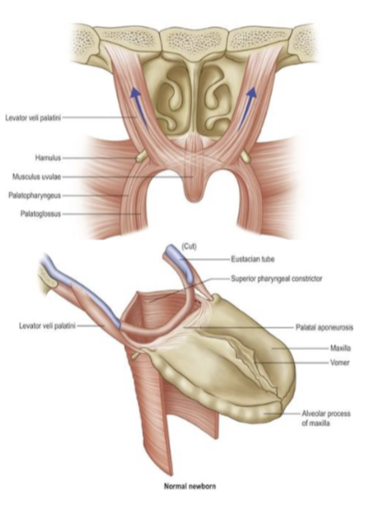
SOFT PALATE ELEVATORS
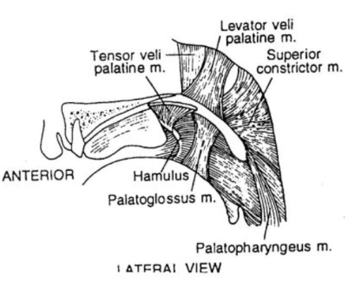
TENSOR VELI PALITINI
 | LEVATOR VELI PALITINI
 |
FURTHER MUSCLES INVOLVED IN SOFT PALATE CLOSURE
Musculus uvulae
Bunches and shortens with contraction
Superior constrictor
Pulls the posterior pharyngeal wall forwards to meet the soft palate
VELOPHARYNGEAL CLOSURE
The role of the velopharyngeal closure is to vary the degree of acoustic coupling between the oral and nasal cavities.
Velopharyngeal closure is necessary for the production of "oral stops" sounds - raising soft palate to posterior pharyngeal wall prevents air from exiting through the nasal cavity.
Impound air pressure within the oral cavity for the production of plosive consonants.
Complete closure of velum against pharynx walls required for wind instrumentalists to avoid air leak through nose.
3 speech sounds require depressed soft palate – m/n/ng.
Inadequate velopharyngeal closure → nasalised vowels and weak plosives
Continuous sphincter type arrangement at the superior pharynx.
Soft palate closes the ‘roof’ above this opening
VELOPHARYNGEAL CLOSURE IS ACHIEVED BY….
Palatal movement
Elevate (LVP) and tense (TVP) soft palate
Anterior movement of posterior pharyngeal wall
Superior constrictor around C1
Inward or medial movement of the lateral walls of the nasopharynx
Superior constrictor, buccinator and orbicularis oris
NOTE =
Small space at beginning of pharynx & end of palate that needs to be closed when there is swallowing procedure happening. Superior constrictor is coordinated by pterygomandibular raphe.
All these muscles act in a coordinated way so that the gap present most superiorly is closed = velopharyngeal closure help superior constrictor come anteriorly so that space is closed.
NERVE SUPPLY OF PALATE - MOTOR
All the muscles of the palate are supplied by Cranial nerve X (vagus)
Except for tensor veli palatini = supplied by mandibular branch of trigeminal(Vmand)
NERVE SUPPLY OF PHARYNX - MOTOR
All the muscles of the pharynx are supplied by Cranial nerve X (vagus)
Except for stylopharyngeus which is supplied by Cranial nerve IX (glossopharyngeal)

LARYNGEAL ELEVATORS
Supra hyoid muscles
geniohyoid, digastric, thyrohyoid (infra), mylohyoid, and stylohyoid muscles
LARYNGEAL DEPRESSORS
Infrahyoid (strap) muscles)
sternohyoid, sternothyroid, omohyoid