Components of the Optometric Examination
 Call Kai
 Learn
Learn Practice Test
 Spaced Repetition
 Match
Match Flashcards
 Knowt Play
 Podcast
1/159

There's no tags or description
Looks like no tags are added yet.
Name | Mastery | Learn | Test | Matching | Spaced | Call with Kai | Chat |
|---|
No analytics yet
Send a link to your students to track their progress
160 Terms
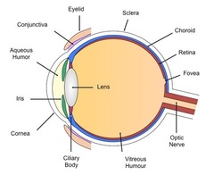
eye's first refractive surface
cornea

aperture in the eye that varies in size to control the amount of light entering the eye (protects inner layer from damage)
pupil

colored muscle of the eye that can contract, controlling the aperture (pupil) in order to vary the amount of light entering
iris

eye's second refractive surface
lens

What are the two refractive surfaces of the eye?
cornea, lens
Which contributes the MOST refractive power to the eye: cornea or lens?
cornea
inside lining of the eye onto which light is focused
retina

area of the retina that generally contains the largest number of cones, where light is most focused
macula
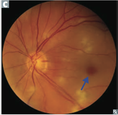
most sensitive area of the retina, found in the macula, where light focuses best; contains largest number cones
**must be intact for good vision
fovea

Which photoreceptors are more sensitive to bright light: rods or cones?
cones
Are rods found in the more central or peripheral portion of the retina?
peripheral
Are cones found in the more central or peripheral portion of the retina?
central
nerve that gathers all the information from the retina and transmits it to the brain
optic nerve
How can you tell from a fundus image which eye you're looking at?
optic nerve is closer to the nose

smooth muscle of the eye that can contract to make the iris aperture (pupil) smaller in bright conditions
iris sphincter (muscle)
smooth muscle of the eye that can contract to make the iris aperture (pupil) larger in dim conditions
iris dilator (muscle)
tough, white, fibrous coat of the eye that serves as a protective layer around the eyeball
sclera
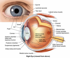
clear, cellophane-like mucous membrane lining the eyelids and covering the anterior portion of the sclera
conjunctiva

What happens in pink eye in regards to the conjunctiva?
conjunctival vessels are inflamed
demarcation between the sclera (with its conjunctival covering) and the cornea; border between sclera and cornea
limbus
What proportion of the eye's total refractive power is attributed to the cornea?
2/3
What proportion of the eye's total refractive power is attributed to the lens?
1/3
Even though the lens contributes less refractive power than the cornea, what makes it special?
it can vary its shape via accommodation
refers to the tightening of the ciliary muscle, allowing the lens to become more rounded for near vision
accommodation
If the eye is unaccommodated, is it prepared for distance or near vision?
distance
In accommodation, do the ciliary muscles relax or contract?
contract

In accommodation, do the zone fibers relax or tighten?
relax

In accommodation, does the lens get thicker or thinner?
thicker

When the eye is NOT accommodated, do the ciliary muscles relax or contract?
relax
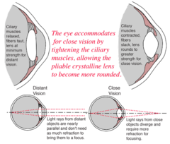
When the eye is NOT accommodated, do the zonule fibers relax or tighten?
tighten

When the eye is NOT accommodated, does the lens get thicker or thinner?
thinner

What distinguishes between the front of the eye and the back of the eye? (i.e. serves as the landmark that separates the eye into anterior and posterior)
iris
refers to the lubricant of the eye surfaces
tear film
space between the cornea and the iris that contains aqueous
anterior chamber
fluid between the cornea and iris in the anterior chamber
**continuously made and drained
aqueous (humor)
gel-like fluid that fills the space between the lens and retina in the posterior segment of the eye
**ages, isn't continuously made and drained
vitreous (humor)
refers to the retinal vasculature - arterioles & venules within the layers of the retina that are visible inside the eye
choroid
portion of the back of the eye that you see with special instruments without dilating the pupil; you see the optic nerve, blood vessels, and the macula
posterior pole
What three things can you see when viewing the posterior pole through an undilated pupil?
optic nerve, blood vessels, macula
portion of the lining of the eye that you can see when you dilate the pupil; has less vasculature
peripheral retina
What does CEE mean?
comprehensive eye examination
CEEs evaluate what 3 important aspects of the eye?
optical, functional, ocular health
What does it mean to evaluate the optical aspects of the eye in a CEE?
determine any refractive errors
normal refractive condition of the eye in which light focuses on the retina normally; 20/20 vision OU
emmetropia
refractive error in which light entering the eye focuses behind the retina and the eye's focusing power is too weak
hyperopia
refractive error in which light entering the eye focuses in front of the retina and the eye's focusing power is too strong
myopia
Why do we correct hyperopia with + power lenses?
increase the too weak focusing power
Why do we correct myopia with - power lenses?
decrease the too strong focusing power
What does it mean to evaluate the functional aspects of the eye in a CEE?
check for appropriate eye tracking, eye teaming, and eye focusing
general term that refers to eye strain; occurs when the eyes don't point correctly at a target or the focusing system doesn't work correctly; causes blurred vision after prolonged work, double vision, eyestrain, and headaches
asthenopia
What does it mean to evaluate ocular health in a CEE?
check for anatomical defects or medical conditions
What are two examples of systemic medical conditions that affect the eye that may be detected in a CEE?
hypertension, diabetes
What are the 5 components of a comprehensive eye examination?
case history, entrance testing, refraction, near testing, ocular health
Why is taking a patient's case history an important first step of a CEE?
so you can learn of current medical problems that may affect the eye or past eye and vision problems
After completing the case history, the doctor may choose to follow the standard testing as part of a CEE, called what?
database
After completing the case history, the doctor may choose to evaluate the patient's visual system by focusing on areas that may need more than a routine evaluation, called what?
problem focused testing
The doctor thinks of a patient's problems in terms of ____ ____; the doctor develops a list of possible causes for the patient's concerns/symptoms. The CEE that follows then attempts to identify the cause of the patient's symptoms.
differential diagnoses
Why do optometrists perform entrance (screening) tests?
quickly provide general information about a patient's visual system to assess whether each component functions properly
What are 8 different entrance tests in a CEE?
visual acuity, pupil testing, cover test, version system, confrontation visual field, color vision, stereopsis, near point of convergence
entrance test that evaluates the resolving ability of the eye
visual acuity
What does sc mean?
without correction
What does cc mean?
with correction
What do you do immediately after the case history but prior to any other testing?
visual acuity
How do you evaluate visual acuity for a new patient?
**For insurance purposes, if a patient has not been to your office in more than three years, you can bill the encounter as a new patient
unaided and aided at distance and near
How do you evaluate visual acuity for an established patient?
aided distance and near
How do you evaluate visual acuity for a patient returning for follow-up care (f/u)?
**less than 1 year since a CEE
aided distance vision only
entrance test that evaluates the ability of the eyes to respond to a light stimulus (part of the eye health screening)
pupil testing
What three things does a doctor assess in pupil testing?
pupil size, shape, and ability to react to light
test that compares the pupil response to light in one eye to the other eye
Marcus Gunn (test)
When do you perform pupil testing? CEE or f/u? New or existing patient?
every visit
(all of those listed)
entrance test that evaluates the ability of the two eyes to point at a target simultaneously; tests eye alignment (part of functional vision screening)
cover test
Is the cover test done with correction?
yes
If a patient is presenting for a CEE, how do you perform the cover test?
distance and near
If a patient is presenting for f/u care, how do you perform the cover test?
distance only
entrance test that evaluates the ability of the eye muscles to move the eyes in different positions of faze and also to track a moving target (eye health screening)
version system
When do you perform version system testing? CEE or f/u? New or existing patient?
every time
(all of those listed)
entrance test that evaluates peripheral (side) vision (central visual field)
confrontation visual field
Is confrontation visual field performed with correction?
no
When do you perform confrontation visual field?
every visit unless patient is returning for automated visual field test
Is color vision testing performed with correction?
yes
(best near correction)
What is the doctor usually screening for with color vision testing?
congenital, genetic color vision deficiencies
Do inherited color vision deficiencies usually indicate a problem with eye health?
no
When do you perform color vision testing?
only for new patients
entrance test that evaluates depth perception - the ability of the two eyes to simultaneously point at the same location and the brain's ability to combine the two things into one
stereopsis
Is stereopsis testing performed with correction?
yes
(best near correction)
When do you perform stereopsis testing?
only for new patients
entrance test that evaluates the ability of the eyes to move inward or outward in a coordinated manner; test the ability of the two eyes to track an object toward the face while maintaining a single image
near point of convergence
When do you perform testing for near point of convergence?
every pediatric visit, only if adults complain
Ultimately, what is the goal of entrance testing?
to help you refine your original differential diagnoses
(for example, a 65 y/o presents with blurry vision, and you might initially think that they need a different Rx or have cataracts, but the entrance test might reveal visual field loss, which isn't caused by refractive error or cataracts)
What are the 4 methods of refractive testing?
lensometry, keratometry, retinoscopy, manifest (subjective) refraction
term for right eye
OD
term for left eye
OS
term for both eyes together
OU
refractive testing technique that measures the power of a spectacle correction
lensometry
instrument used to manually focus a pattern (mire) to determine the power of a lens
lensometer

two terms for refractive testing technique that measures the curvature of the cornea
keratometry, ophthalmometry
instrument that measures the curvature of the cornea and thus provides information about both refractive and ocular health aspects of a patient's eye
keratometer

The curvature of the front of the eye, determined with keratometry, gives you information about what condition?
**with this, there is not a single point of focus of light in the eye, so everything is a blur
astigmatism
A keratometer may give measurements such as 43.75 @ 165 and 44.62 @ 075. What does that mean?
if the two numbers are different, this means there is astigmatism to some degree
condition in which the surface of the eye is abnormally thin in the center, resulting in a cone-shape
this disrupts the cornea's ability to act as an accurate refractive surface and can cause the cornea to scar, which disrupts the clarity of the surface
keratoconus