Anatomy Exam 1 LOCK IN
 Call Kai
 Learn
Learn Practice Test
 Spaced Repetition
 Match
Match Flashcards
 Knowt Play
1/171

There's no tags or description
Looks like no tags are added yet.
Name | Mastery | Learn | Test | Matching | Spaced | Call with Kai | Chat |
|---|
No analytics yet
Send a link to your students to track their progress
172 Terms
The “brain box”
Neurcranium
Cranial Base - Structure
The bottom of the inside of the skull - like a bowl
Calvary
the “skull cap”
Cranial Cavity
Depression, or empty space inside of the skull
Anterior cranial fossa
The front portion of the cranial base depression
Middle cranial fossa
The center portion of the cranial base depression
Posterior Cranial Base
The back portion of the cranial base depression
Viscerocranium
The facial skeleton
Orbits
The “eye sockets”
Nasal Cavities
probe inside the nose
Bony Nasal Septum - structure
Separates left and right nostril
Piriform Aperture
Tracing the rim of the nose
Hard palate
Red and purple bone - roof of the mouth
Zygomatic Arch - Structure
yellow and brown bone - the “bridge” on the side of the face
Infratemporal Fossa
Inside the gap between the zygomatic arch and the rest of the skull
Coronal Suture
The front suture - where a tiara would sit
Lambdoidal Suture
On the back of the head, looks like a greek letter
Sagittal Suture
Down the middle of your skull, splits you at the ____ plane
Squamous Suture
On the side of your head, piecing together temporal bones
Frontal bone - Structure
Forehead bone
Supraorbital Margin - Feature
Eyebrow bones, above the orbits
Parietal Bones - Structure
The bones on the top of your head
Occipital Bone
the bones making up the back of your head
External occipital protuberance
the bun on the back of your head
Occipital Condyles
Two bumps in front of the foramen magnum

Nuchal Lines
The faint lines underneath the external occipital protuberance
Temporal Bones
the brown bones, or side of the skull - where you rub when you are annoyed
Mastoid Process of the Temporal Bone - Projection
rugged backing of the brown bone, breast shaped
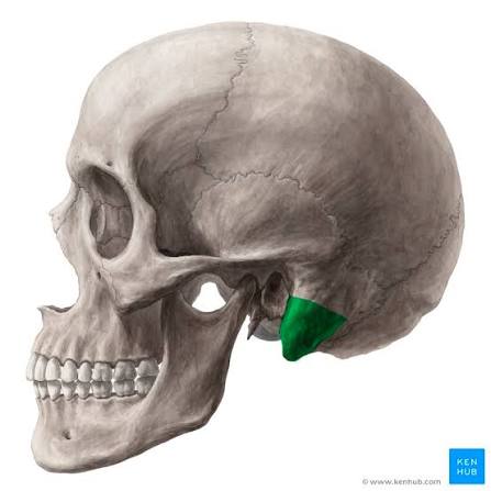
Styloid Process of the Temporal Bone
White thing sticking out in front of the mastoid process

Zygomatic Process of the Temporal Bone
Back part of the “bridge”

Petrous Portion of the Temporal Bone
Faint triangle impression above the mastoid process, just past the auditory meatus
Mandibular Fossa
depression, where the mandible fits in


Ethmoid Bone
F*CK THIS BONE AHHHHH
Crista Galli
The “cock’s comb”, fin at the top of the ethmoid bone

Cribriform Plate
Where the olfactory nerves go through in the ethmoid bone, Brandon can’t pronounce this
Perpendicular Plate of the Ethmoid Bone
Forms the upper part of the bony nasal septum, the “tail” of the ethmoid bone

Orbital Plate of the Ethmoid Bone
the outer casings of the ethmoid bone

Superior and Middle conchae of the ethmoid bone
one looks like the ballsack of the ethmoid bone from the posterior view, the other is the spot right above it
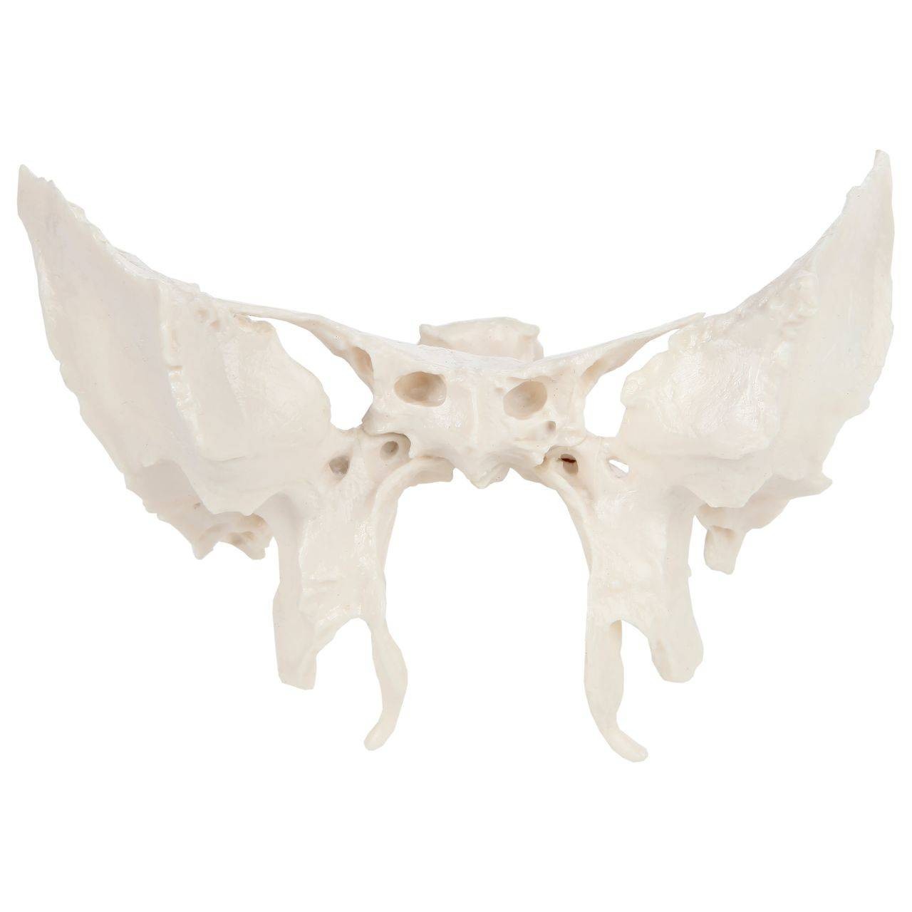
Sphenoid Bone
Looks like a big butterfly, sits in the center of the skull

Greater Wing of the Sphenoid Bone
upper wings of the butterfly

Lesser Wing of the Sphenoid Bone
tiny wings above the greater wing

Sella Turcica
like the saddle on the back of the butterfly, visible from the top of the skull

Pterygoid Processes
The legs of the butterfly

The medical and lateral plates of the pterygoid process of the sphenoid bone
The individual legs of the butterfly (in sets of 2)
Mandible
Makes up the lower jaw
Body of the mandible
The whole lower half
Ramus of the mandible
The back top part of the mandible, where it connects to the mandibular fossa

Dental Alveoli of the Mandible
“tooth sockets” in the lower half of your jaw
Angle of the mandible
Where you point when mogging
Coronoid Process of the mandible
The front bump on the ramus, the side closer to the rest of the mandible
Condylar Process of the Mandible
The back projection on the ramus
The head of the condylar of the mandible bone
the very top of the back projection on the ramus
Maxillae
Upper jaw
Body of maxilla
The entire area around the bottom of the nose and upper lip

Dental alveoli of the maxilla
Depressions for teeth- “tooth sockets” of the upper jaw
Infraorbital Margin
Lower rim of the orbits
Palatine Process
Purple part of the roof of the mouth, front portion of the roof of your hard palate

Zygomatic Bones
“cheek bones”
Temporal Process of the Zygomatic bones
The front portion of the zygomatic arch, connected to the zygomatic bones

Nasal Bones
structure, white bones of the nose, fractures produce a “broken nose”
Palatine Bones
L-shaped bones
Horizontal Plate of the Palatine Bones
the part of the palatine bones that contributes to the hard palate

Perpendicular Plate of the Palatine Bones
On either side of the vomer, the lateral walls of the nasal cavity

Vomer
yellow divider in the nose, makes up the bottom part of the bony nasal septum
Lacrimal Bones
orange parts inside the eye sockets
Opening for nasolacrimal duct
“This will bring tears to your eyes,” the space between the orange and purple
Inferior Nasal Conchae
yellow on either side of the nose, projects into the nasal cavities
Supraorbital Foramen
The little holes in the skull above the eye socket, around the supraorbital margins

Infraorbital Foramen
The little holes in the skull right below the eye socket, on the tops of the cheeks
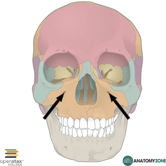
Mental foramen
the holes in the chin

Optic Canal
where the optic nerve passes through the orbit, the hole at the back of the eye socket, closer to the nose

Superior Orbital Fissure
Space by the optic canal, looks like an amoeba more than a circle

Inferior Orbital Fissure
Looks connected to the superior orbital fissure but isn’t, bottom space
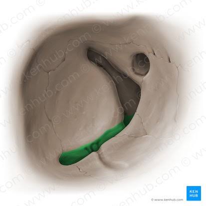
External Acoustic Meatus
The divet space between the styloid process and the mastoid process

Cribriform Foramina
the little holes around the nose area - on the cribriform plate - holes are for the olfactory nerves

Optic Canals - Anterior View
holes right underneath the saddle horns

Foramen Rotundum
will be labeled with a pink pipe cleaner, more anterior than the formamen ovale, very small

Foramen Ovale
Used as our “North Star”, largest and most distinct middle cranial fossa
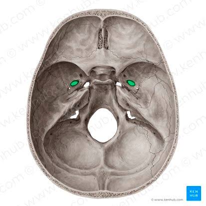
Foramen Spinosum
Tiny hole right below the foramen ovale

Foramen Laceram
More medial than the foramen ovale

Internal Acoustic Meatus
posterior to foramen ovale and more medial, along the side of the ridge

Jugular Foramen
holes at the bottom of the skull, just outside the occipital condyles, kind of like a cave more than a hole

Hypoglossal canal
just along the upper rim of the foramen magnum

Foramen magnum
Large hole for the spine/neck

Carotid canal
The hole just above the jugular foramen

Stylomastoid Foramen
right between the styloid and mastoid processes
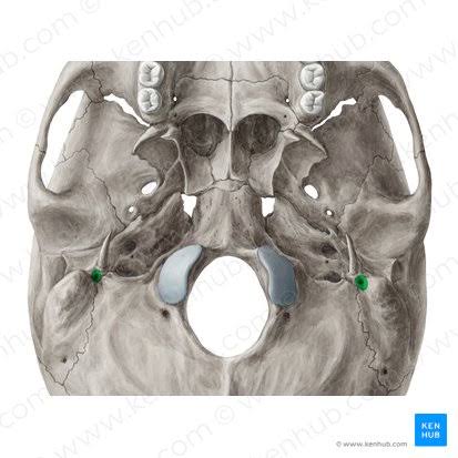
Incisive Fossa
The whole right behind the upper incisor teeth, the front teeth

Greater palatine foramen
near the upper molar teeth

Mandibular foramen
holes in the upper ramus of the mandible

Frontal sinuses
space inside the inner corner of the eyebrows, lined by mucous membranes in the living
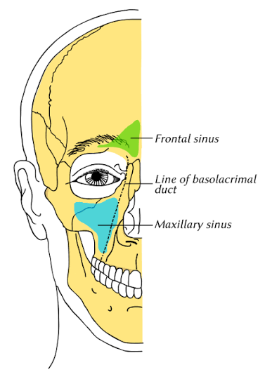
Maxillary sinuses
the space inside the cheekbones - the largest
Ethmoid air cells
in the bridge of our nose, where we pinch when we have headaches
Cervical Vertebrae Region
The top 7 vertebrae

Thoracic Vertebrae Region
The middle 12 vertebrae

Lumbar Vertebrae Region
The lowest 5 vertebrae

Sacrum
fused bit of bone at the bottom of the vertebral column

Coccyx
Tailbone

Thoracic Kyphosis
the second curvature, the hump of your back - one of the primary curvatures

Sacral Kyphosis
the last curvature, curves out and back around the sacrum, one of the primary curvatures of the spine

Cervical Lordosis
The first curvature, curves in with the neck, one of the secondary curvatures
