PAS 407- Hip and Knee Joints
1/48
There's no tags or description
Looks like no tags are added yet.
Name | Mastery | Learn | Test | Matching | Spaced | Call with Kai |
|---|
No analytics yet
Send a link to your students to track their progress
49 Terms
What are the three bones of the os coxae?
What do they form when fused?
ilium, ischium, and pubis
acetabulum
What is the angle of inclination?
the angle the femoral neck makes with the femoral shaft as weight is redistributed medially
What is Ricketts?
What causes it?
What does it result in?
condition characterized by bone demineralization
caused by vitamin D deficiency
results in excessive bowing of legs in response to stress of weight bearing
What type of joint is the hip joint?
What is articulating?
ball and socket joint
femoral head articulates with lunate surface within superior margin of acetabulum
What does the acetabular labrum do?
What is it continuous with?
increases surface area of acetabulum and deepens the joint
continuous with transverse acetabular ligament
What is the joint capsule attached to in the hip joint?
What does it resist?
What does the weak posterior capsule allow?
attaches outer rim of acetabulum to intertrochanteric line anteriorly
resists hyperextension
allows synovial membrane to protrude posteriorly and form bursa to cushion overlying muscle
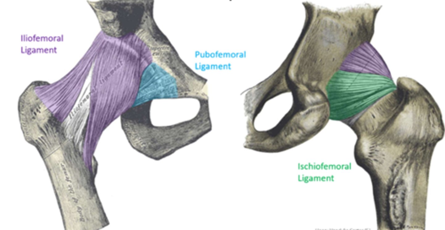
What are the intrinsic/capsular ligaments in the hip joint?
1) iliofemoral ligament
2) ischiofemoral ligament
3) pubofemoral ligament

What is the strongest ligament in the body?
iliofemoral ligament
What does the iliofemoral ligament connect?
What does is its function
What are its two bands?
connects AIIS and acetabular rim to intertrochanteric line
resists hyperextension through corkscrew mechanism
transverse and descending bands
**purple in picture

What does the ischiofemoral ligament connect?
What does it blend with?
What is its function?
connects posterior acetabular rim to intertrochanteric line
blends with fibers of transverse band of iliofemoral ligament
resists hyperextension through corkscrew mechanism
**green in picture

What does the pubofemoral ligament connect?
What is its function?
connects obturator crest of pubic bone to intertrochanteric line
prevents hyperabduction of joint and dislocation of femoral head (e.g. during splits)
**blue in picture
What does the ligamentum teres femoris connect?
What is it surrounded by?
What is its function?
acetabular notch to fovea capitis on the head of the femur
surrounded by synovial membrane
protects the artery to the head of the femur
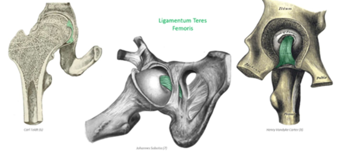
What would happen if the artery to the head of the femur was disrupted?
avascular necrosis and degeneration of the femoral head
What movements does the hip joint allow?
How are extension and flexion limited?
flexion, extension, abduction, adduction, circumduction, and rotation
extension limited by ligaments
flexion limited by soft tissue (thigh against abdomen)
What 3 bones are involved in the knee joint?
femur, tibia, and patella
*fibula not involved
Explain how weight distribution is directed
initially directed laterally into proximal region of femur and then redirected medially
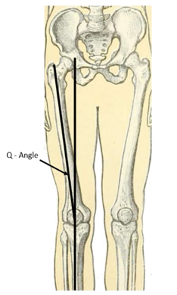
What is Q-angle?
How is it found?
What is its typical range?
Who is greater in? Why?
measure of obliqueness of femoral shaft relative to tibial shaft
found by taking the angle between a vertical line through knee and line connecting ASIS to patella
10-15 degrees
greater in females because of a wider pelvis
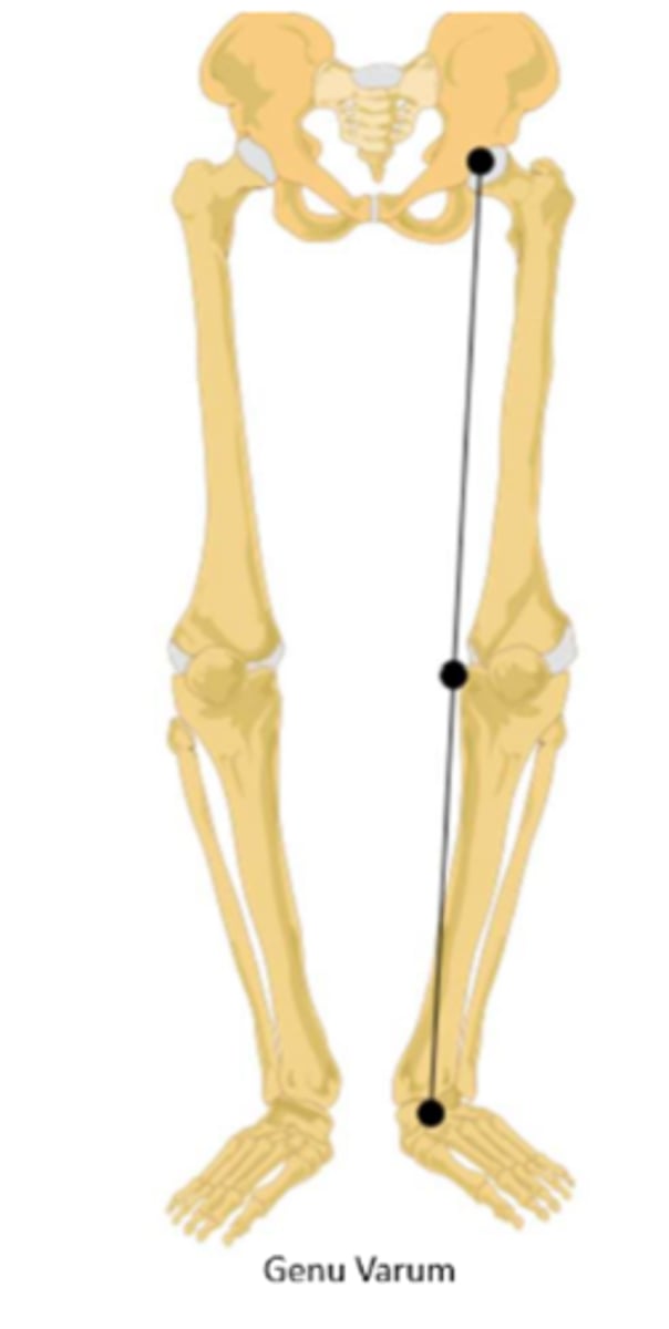
What is genu varum?
What structure receives the increased load?
"bowlegged" ; femoral shaft is more vertical and results in a low Q angle (5 degrees)
medial condyles take increased load
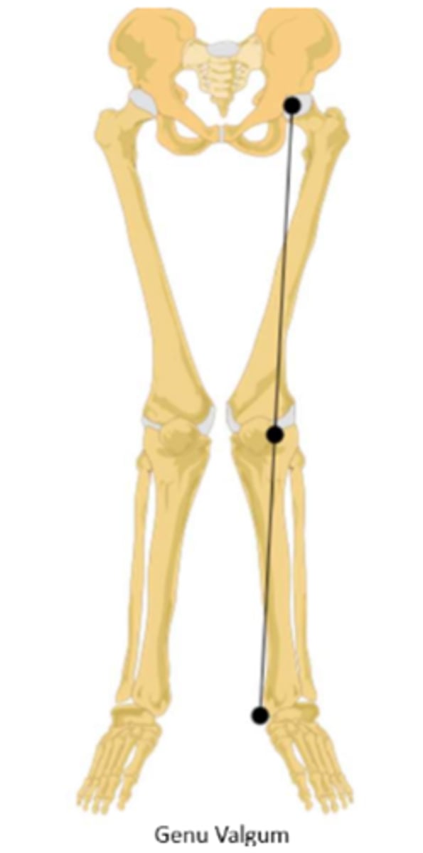
What is genu valgum?
What structure receives the increased load?
What does it increase the susceptibility to? Why?
"knock-knee"; femoral shaft lies more obliquely and results in a Q-angle greater than 15 degrees
lateral condyles take increased load
increase susceptibility to patellofemoral syndrome because quadriceps tendon may pull patella laterally
How many separate articulations are found in the knee joint?
3
What are the different articulations in the knee joint?
lateral and medial condyles of femur and tibia = tibiofemoral joints
anterior intercondylar surface of femur and patella = patellofemoral joint
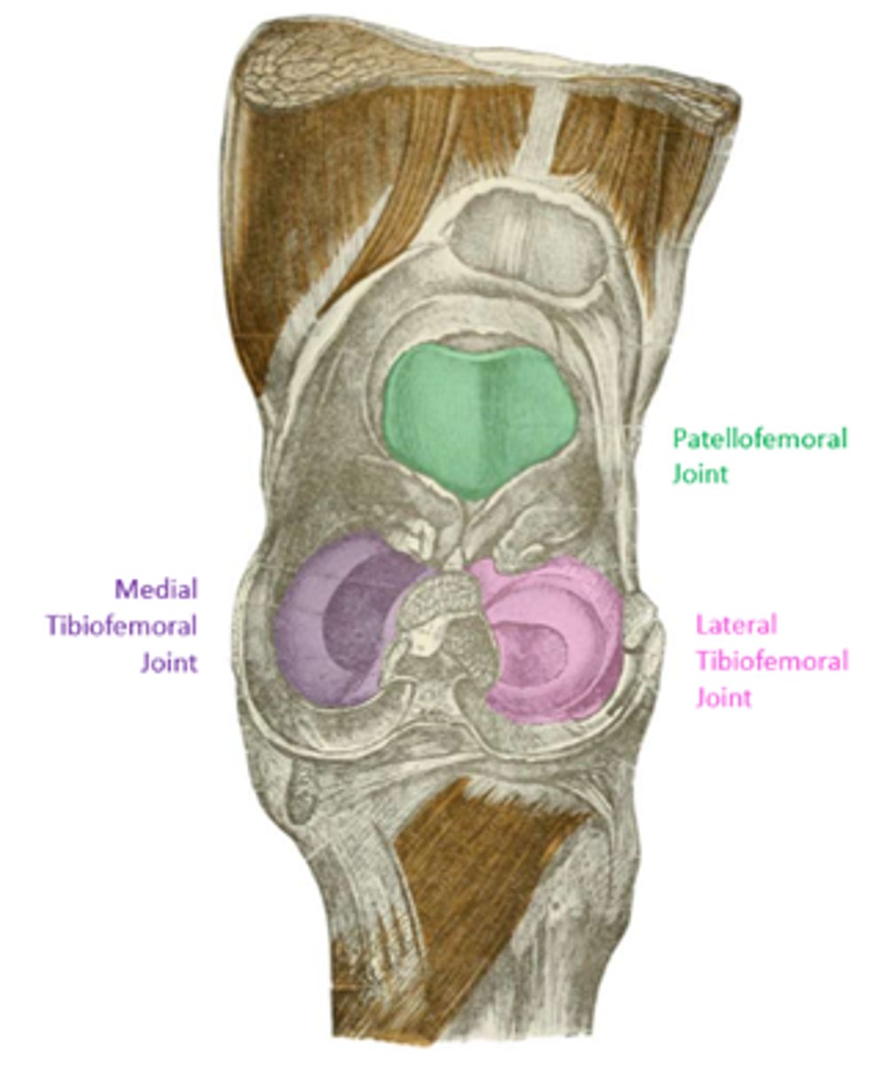
Describe the shape of the tibiofemoral joints
What structure improves the surface area?
In what position do these joints have the best surface contact?
incongruent shape; femoral condyles are condyloid and tibial condyles are planar
cartilaginous discs (menisci)
extended position
What does the knee joint capsule connect?
What structures does it fuse with?
What passes through it?
connect femur to tibia peripheral to articular surfaces
fused with patellar tendon superiorly and patellar ligament inferiorly
allows passage of popliteus tendon due to incomplete covering over lateral epicondyle
What does the synovial membrane of the knee joint line?
What does it extend superiorly as?
lines all non-articulating surfaces internal to capsule; lines cruciate ligaments and infrapatellar fat pad
extends superiorly as suprapatellar bursa
What are intrinsic ligaments?
What are the intrinsic ligaments of the knee joint?
thickening of the joint capsule around the knee
patellar ligament
tibial/medial collateral ligament
oblique popliteal ligament
arcuate popliteal ligament
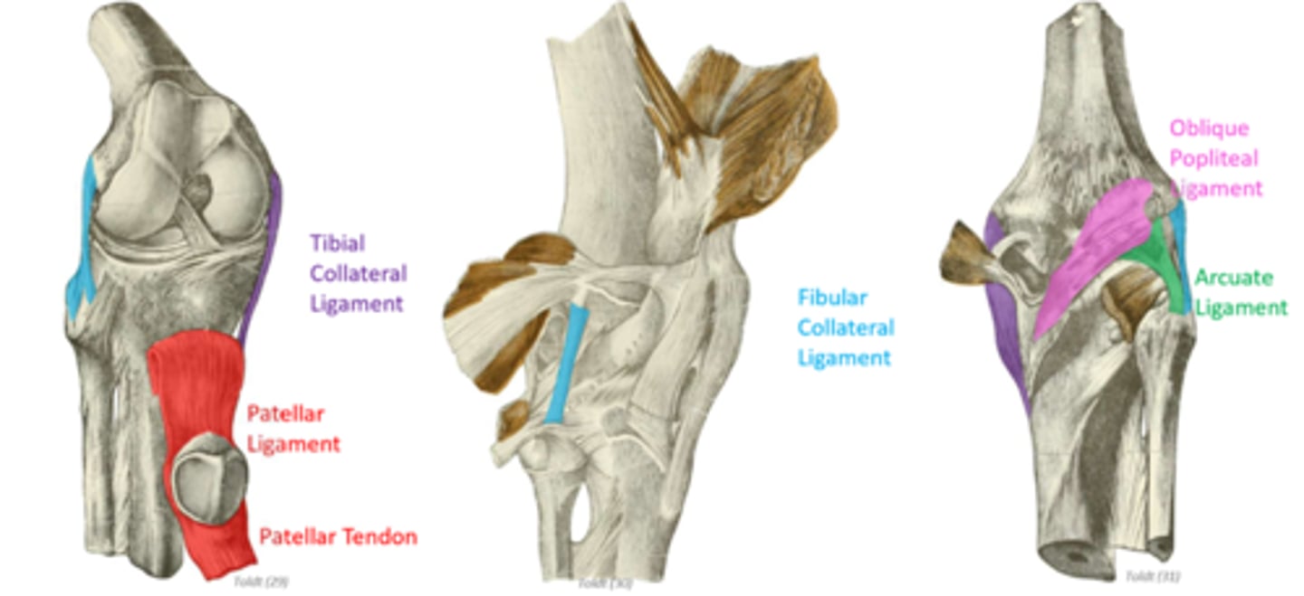
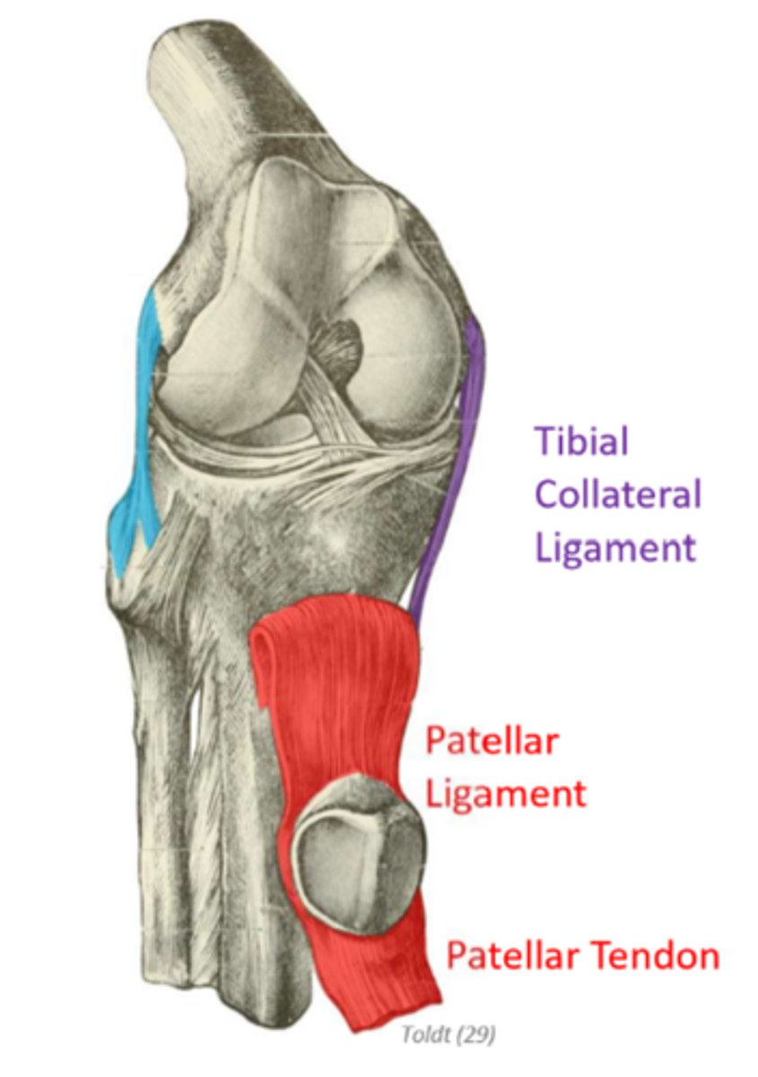
What is the patellar ligament?
Where does it insert?
continuation of the patellar/quadriceps tendon beyond the patella
inserts on tibial tuberosity

What is the tibial/medial collateral ligament (MCL)?
What does it connect?
Where do its deep fibers insert?
What is its function?
strong, flat band on medial surface of joint capsule
connects medial epicondyle of femur to medial condyle of tibia
deep fibers insert on medial meniscus
resists valgus forces that try to abduct the knee and lateral rotation
What is the fibular/lateral collateral ligament (LCL)?
What does it connect?
What is its function?
extracapsular ligament; strong cord on lateral surface of joint
connects lateral epicondyle of femur to fibular head
resists varus forces that try to adduct the knee and lateral rotation
*blue in picture

What structure does the anterolateral ligament (ALL) originate with?
Where does it insert?
originates with LCL
inserts on tibia
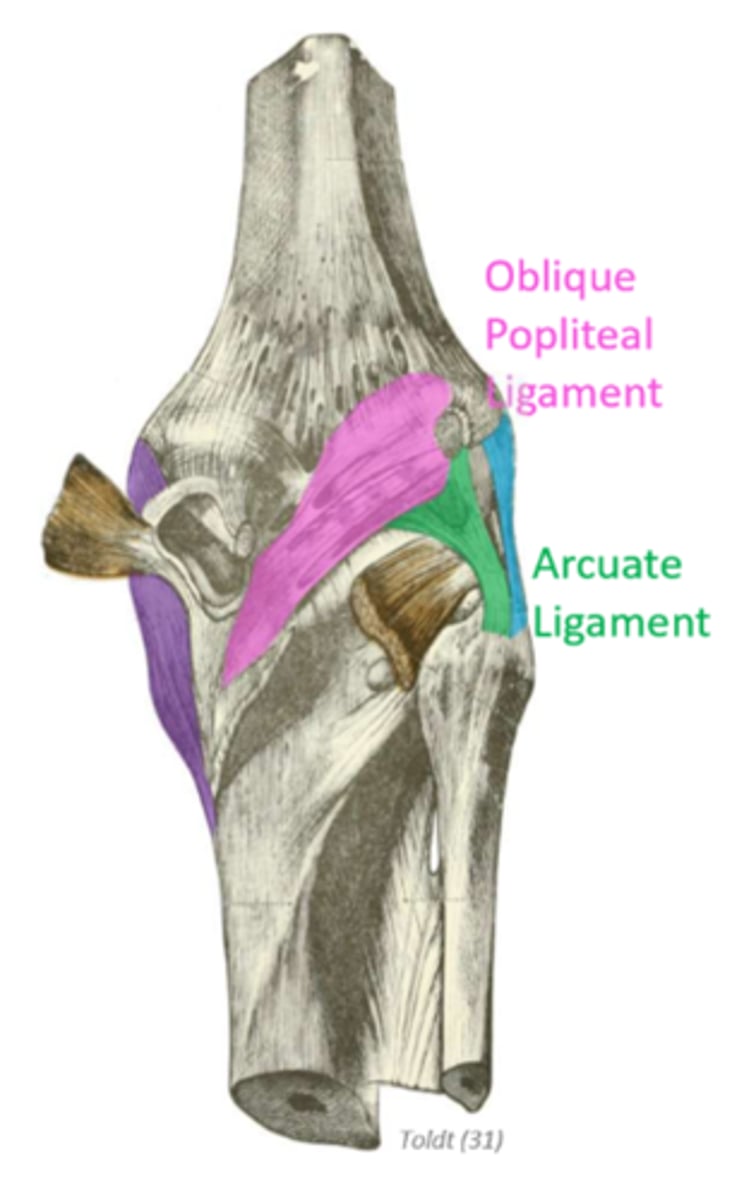
What is the oblique popliteal ligament?
What is its function?
extension of semimembranosus tendon that blends into the knee joint capsule
provides posterior reinforcement to resist hyperextension

What does the arcuate popliteal ligament connect?
What is its function?
connects head of fibula to intercondylar area of femur; arches over popliteal muscle
provides posterior reinforcement to resist hyperextension
Where are the cruciate ligaments located?
located within intracondylar region of joint
internal to joint capsule and external to synovial
Explain how the cruciate ligaments move around each other during medial and lateral rotation of the knee
wind tight during medial/internal rotation; limiting to 10 degrees
unwind during lateral/external rotation; allowing 60 degrees in flexed knee position
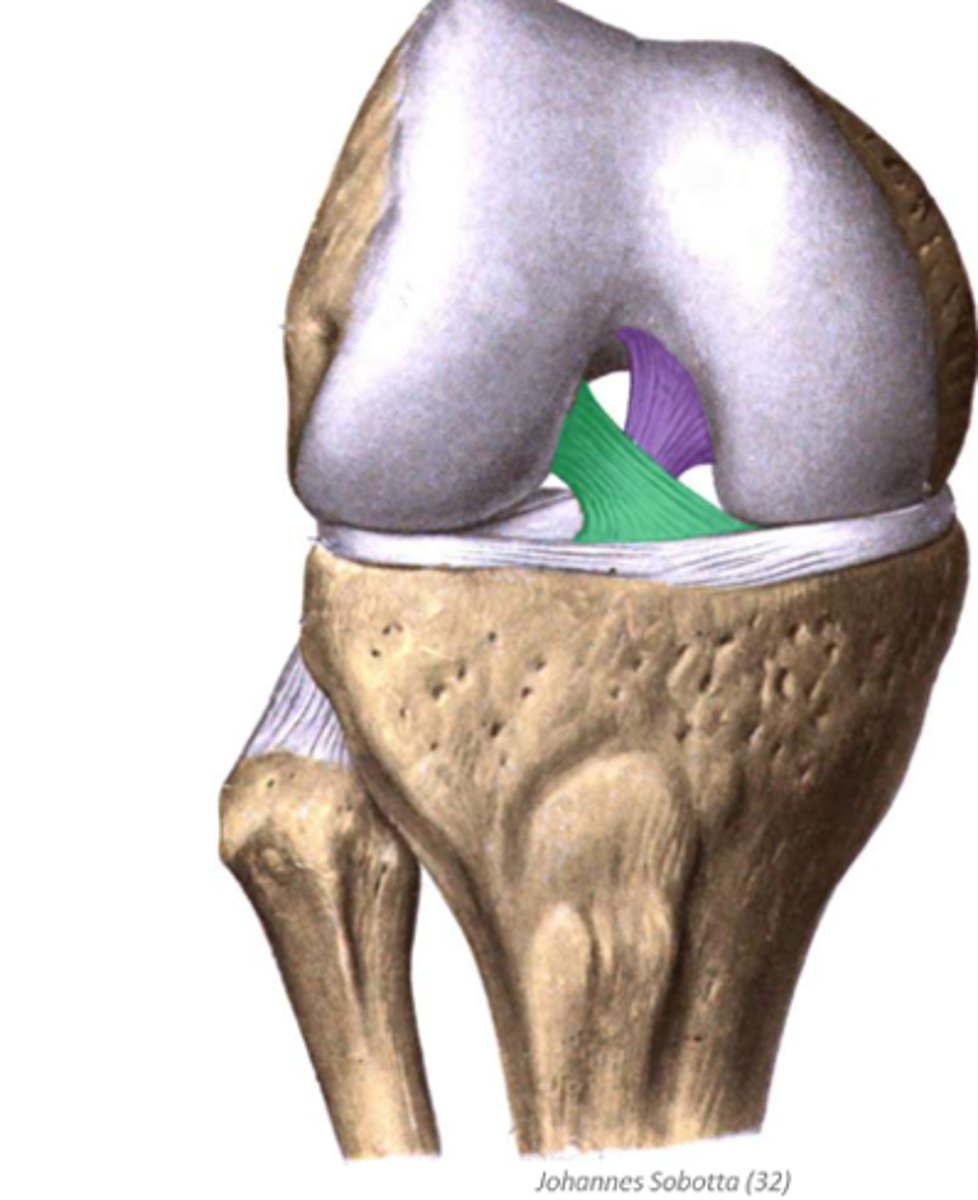
How does the anterior cruciate ligament (ACL) connect? What does it connect?
What is its function?
attaches on anterior aspect of intercondylar eminence of tibia, projects posteriorly and lateral to PCL to insert just medial to the lateral condyle of the femur
prevents anterior glide of tibia on femur and limits medial rotation
*green in image

How does the posterior cruciate ligament (PCL) connect? What does it connect?
What is its function?
attaches on posterior aspect of intercondylar eminence of tibia, projects anteriorly and medial to ACL to insert just lateral to the medial condyle of the femur
prevents posterior glide of tibia on femur and limits medial rotation
*purple in image
What are the menisci?
What is their shape?
What is their function?
fibrocartilaginous discs anchored to the tibia through coronary ligaments
crescent superior surface to accommodate femoral condyles and flat inferior surface to accommodate tibia
increase surface area contact between articulating surfaces and absorbs shock during foot strike
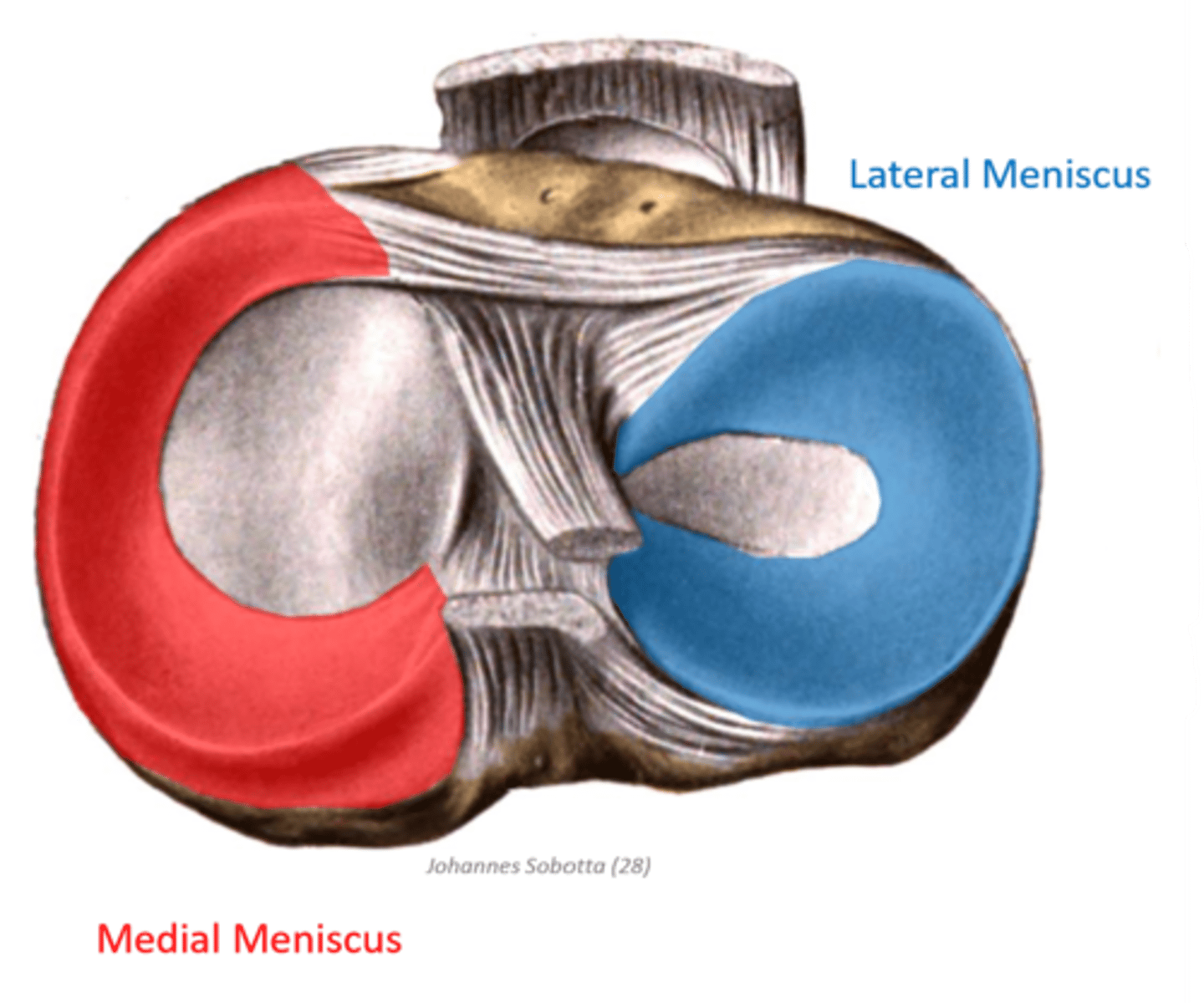
Where is the medial meniscus located?
What is its shape?
What is it anchored to?
located within medial tibiofemoral portion of knee joint
C-shape
lateral margin anchored to MCL

Where is the lateral meniscus located?
What is its shape?
What is it anchored to?
located within lateral tibiofemoral portion of knee joint
circular shape
anchored to popliteus tendon and PCL

Which menisci is more mobile?
Why?
lateral meniscus
less anchorage to tibia through coronary ligaments
How do the knee ligaments function during knee flexion?
collaterals and posterior joint capsule become loose due to decreased distance between attachment points = greater degree of rotation and dependence on cruciate ligaments
cruciate ligaments maintain some tautness because they run obliquely
How do the knee ligaments function during knee extension?
How does the tibia move relative to the femur?
all ligaments become taut but collateral ligaments have some slack to permit a small amount of varus and valgus stresses
lateral rotation of tibia on femur
Explain how "locking knee" occurs
ligaments in knee become taut and tibia externally rotates
minimizes energy expenditure from muscular stabilization
*greater susceptibility to ligament sprains because of the tension in ligaments and rigidity in knee joint
Explain how collateral ligament sprains occur
MCL: tearing during pivoting or valgus stressors (blow to lateral knee)
LCL: tearing during varus stresses; not common
Explain how cruciate ligament sprains occur
ACL: tearing during hyperextension or twisting motions; may also tear with MCL
PCL: tearing when fall on flexed knee (pushes tibia back); uncommon
How would a patient with an ACL tear present?
patient experiences a pop followed by rapid onset of swelling
joint instability/slipping after initial swelling subsides
Explain how meniscal tears occur
medial: more likely to be affected in isolated tear because it is less mobile; most commonly torn from shearing forces of femur; may also tear with MCL
lateral: most commonly torn from compression forces of femur during valgus stressor; less commonly torn with rotational movements because it is less anchored
How would a patient present with an acute meniscal tear?
point tenderness along tibial plateau
intercapsular swelling
knee catching/locking up
What is the "unhappy triad"
tearing of MCL, ACL, and meniscus from mechanical trauma with excessive valgus force
What is osteoarthritis?
What is it associated with?
What does it result in?
breakdown of joint cartilage and underlying bone
associated with history of mechanical trauma at affected joint
results in bone spurs, disfiguration, stiffness, and pain