Med Surg Exam 2 - Neurosurgical & Neurological Disorders
 Call Kai
 Learn
Learn Practice Test
 Spaced Repetition
 Match
Match Flashcards
 Knowt Play
1/130

There's no tags or description
Looks like no tags are added yet.
Name | Mastery | Learn | Test | Matching | Spaced | Call with Kai | Chat |
|---|
No analytics yet
Send a link to your students to track their progress
131 Terms
I = benign through IV = highly malignant
What do the WHO tumor classes mean?
yes (can cause edema that shifts structures, and increases ICP)
Are benign brain tumors problematic?
gliomas
Brain tumors that develop from glial cells
astrocytoma
type of glioma formed from astrocytes
graded I-IV
I = can be treated with craniotomy
What is a class I astrocytoma?
glioblastoma multiforme (class IV)
Most common and fastest growing brain tumor glioma
No cure, average survival is 12-18 months
ependyoma
brain tumor that originates in the lining of the ventricles
usually benign
treated with radiation
meningeal tumor (meningioma)
Brain tumor that arises from the meninges, the layers of tissue covering the brain and spinal cord
Usually slow-growing and often benign
Treated with surgery
lung or breast cancer
Which type of cancers most commonly cause metastatic brain tumors?
biopsy
What is needed for definitive diagnosis of brain tumors?
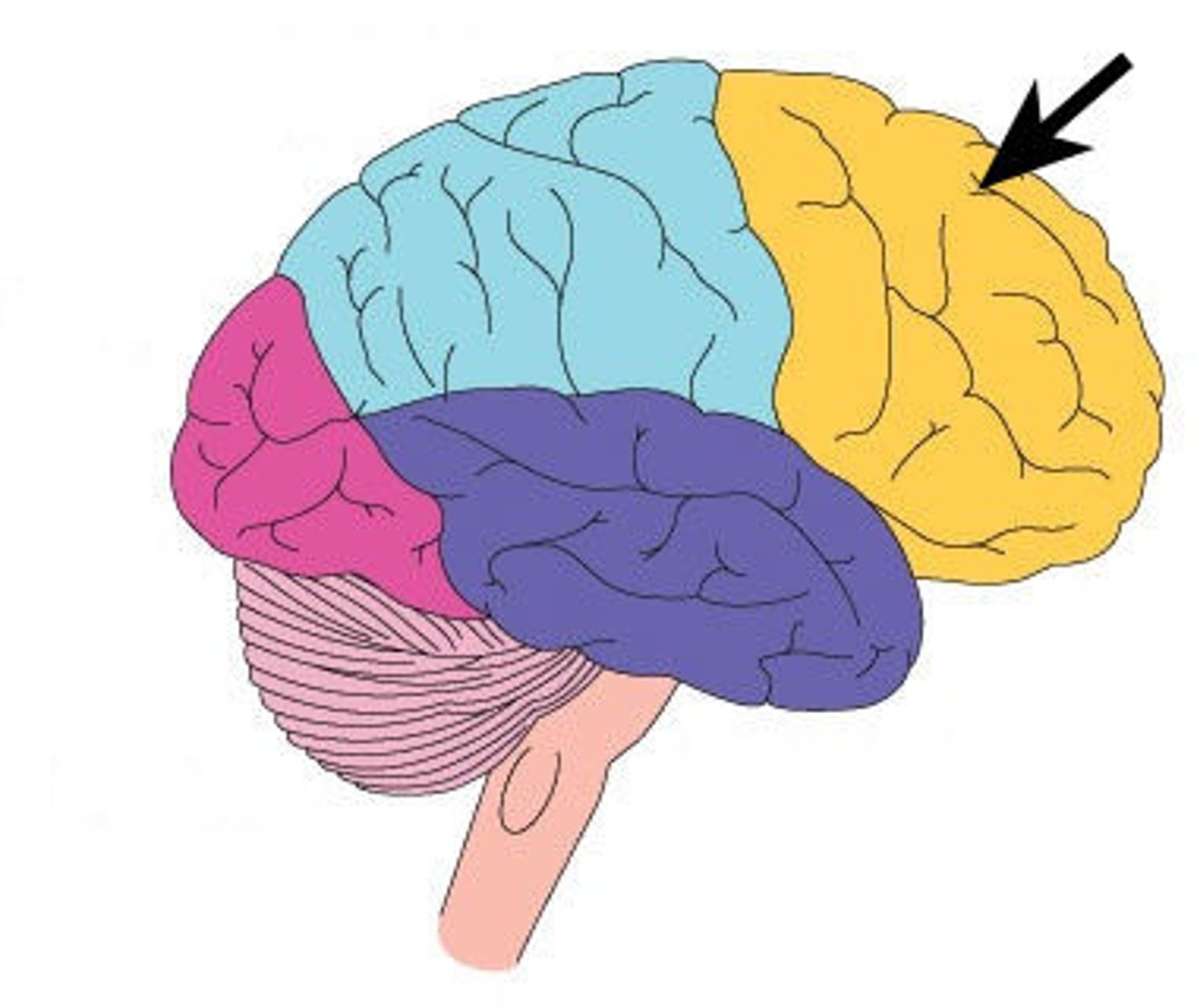
frontal lobe
A region of the cerebral cortex that has specialized areas for movement, abstract thinking, judgment, and personality
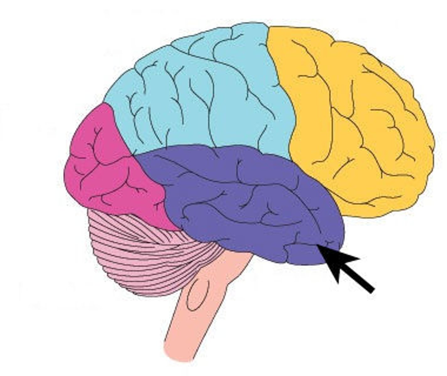
temporal lobe
A region of the cerebral cortex responsible for hearing and language.
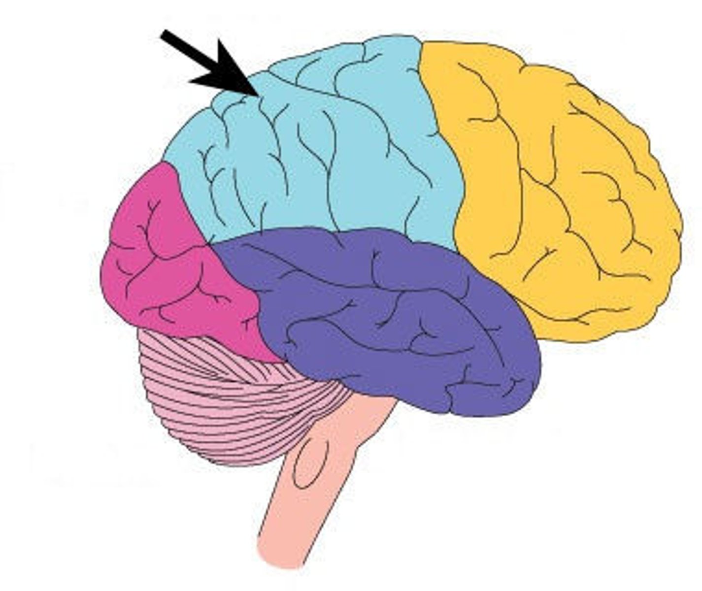
parietal lobe
A region of the cerebral cortex whose functions include processing information about touch (sensory).
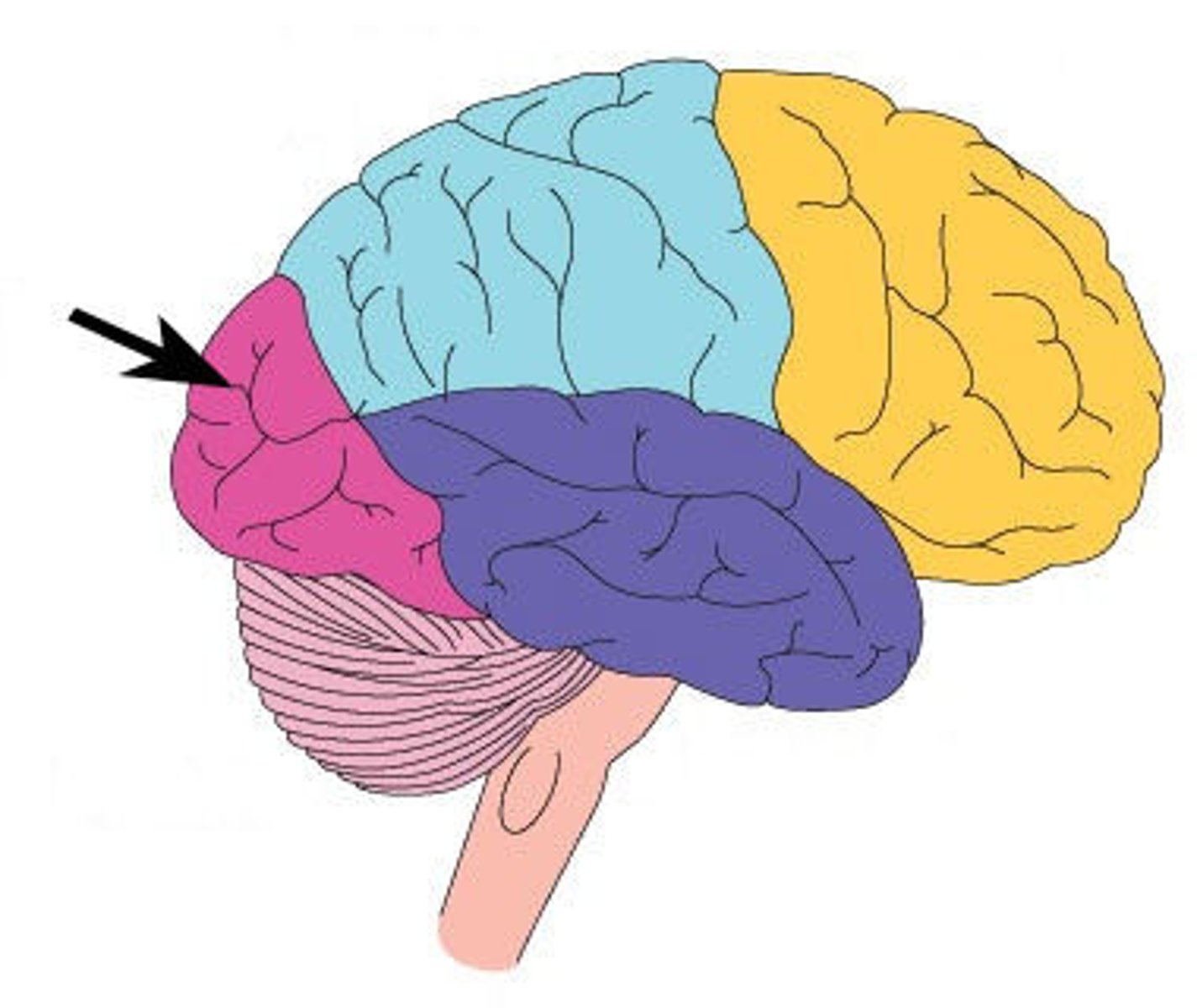
occipital lobe
A region of the cerebral cortex that processes visual information
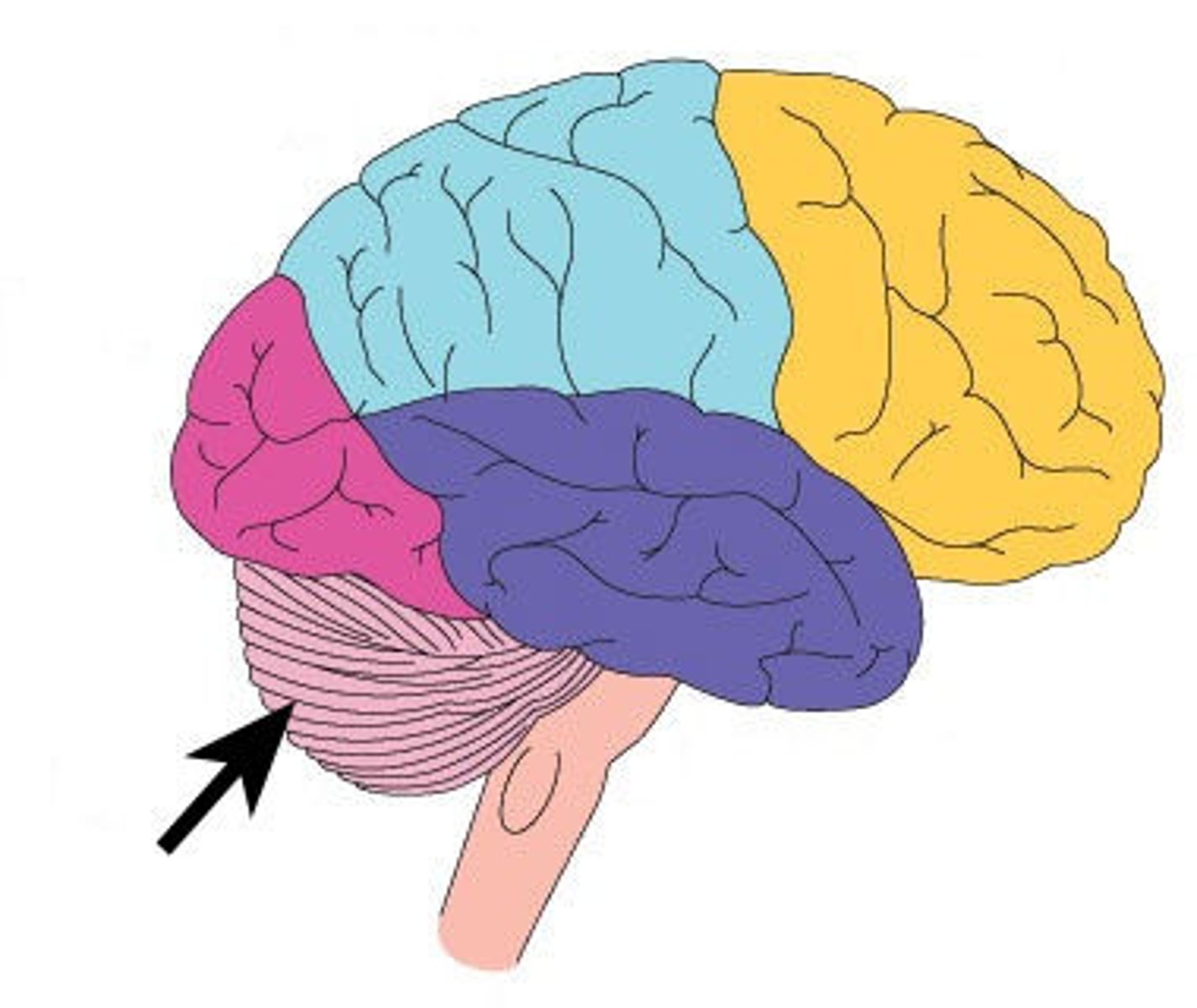
cerebellum
A large structure of the hindbrain that controls fine motor skills (coordination)
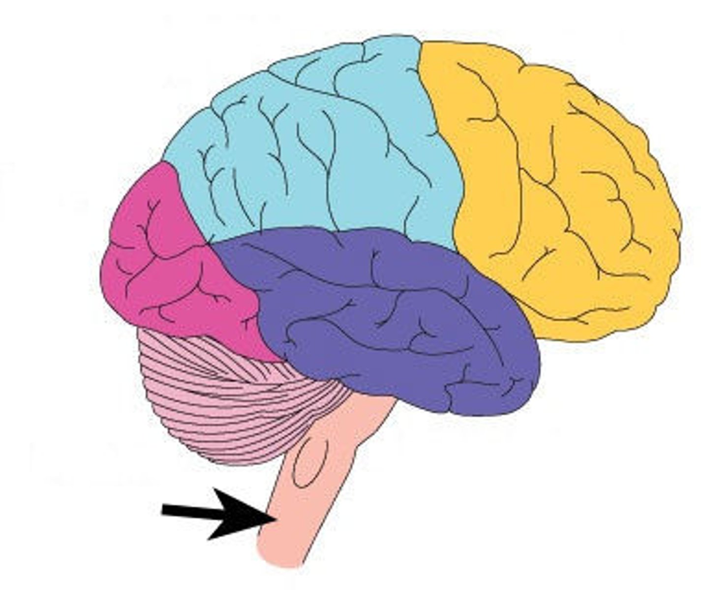
brainstem
the lower part of the brain that connects the brain to the spinal cord. It controls many basic life functions and acts as a relay center between the brain and the body.
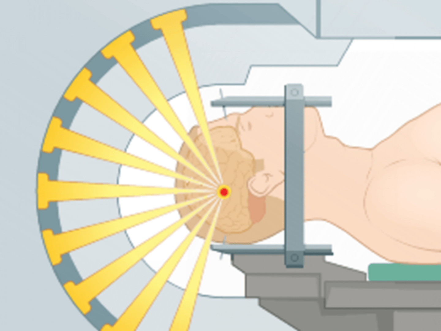
stereostatic surgery
a minimally invasive surgical technique that uses a three-dimensional (3D) imaging system to precisely target specific areas in the brain or other parts of the body. It enables surgeons to reach deep areas with great accuracy, often without making large incisions
no
Can chemo cross the BBB?
biodegradable wafer, ommaya reservoir
How is chemo delivered to the brain?
biodegradable wafer
a small, dissolvable disk that contains chemotherapy drugs. It is placed directly in the brain at the tumor site during surgery, allowing for a targeted release of chemotherapy to treat brain tumors
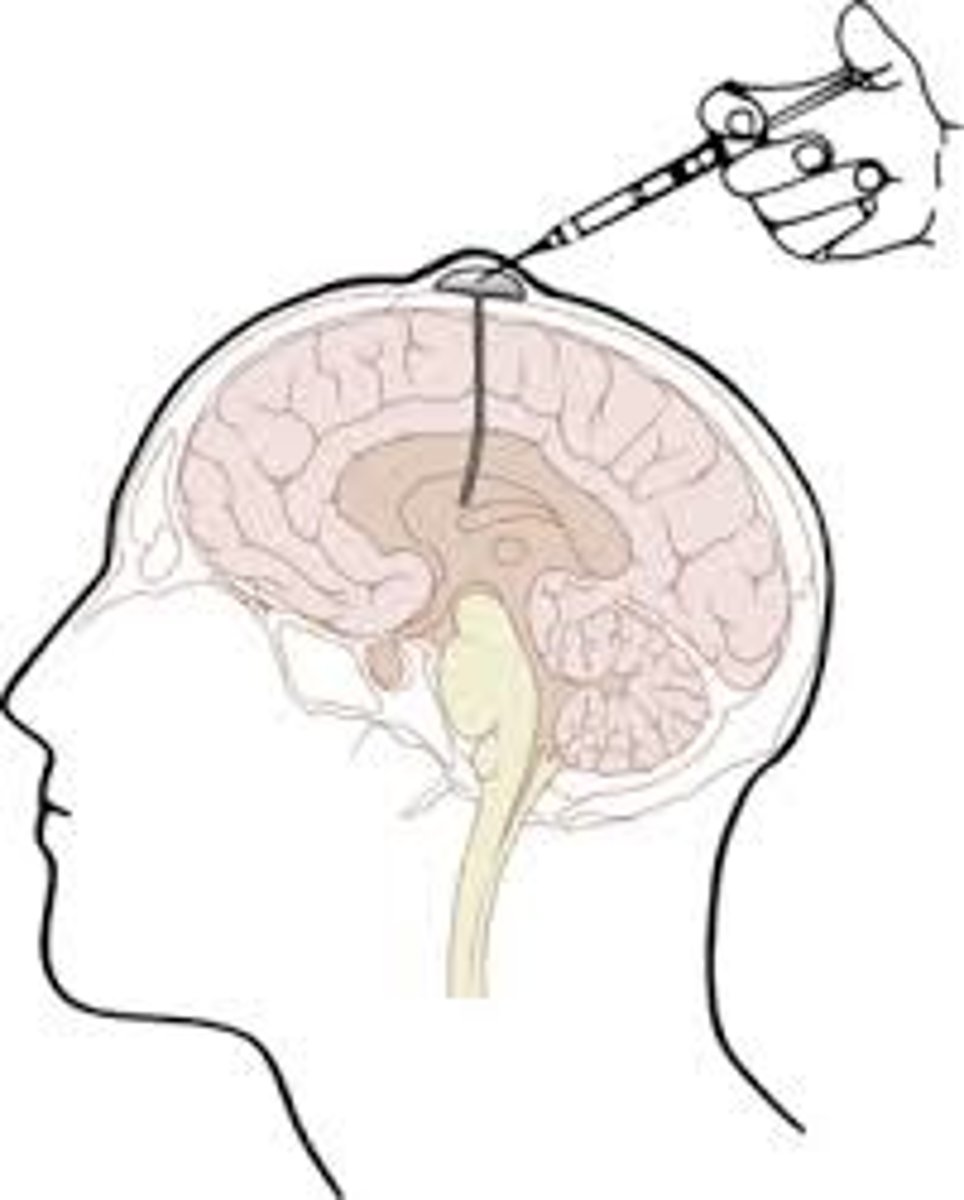
Ommaya reservoir
a device used to deliver chemotherapy directly into the cerebrospinal fluid (CSF) around the brain and spinal cord. It allows chemotherapy to reach areas of the central nervous system (CNS) that might otherwise be difficult to treat due to the blood-brain barrier
increased ICP, infection, sodium imbalances, intracranial hemorrhage, seizures, DVT/PE, gastric ulcer, cognitive dysfunction
What are some complications of brain tumors?
craniotomy
part of the skull is removed to access the brain
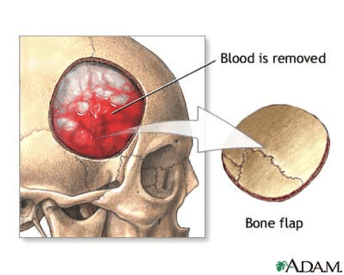
ultrasound, stereotaxy, endoscopic/keyhole surgery, cortical mapping, neurendoscopy
What are some ways that craniotomy surgery is guided?
endoscopic/keyhole surgery
a hole is made into the skull of through the lip or nose so that endoscopic instruments can be used for the procedure
cortical mapping/somatosensory evoked potentials
language, motor, and sensory areas of the brain are identified during surgery through EMG, somatosensory response, and direct stimulation
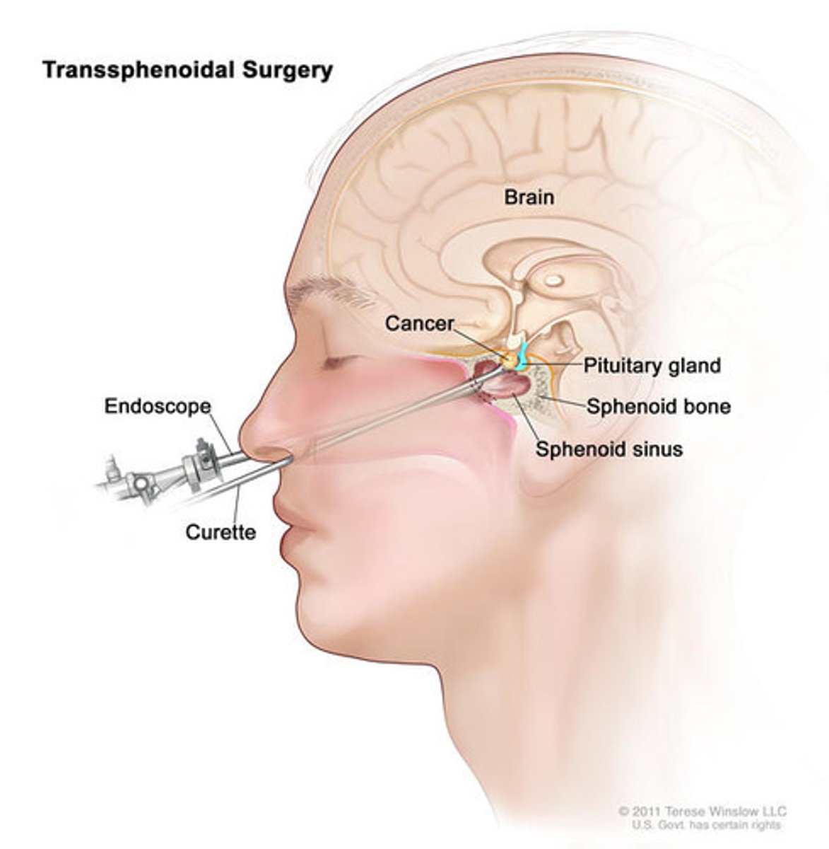
transsphenoidal hypophysectomy
endoscopic procedure to surgically remove a pituitary tumor through an incision in the sphenoid sinus without disturbing brain tissue
do not remove (sometimes made from fat from abdomen or thigh)
What is important nursing care for nasal packing after a transsphenoidal hypophysectomy?
CSF lead, pneumocephalus
What are some complications of transsphenoidal hypophysectomy?
blood stain surrounded by a yellowish stain; highly suggestive of a cerebrospinal fluid leak
What is a "halo sign"?
avoid blowing nose, coughing, sneezing, drinking with a straw, bending over, or straining for 4 weeks
What is an important teaching for a patient after a transsphenoidal hypophysectomy?
must take hormone replacements for life
What does a patient need to know if they had their entire pituitary gland removed?
place a dressing and turn to the side of the leak
What should you do if your patient has a CSF leak from their ear?
place a dressing under their nose
What should you do if your patient has a CSF leak from their nose?
suction nose or use nasal packing, put fingers in the nose or ears
What should you not do if a patient has a CSF leak from their nose or ears?
encourage participation in care and rehab
What is an important psychological teaching for a patient after neurosurgery?
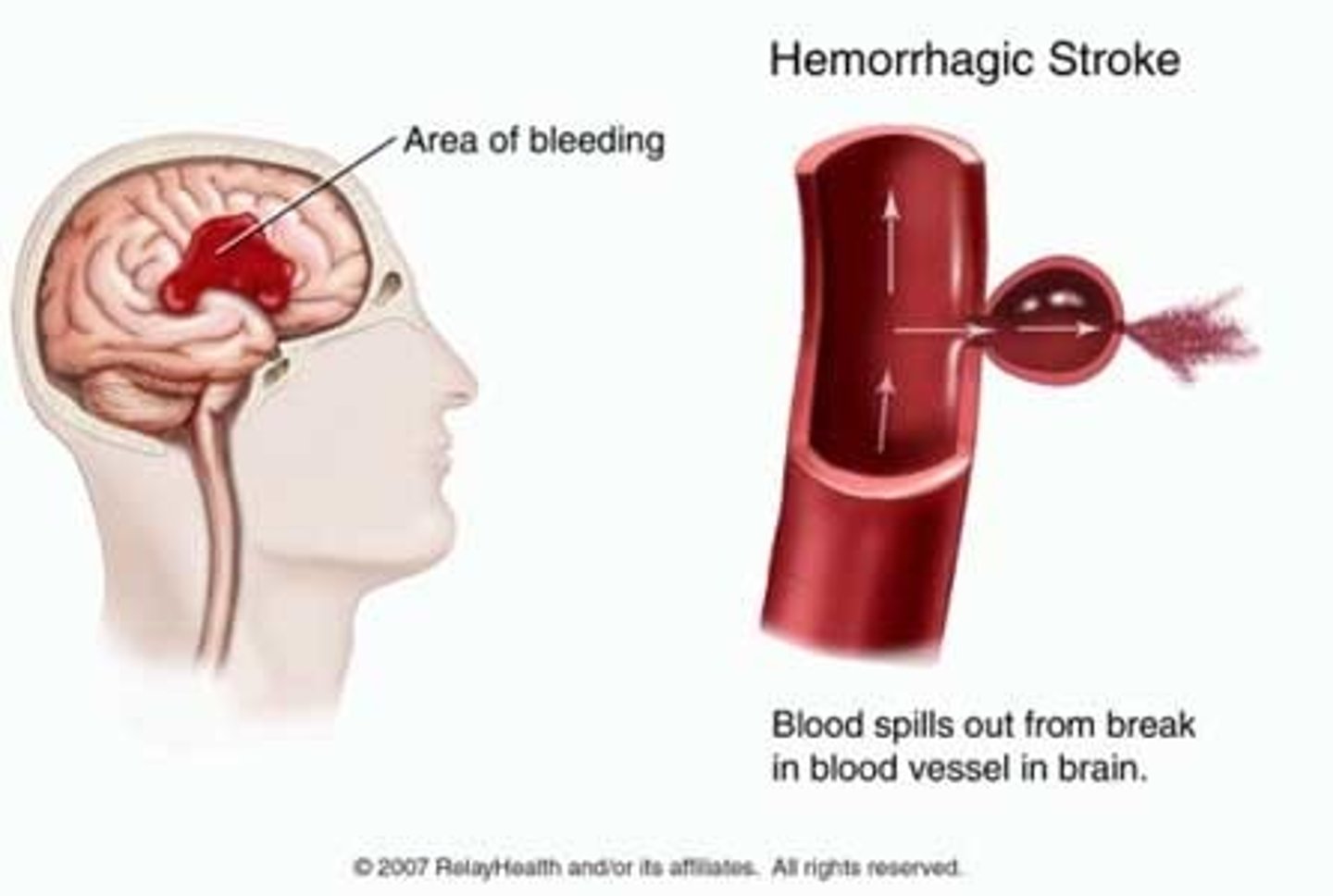
hemorrhagic stroke
when a blood vessel in the brain bursts, causing bleeding (hemorrhage) in or around the brain. This bleeding increases pressure on brain tissue, leading to cell damage and loss of brain function
brain tumors, aneurysms, arteriovenous malformations, traum
What are some causes of hemorrhagic stroke?
anticoagulant use
What is a major risk factor for hemorrhagic stroke?
intracranial aneurysm
weakening of the vessel wall causes dilation or ballooning, which may rupture or put pressure on surrounding brain tissue
subarachnoid space
Where do intracranial aneurysms primarily rupture?
HTN, smoking, ETOH use, females, congenital weakness in vessel walls, genetics
What are risk factors for intracranial aneurysms?
rupture or vasospasms
What are the major issues that can occur from an intracranial aneurysm?
sudden, severe headache (thunderclap headache), nuchal pain and rigidity, photosensitivity
What are clinical manifestations that an intracranial aneurysm has ruptured?
CT
What is the primary diagnostic test for intracranial aneurysms?
yellow-ish (color change occurs as red blood cells break down in the CSF, releasing hemoglobin and causing a yellow hue)
What does the CSF from an LP on a patient with a ruptured intracranial aneurysm look like?
control hypertension (very important), treat electrolyte abnormalities (hypo-natremia, calcemia, kalemia, and magnesemia), stool softeners, analgesics (limit narcotics)
How are intracranial aneurysms treated with medicine?
clipping or coiling
How are intracranial aneurysms treated surgically?
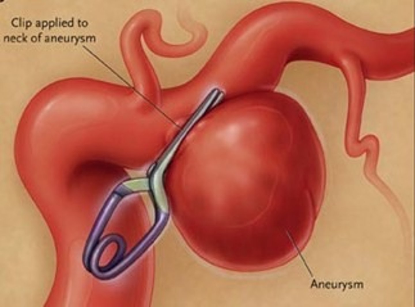
aneurysm clipping
surgical method to isolate an aneurysm from the rest of the circulatory system by placing a small clip across its neck
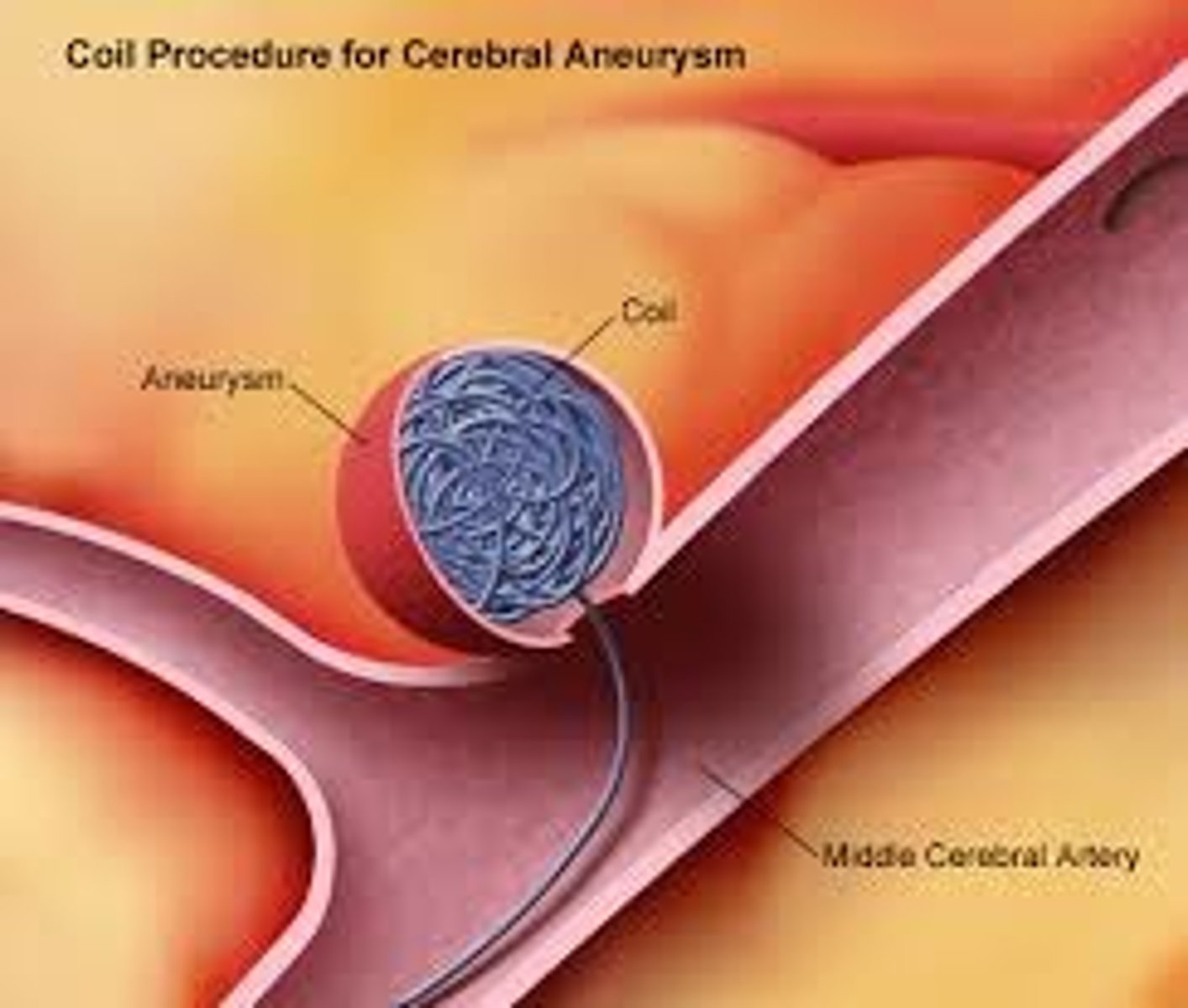
aneurysm coiling
Placement of catheter via femoral artery to area of aneurysm. Multiple small coils are placed/packed eventually leading to clot formation preventing continued bleeding
delayed cerebral ischemia
a condition where blood flow to the brain is reduced, typically occurring 5-9 days after a subarachnoid hemorrhage (SAH) due to a ruptured aneurysm. Reduced blood flow can lead to brain tissue damage and neurological decline
the excess calcium from hemorrhaged blood irritates the vessels and cause vasospasms or just by inflammation
What are the causes of delayed cerebral ischemia?
change in LOC and new neuro deficits
What are the causes of delayed cerebral ischemia?
nimodipine (calcium channel blocker that can cross the BBB to treat and prevent vasospasms), increase BP to >20mmHg above baseline, angioplasty
What are some ways delayed cerebral ischemia is treated?
arteriovenous malformation
an abnormal tangle of blood vessels connecting arteries and veins in the brain or spinal cord. This tangle disrupts normal blood flow, increasing the risk of bleeding and other complications.
rupture
A complication of arteriovenous malformations is that 40-70% of them _______________.
CT or MRI
How are arteriovenous malformations diagnosed?
surgery, radiation, or embolization
How are arteriovenous malformations treated?
traumatic brain injury (TBI)
a blow to the head or a penetrating head injury that damages the brain
alcohol or drug use, failure to use safety devices (seatbelts/helmets)
What are some contributing factors to TBIs?
acceleration-deceleration TBI
a type of brain injury caused by sudden forward and backward or side-to-side movement of the head. This movement causes the brain to collide with the skull, resulting in damage to brain tissue
head does not collide with another object
coup-countercoup injury
a type of brain injury that occurs when the brain strikes one side of the skull (coup) and then bounces back to hit the opposite side (contrecoup). This causes damage to both the initial impact site and the opposite side of the brain
rotational TBI
when the head experiences a sudden twisting or rotational force, causing the brain to rotate inside the skull. This rotational movement stretches and shears brain tissue, particularly the nerve fibers (axons) that connect different brain regions
penetrating TBI
when an object, such as a bullet, knife, or shrapnel, pierces the skull and directly damages brain tissue. This type of injury can cause localized and severe brain damage
0-30 mintues
For a mild TBI, how long are there LOC changes?
30 min - 24 hours
For a mild moderate, how long are there LOC changes?
>24 hours
For a severe TBI, how long are there LOC changes?
a moment up to 24 hours
For a mild TBI, how long is there alteration of consciousness/mental state change?
>24 hours
For a moderate or severe TBI, how long is there alteration of consciousness/mental state change?
post-traumatic amnesia (PTA)
a state of confusion and memory loss following a traumatic brain injury (TBI). The person may not remember the accident or events after it, and they may have difficulty forming new memories
0-1 day
For patients with mild TBIs, how long can PTA last?
1-7 days
For patients with moderate TBIs, how long can PTA last?
>7 days
For patients with severe TBIs, how long can PTA last?
13-15
What is the GCS score of a patient with a mild TBI?
9-12
What is the GCS score of a patient with a moderate TBI?
<9
What is the GCS score of a patient with a severe TBI?
primary brain injury
brain injury that is a direct result of the initial injury
sutures, staples, or glue
How are scalp lacerations treated?
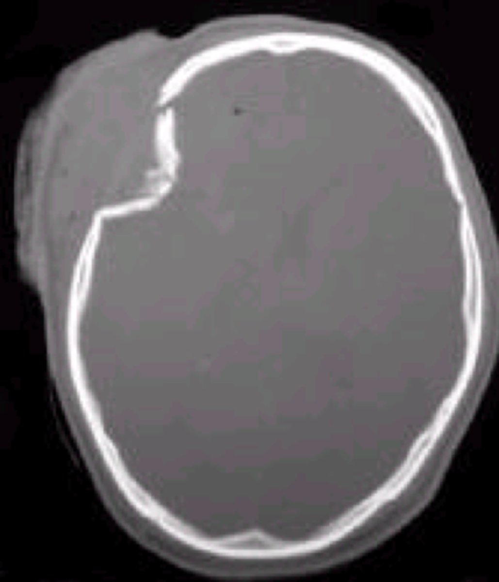
depressed skull fracture
type of skull fracture where a portion of the skull is pushed inward toward the brain due to trauma, often from a direct blow or impact to the head. The fracture can cause the bone to press against the brain tissue, leading to potential injury
meningitis
What condition is a patient at an increased risk for if they have a depressed skull fracture?
basilar skull fracture
a break in the bones at the base of the skull, often involving the bones around the brainstem, inner ear, and the cervical spine
patient must not blow nose, no NG or nasotracheal tubes, no BiPAP (the bones to stop air/foreign objects from going into brain is broken)
Which things must be avoided if a patient has a basilar skull fracture?
Battle's sign, raccoon eyes
What are two signs of a basilar skull fracture?
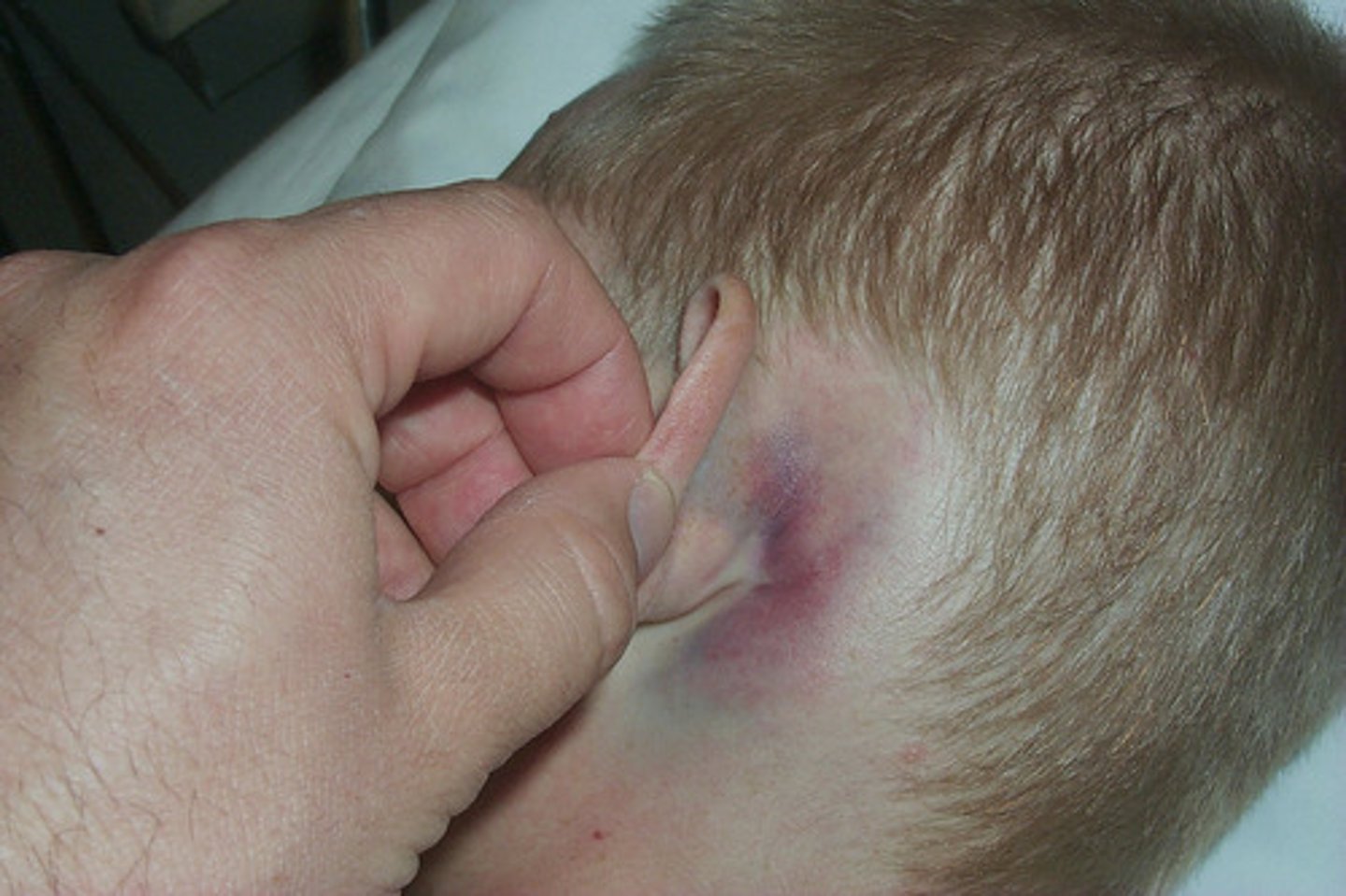
Battle's sign
Bruising behind the ears, indicative of a basilar skull fracture
raccoon eyes
Bruising around the eyes, indicative of a basilar skull fracture

concussion
a mild traumatic brain injury (TBI) that results from a blow or jolt to the head, causing the brain to move within the skull. It can temporarily affect brain function, including cognition, balance, and memory
brain rest (no hard "thinking," dark room)
What is the treatment for concussions?
short-term memory loss, HA, decreased attention, dizziness, irritability, difficulty with executive functioning, emotional lability, nausea
What are some manifestations of a concussion?
post-concussion syndrome
lingering symptoms from a concussion that last for an extended period of time (for a week up to a year)
amitriptyline
Which medication is used to treat headaches associated with post-concussion syndrome?
brain contusion
brain tissue bruise
cerebral edema can cause increased ICP 24-72 hours post injury
What is a complication of brain contusions?
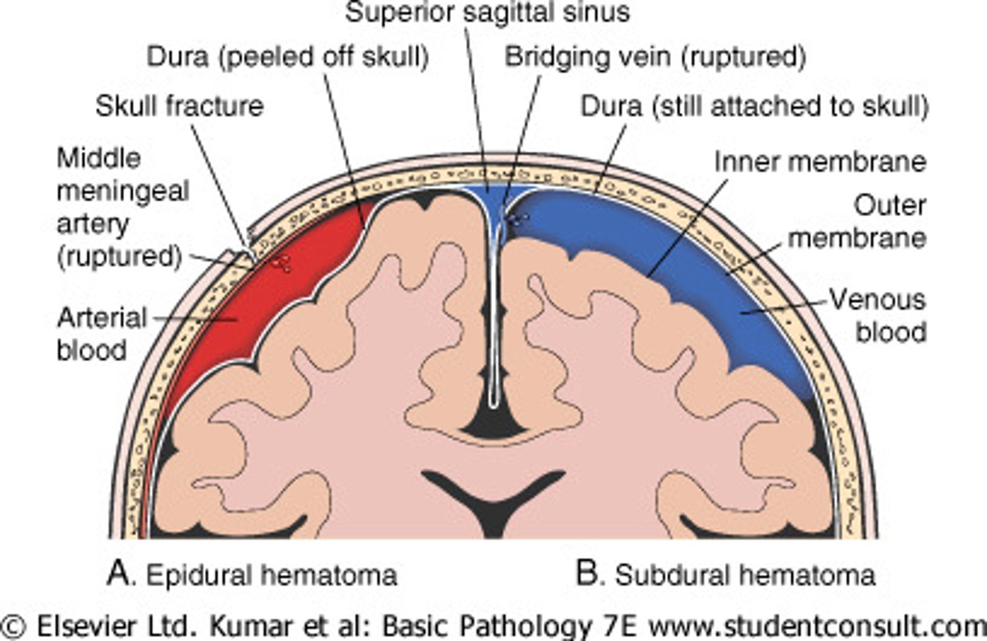
epidural hematoma
a collection of blood between the skull and the dura mater
arterial (bleed very quickly - can rapidly expand and cause herniation)
Are epidural hematomas caused by arterial or venous bleeds?
6-8 hours
How quickly do epidural hematomas reach their peak size?
"Talk and die syndrome" (patients seem normal for neuro assessments then rapidly decline)
Epidural hematomas are also called:
15-20%
What percentage of epidural hematomas are fatal?
subdural hematoma
a collection of blood between the dura mater (the outer layer of the brain's protective covering) and the arachnoid mater (the middle layer). It typically results from a tear in the veins that drain blood from the brain to the dural sinuses, causing blood to pool in the subdural space
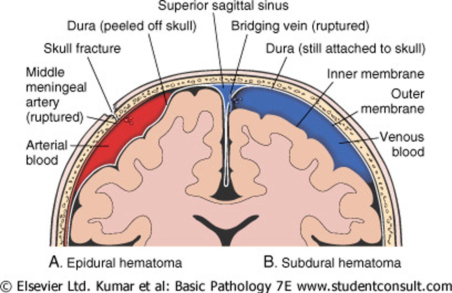
venous (slower bleeding)
Are subdural hematomas caused by arterial or venous bleeds?
24-48 hours post trauma
When do acute symptoms of a subdural hematoma show?