pediatric emergencies
 Call Kai
 Learn
Learn Practice Test
 Spaced Repetition
 Match
Match Flashcards
 Knowt Play
1/103

There's no tags or description
Looks like no tags are added yet.
Name | Mastery | Learn | Test | Matching | Spaced | Call with Kai | Chat |
|---|
No analytics yet
Send a link to your students to track their progress
104 Terms
Early/compensated shock: Tachycardia, cool extremities, delayed cap refill, weak peripheral pulses
Late/decompensated shock:
Hypotension
→ altered mental status
→ bradycardia/cardiac arrest
late ominous sign of circulatory failure in peds
Severe distress: nasal flaring, head bobbing, inability to speak/cry, cyanosis.
signs of airway distress
perform CPR for 2 min, get AED
⭐ PALS board rule: If HR <60/min + poor perfusion despite adequate oxygenation/ventilation → START CPR (chest compressions).
Think: Pediatric bradycardia = usually hypoxia → pre-arrest sign.
pulse less than 60 in child inn distress indcates
UNSTABLE: atrial fibrillation/flutter
UNSTABLE: supra ventricular tachycardia (SVT)
ventricular tachycardia with a pulse (w/o pulse = defib)
synchronized cardioversion is the electrical management of choice for
Supraventricular tachycardia
-most common tachyarrhythmia
-may be stable or unstable
-abrupt onset, no rate variability
-no P waves, short QRS
->220 bpm in infants, >180 in older children
clinical signs of SVT
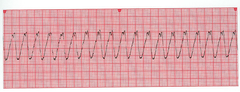
ventricular tachycardia
A rapid heart rhythm in which the electrical impulse begins in the ventricle (instead of the atrium), which may result in inadequate blood flow and eventually deteriorate into cardiac arrest.
-IV access (±adenosine)
-synchronized cardioversion
🟢 Stable SVT → adenosine after vagal maneuvers.
🔴 Unstable SVT → synchronized cardioversion FIRST (0.5–1 J/kg, then 2 J/kg).
In unstable SVT, adenosine can be considered if IV/IO access is already available, especially for regular narrow-complex SVT, but do not delay cardioversion.
management of unstable SVT
perform synchronized cardioversion if pulse is present
defib if no pulse!
management of unstable ventricular tachy
vagal maneuvers----> consider adenosine IV
management of stable SVT
adenosine if the rhythm is regular and uniform QRS
PER PALS:
Situation | Management |
|---|---|
Stable VT, pulse present | Antiarrhythmic + expert consultation |
Medication options | Amiodarone OR procainamide |
Becomes unstable | ⚡ Synchronized cardioversion |
Becomes pulseless VT | ⚡ Defibrillation + CPR |
PALS doses:
Amiodarone: 5 mg/kg IV/IO over 20–60 min
Procainamide: 15 mg/kg IV/IO over 30–60 min
⚠ Do NOT routinely give amiodarone and procainamide together → risk of hypotension/QT prolongation.
⭐ Boards:
Stable VT + pulse → amiodarone/procainamide
Unstable VT + pulse → synchronized cardioversion
Pulseless VT → defibrillation + CPR
management of stable ventricular tachycarddia
(stable = pulse+ & no cardiopulm compromised)
1 rescuer → 30:2 compressions:breaths
2 rescuers → 15:2
ratio for CPR in pediatrics if no pulse of HR
Shock phenotype | What you see | Preferred vasoactive agent |
|---|---|---|
🥶 Cold shock | Low cardiac output, ↑ SVR → cool extremities, delayed cap refill, weak pulses | Epinephrine |
🔥 Warm shock | ↓ SVR → warm/flushed extremities, bounding pulses, wide pulse pressure | Norepinephrine |
Epinephrine → β₁ → ↑ contractility + cardiac output → helps cold/low-output shock
Norepinephrine → α₁ → ↑ vasoconstriction/SVR → helps warm/vasodilatory shock
adrenergic treatment for cold vs warm shock
1st dose: 2 J/kg
→ 2nd dose: 4 J/kg
→ ≥4 J/kg for subsequent (max 10 J/kg or adult dose).
dose of defribilllators
croup, diptheria
Croup | Diphtheria | |
|---|---|---|
Cause | Parainfluenza virus most commonly | Corynebacterium diphtheriae |
Classic age | 6 months–3 years | Usually unvaccinated/under-vaccinated children |
Cough | ⭐ Barking “seal” cough | Not typically barking |
Stridor | Inspiratory stridor | Can occur if severe obstruction |
Voice | Hoarse | Hoarse/muffled |
Throat | No pseudomembrane | ⭐ Gray, adherent pseudomembrane that bleeds if scraped |
Neck | Usually normal | ⭐ “Bull neck” cervical LAD/edema |
X-ray | ⭐ Steeple sign | Upper-airway narrowing may occur |
Treatment | Dexamethasone ± nebulized epinephrine | Antitoxin + erythromycin/penicillin + isolation |
Prevention | No routine parainfluenza vaccine | DTaP/Tdap |
Important: Diphtheria toxin inhibits EF-2 → stops protein synthesis and can cause myocarditis and neuropathy.
Ddx for pt with barking cough, hoarse voice
IM Epinephrine (0.01 mg/kg, max 0.3-0.5 mg) into lateral thigh immediately.
Airway support + O₂.
IV fluids for hypotension.
Adjuncts: antihistamines, corticosteroids, inhaled bronchodilators.
Observe for biphasic reaction (≥4-6 hr).
management of anaphylaxis
Severe asthma unresponsive to initial bronchodilator.
Tachypnea, prolonged expiratory phase, wheezing → silent chest (ominous).
Hypoxemia, accessory muscle use.
presentation of status asthmaticus
Supportive: Nasal suction, O₂ if sats
management of bronchiolitis
Complete obstruction:
Infants <1 yr: back blows + chest thrusts.
Children >1 yr: abdominal thrusts (Heimlich).
Partial obstruction: Allow spontaneous coughing, avoid blind sweeps.
Rigid bronchoscopy = definitive removal.
management of foreign body aspiration
Mild: Humidified air, oral dexamethasone.
Moderate-severe (stridor at rest): Dexamethasone + nebulized racemic epinephrine.
Intubation if severe obstruction unresponsive to treatment.
management of croup
diabetic ketoacidosis
Kussmaul breathing = deep, rapid, labored breathing caused by severe metabolic acidosis.
The body is trying to blow off CO₂ → ↓ CO₂ → helps raise the pH.
Example presentation:
Diabetic patient + hyperglycemia + dehydration + fruity/acetone breath + deep rapid breathing → DKA with Kussmaul respirations
DKA → ketoacids ↑ → HCO₃⁻ ↓ → pH ↓ → Kussmaul breathing → CO₂ ↓.
Polyuria, polydipsia, weight loss, abdominal pain, vomiting.
Kussmaul respirations, dehydration, altered mental status.
Labs: hyperglycemia, metabolic acidosis (low bicarb, low pH), ketonemia/ketonuria
Airway, IV access.
Fluids: 10-20 mL/kg isotonic saline bolus → careful correction to avoid cerebral edema.
Insulin infusion: 0.1 units/kg/hr (after fluids).
Electrolyte monitoring: Replace K⁺ once urine output confirmed (even if serum normal/low)
management of DKA
If symptomatic: IV dextrose bolus (0.5-1 g/kg)
(D10 for infants, D25-50 for children).
glucagon if unable to get iv access
If mild and alert: oral glucose if able to swallow.
Investigate underlying cause (endocrine, metabolic, insulin excess).
management of hypoglycemia
Elevate head of bed, maintain normoxia and normocapnia.
3% HYPERtonic saline or mannitol if acute decompensation.
Neurosurgery consult for shunt/hematoma evacuation.
Avoid hypotension, hypoxemia, and excessive fluids.
management of increased intracranial pressure
tachycardia
earliest sign shock
Benzodiazepines
tx for stimulant induced seizures
irritability, tremors, yawning, diarrhea, poor feeding
signs of opioid withdrawal
tremors, seizures, agitation.
signs of alcohol/benzo withdrawal
BRUE = Brief Resolved Unexplained Event 👶
Sudden, brief, now resolved episode in an infant <1 yr, characterized by ≥1 of:
Cyanosis/pallor
Absent, decreased, or irregular breathing
Marked change in tone (hyper- or hypotonia)
Altered responsiveness
By definition, infant returns to baseline after event
(dx of exclusion)
term, >60 days, first event, short duration, no CPR, no concerning hx/exam
-manage with reassurance
criteria of a low risk BRUE
premature, recurrent, abnormal exam, family history of sudden death
-admit these pts for monintoring, address underlying cause
criteria of a high risk BRUE
GER (physiologic): Spit-ups in infants, no distress, normal growth.
GERD (pathologic): Poor weight gain, irritability, feeding refusal, respiratory symptoms (apnea, wheeze, aspiration).
differentiate GER and GERD
bronchiolitis, pneumonia
examples of LRTI
seizure >5min or recurrent without recovery
define status epilepticus
1st line: IV/IM/PR benzodiazepine (lorazepam, diazepam, midazolam).
2nd line: IV fosphenytoin, valproate, levetiracetam.
3rd line: Intubation + anesthetic agents (midazolam, pentobarbital).
medical management of status epilepticus
Sudden, severe intermittent colicky abdominal pain (knees to chest).
"Currant jelly stool" (blood + mucus).
Palpable "sausage-shaped" abdominal mass.
Vomiting (may be bilious).
clinical presentation of intussusception
Immediate stabilization (IV fluids, NG decompression if needed).
1st image: U/S —> target/donught sign
Diagnostic + therapeutic air/contrast enema (if stable, no perforation).
Surgery if enema fails, signs of perforation, or peritonitis.
treatment of intussusception
Painless rectal bleeding (melena or bright red blood).
Sometimes presents with obstruction, diverticulitis, or mimics appendicitis.
Cause: Failure of the vitelline (omphalomesenteric) duct to completely obliterate during embryologic development.
Normally: Vitelline duct disappears → if proximal portion persists → Meckel diverticulum.
Occurs due to ectopic gastric mucosa secreting acid —> adjacent ileal ulceration —> bleeding
presentation of meckel's diverticulum
Meckel's scan (technetium-99m pertechnetate).
diagnostic modality of meckel's diverticulum
Intermittent painless rectal bleeding.
Often benign juvenile polyps (age 2-10).
Rarely, multiple polyps → polyposis syndromes with malignancy risk.
presentation of juvenile polyps
Protrusion of rectal mucosa through anus, esp. during straining.
Associated with constipation, cystic fibrosis, chronic diarrhea, malnutrition
presentation of rectal prolapse
Acute episode: Manual reduction (lubrication, gentle pressure).
Recurrent/prolonged: Surgical repair.
management of rectal prolapse
infection (esp. Pasteurella, anaerobes), rabies, tetanus.
main infectious risks from animal bites
Face 🙂 | Extremity 🖐🦵 | |
|---|---|---|
Irrigation | ✅ Copious irrigation | ✅ Copious irrigation |
Debridement | As needed | As needed |
Primary closure | ✅ Usually close for cosmetic benefit | ⚠ Usually leave open if infection risk is significant |
Infection risk | Lower due to excellent blood supply | Higher, especially hand bites |
Antibiotic prophylaxis | Often indicated for moderate/severe facial bites | Especially indicated for hand, deep/puncture, high-risk wounds |
First-line antibiotic | Amoxicillin-clavulanate | Amoxicillin-clavulanate |
face vs extremity wound care from animal bite
History of head trauma with:
Headache, confusion, dizziness, amnesia, nausea/vomiting, photophobia.
NO focal neuro deficits, seizure, or prolonged loss of consciousness.
typical presentation of concussion
prolonged LOC, worsening headache, repeated vomiting, seizure, focal deficit →
^^^ if present → CT scan.
red flags of concussion
Physical & cognitive rest for 24-48 hrs.
Gradual return to school & sports once symptom-free, under medical supervision. STEP-WISE APPROACH
Step | Management |
|---|---|
1. Rule out emergency | Assess ABCs, neuro exam, GCS; look for signs of intracranial injury |
2. Imaging? | CT is NOT routine; use clinical decision rules (e.g., PECARN) when indicated |
3. First 24–48 hr | Relative physical + cognitive rest; avoid activities that significantly worsen symptoms |
4. Symptoms | Acetaminophen can be used for headache; maintain hydration and normal sleep |
5. Return to school | Gradual return within ~1–2 days as tolerated; accommodations if symptomatic |
6. Return to sports | No same-day return to play. Gradual stepwise return only after return to regular activities/school and symptoms have resolved |
7. Follow-up | Monitor symptoms and recovery; reassess if prolonged/worsening |
management of concussion
MI, left ventricular wall rupture, acute VSD, CHF, massive PE, over dose on beta blockers
causes of cardiogenic shock
neurogenic shock
occurs when there is damage to the brain or spinal cord that inhibits transmission of neural stimuli to arteries and arterioles, which reduces vasomotor tone
spinal cord injury, ANS toxins, spinal anesthesia, GBS
causes of neurogenic shock
IV fluids
O2 therapy
vasopressors and inotropes
tx of cardiogenic shock
hypotension, tachycardia, shallow respirations, oliguria, confusion, cool, clammy skin
signs of cardiogenic shocck
IV fluid therapy
O2 therapy
blood transfffusion
tx of hypovolemic shock
hypotension, tachycardia, shallow respirations, oliguria, oligguria restlessness, cool./clammy skin
siggns of hypovolemic shock
hypotension, tachycardia, increased respirations, oliguria, fever
flushed and warm skin
signs of septic shock
hypotension, tachycardia, bronchoconstriction, oliguria, LOC, hives localized edema
signs of anaphylactic shock
IV fluid therapy, O2 therapy, inotropic agennts
-atropine if severe bradycardia
tx of neurogenic shock
hypotension, bradycardia, no bladder control, LOC
dry, warm skin
signs of neurogenic shock
neurogenic, anaphylactic, septic
types of shock associated with warm skin
obstructive shock
Shock that occurs when there is a block to blood flow in the heart or great vessels, causing an insufficient blood supply to the body's tissues.
Pulmonary embolism, tension pneumo, pericardial tamponade, restrictive cardiomyopathy
causes of obstructive shock
distributive shock
shock due to severe peripheral vasodilation
-peripheries are warm
septic shock, anaphylaxis, neurogenic
causes of distributive shock
cardiogenic shock
Tachycardia, hypotension
Poor perfusion (cool extremities, delayed cap refill)
Signs of fluid overload: hepatomegaly, pulmonary rales, gallop rhythm
Common causes: myocarditis, congenital heart disease, arrhythmia
Exam: muffled heart sounds, distended neck veins (tamponade); unilateral decreased breath sounds, tracheal deviation (pneumothorax).
exam findings associated with obstructive shock
dissociative shock
*Type of shock*
Perfusion problems occur because the *blood has a decreased ability to carry oxygen* to the tissues.
Causes: CO poisoning , Anemia , Cyanide poisoning
toddlers 1-3 y/o
age of peak risk for accidental ingestion
dosing errors
risk of poisoning in inffants is usually due to
Blind as a bat (Mydriasis)
Mad as a hatter (Altered mental status)
Red as a beet (vasodilation, flushed)
Hot as a hare (febrile)
Dry as a bone (no secretions/diaphoresis)
Bowel and bladder lose their tone
Heart runs alone (tachycardia)
signs of anticholinergic syndrome
Atropine, antihistamines, scopalamine, antipsychotics
agents of anticholinergic syndrome
SLUDGE(salivation, lacrimation, urination, deification, GI distress, emesis).
Presents with pin point pupils, sweating, vomiting, frothing of mouth, bradycardia, urination, diarrhea
presentation of cholinergic toxidrome
insecticides
agents of cholinergic toxidrome
miosis, sedation, respiratory depression
signs of opioid toxidrome
miosis
constricted pupils
glucagon
-monitor potassium
tx of CCB and beta blocker overdose
sodium bicarb
tx of TCA overdose
Acetylcholinesterase inhibitors
Promote the accumulation of acetylcholine, resulting in prolonged cholinergic effects
DUMBBELSS: Diarrhea, Urination, Miosis, Bronchospasm, Bradycardia, Excitation (CNS), Lacrimation, Sweating, Salivation
cholinergic effefcts
-Atropine & pralidoxime
tx for organophosphate poisoning
Hot, dry skin; flushed; dilated pupils; urinary retention; delirium, hallucinations; tachycardia
signs of anticholinergic toxidrome
Supportive, benzodiazepines for agitation, physostigmine in severe cases (avoid if TCA overdose)
mgmt of anticholinergic toxidrome
CNS depression, pinpoint pupils, respiratory depression, hypotension, bradycardia
signs of opioid toxidrome
Agitation, hypertension, tachycardia, dilated pupils, sweating, hyperthermia
signs of sympathomimetic toxidrome
Cocaine, amphetamines, ADHD meds
causes of sympathomimetic toxidrome
Supportive, benzodiazepines, avoid beta-blockers
mgmt of sympathomimetic toxidrome
Benzodiazepines, barbiturates, ethanol
causes of sedative toxidrome
Supportive, flumazenil rarely (risk of seizures), airway support
tx of sedative toxiddrome
Agitation, clonus, hyperreflexia, hyperthermia, diaphoresis
signs of serotonin synddrome
SSRIs, MAOIs, MDMA, drug interactions
agents of serotonin syndrome
Stop agents, supportive, cyproheptadine for severe cases
mgmt of serotonin syndrome
Lead pipe rigidity, fever, altered mental status, autonomic instability
signs of neuroleptic malignant syndrome
antipsychotics
cause of neuroleptic malignant syndrome
Stop antipsychotics, supportive, dantrolene or bromocriptine
tx of neuroleptic malignant syndrome
Tinnitus, hyperventilation (resp alkalosis → metabolic acidosis), vomiting, confusion.
signs of salicylate poisoning
Serum acetaminophen level at 4 hrs post ingestion
→ Rumack-Matthew nomogram.
best tool to evaluate acetaminophen poisonin
Activated charcoal (early),
alkalinize urine with IV sodium bicarb, fluids,
hemodialysis if severe.
tx of salicylate poisoning
GI phase (vomiting, diarrhea, hematemesis),
latent phase, shock, metabolic acidosis, hepatotoxicity.
presentation of iron overdose
Deferoxamine chelation, supportive, GI decontamination if early.
mgmt of iron overdose
hydrocarbon (gasoline, lamp oil) poisoning
Presentation: Coughing, choking, vomiting, respiratory distress → risk of aspiration pneumonitis.
Management: Supportive only (O₂, ventilation PRN). No gastric lavage/charcoal (risk of aspiration).
Remove exposure; chelation (succimer if moderate, EDTA if severe)
tx of lead poisoning