Lecture 1-10 UCSD Extension Human Anatomy
1/295
There's no tags or description
Looks like no tags are added yet.
Name | Mastery | Learn | Test | Matching | Spaced |
|---|
No study sessions yet.
296 Terms
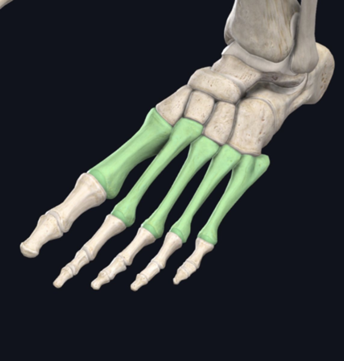
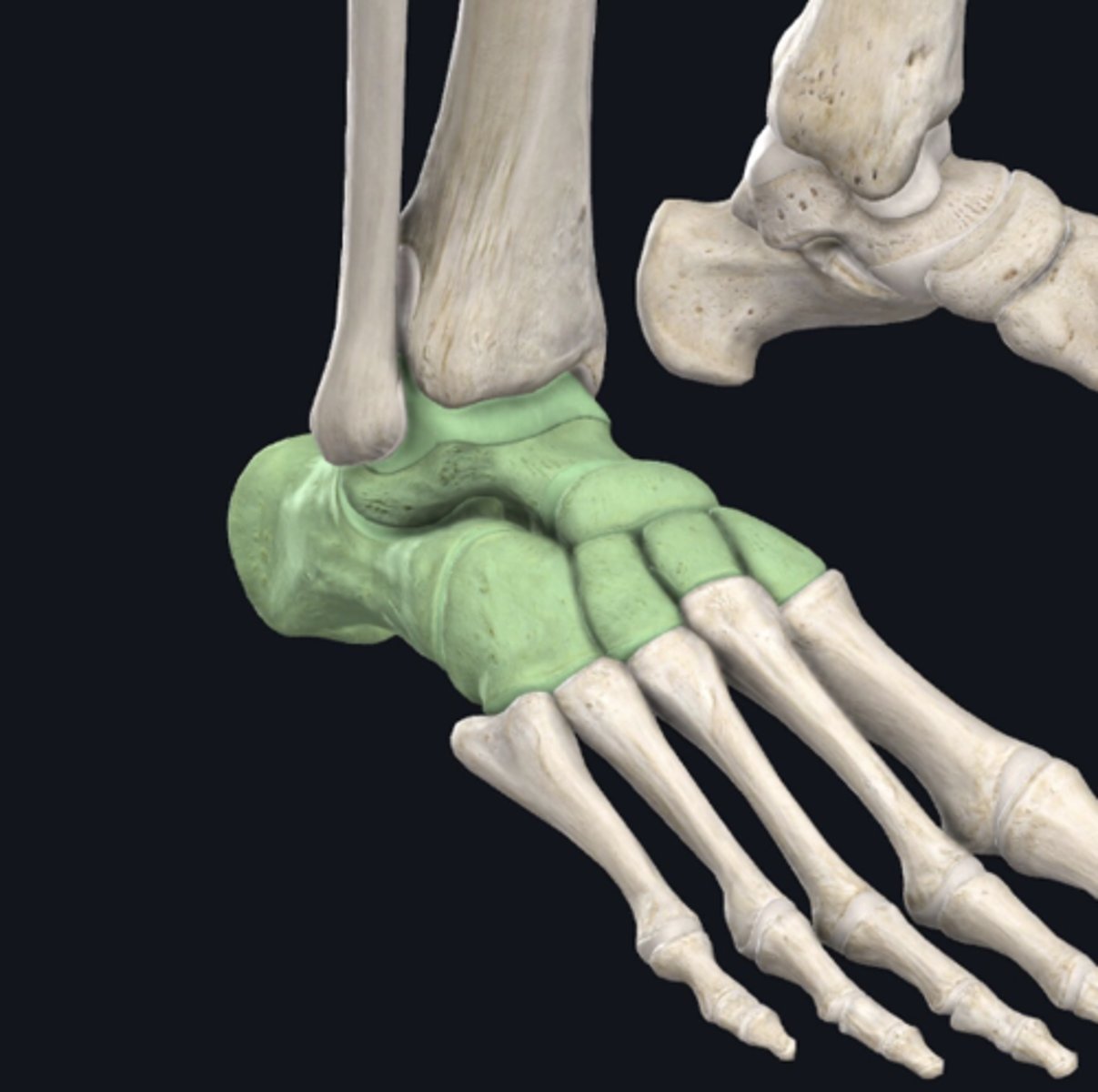
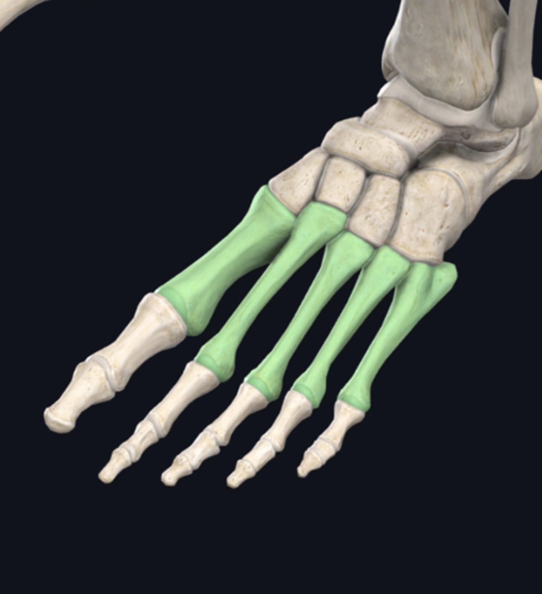
Tarsals
identify the group of bones indicated in green
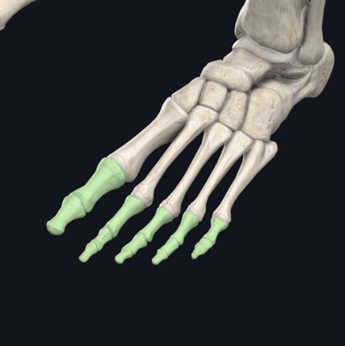
Phalanges
identify the group of bones indicated in green
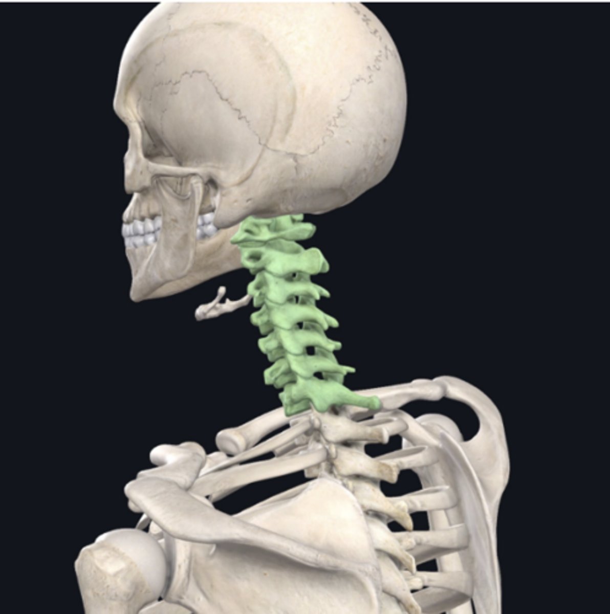
Cervical Vertebrae (C1-C7)
identify the group of bones indicated in green
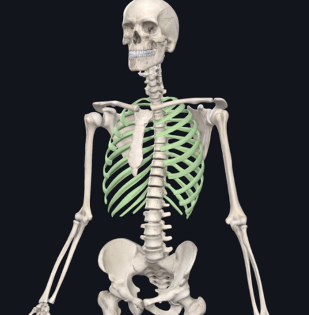
Ribs
identify the group of bones indicated in green
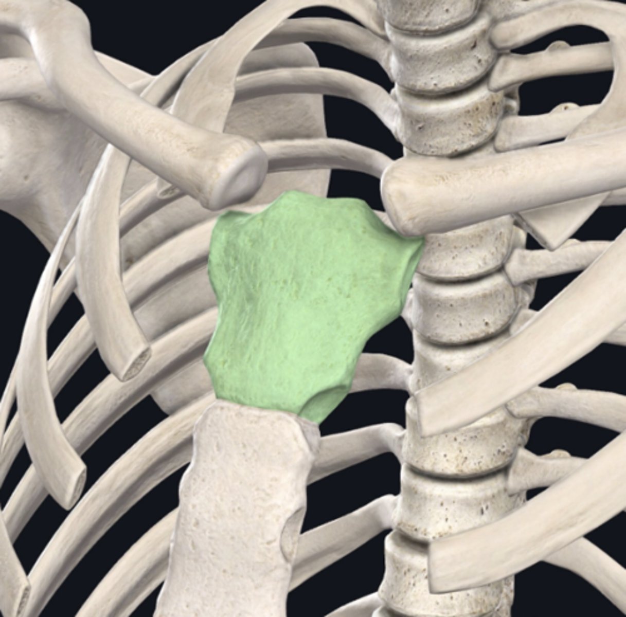
manubrium
identify the bone indicated in green
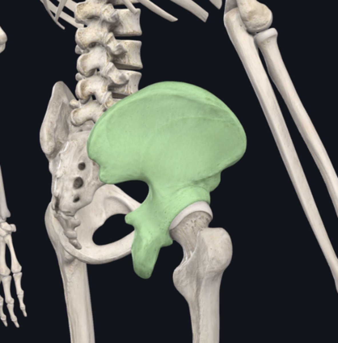
pelvic bone
Identify the bone indicated in green (you don't have to name the individual bone, but that would count too):
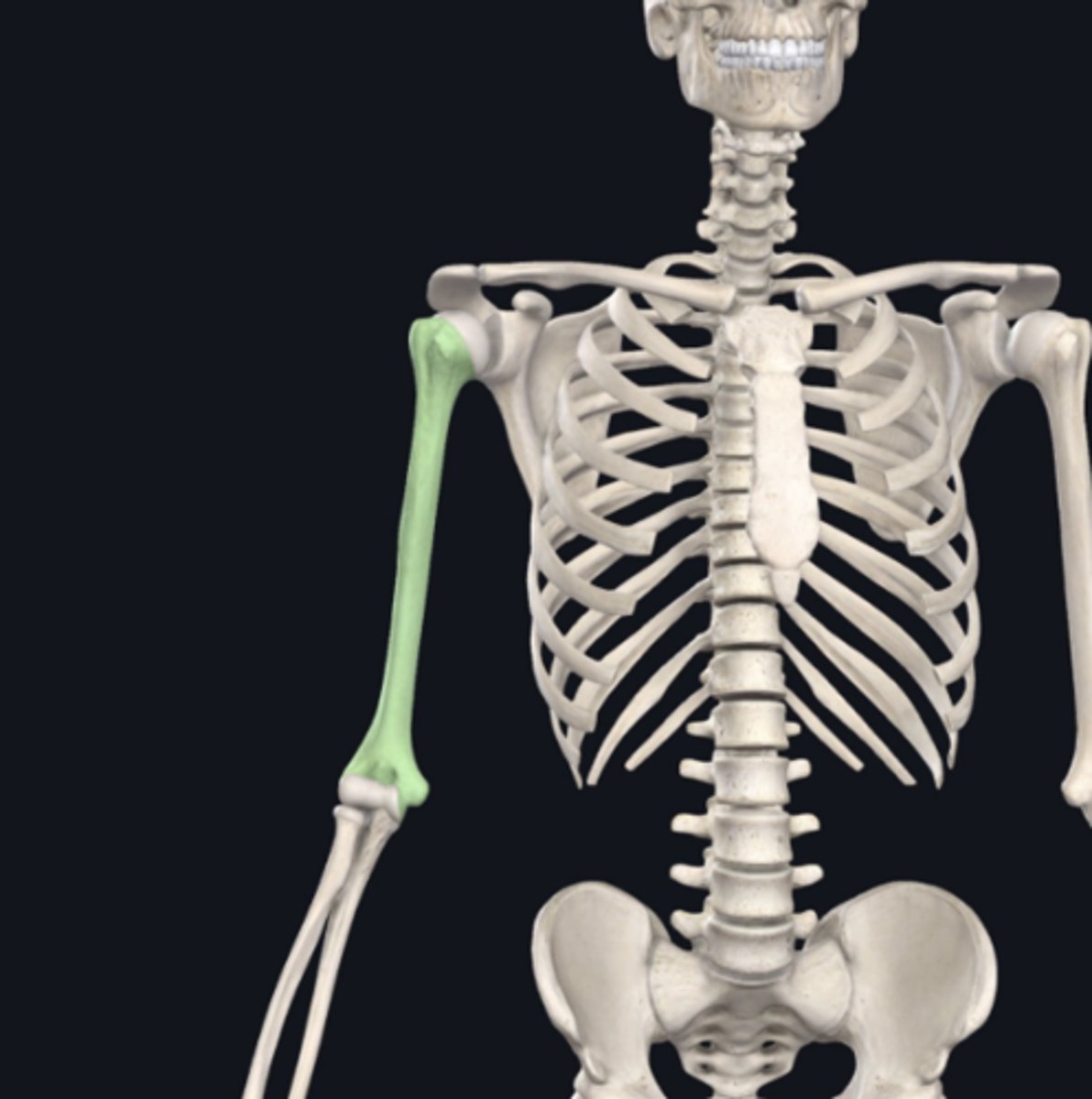
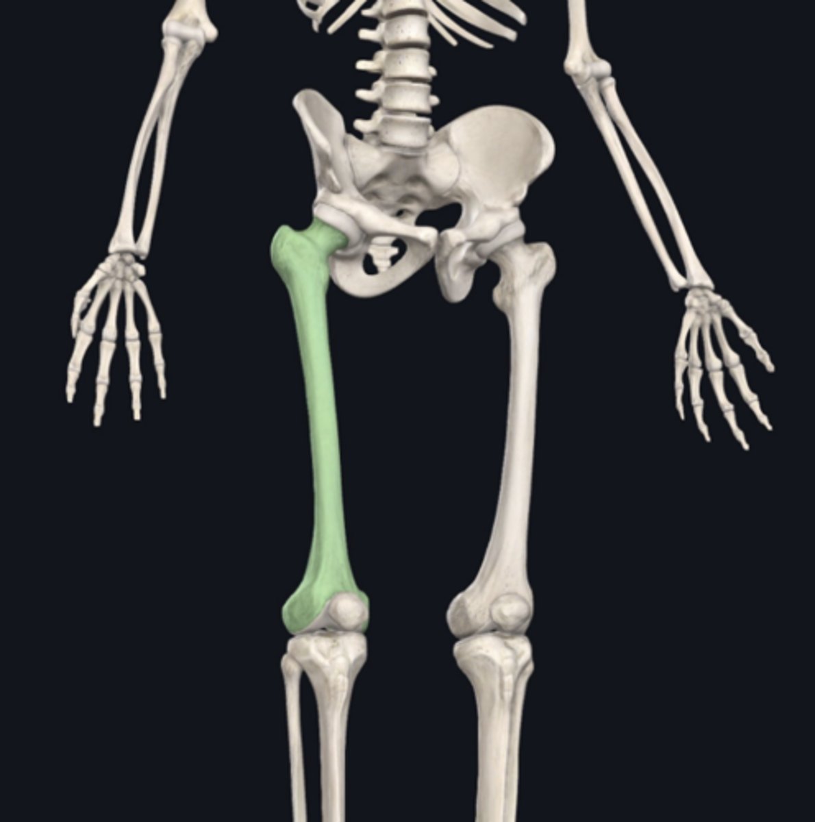
Humerus
Identify the bone indicated in green:
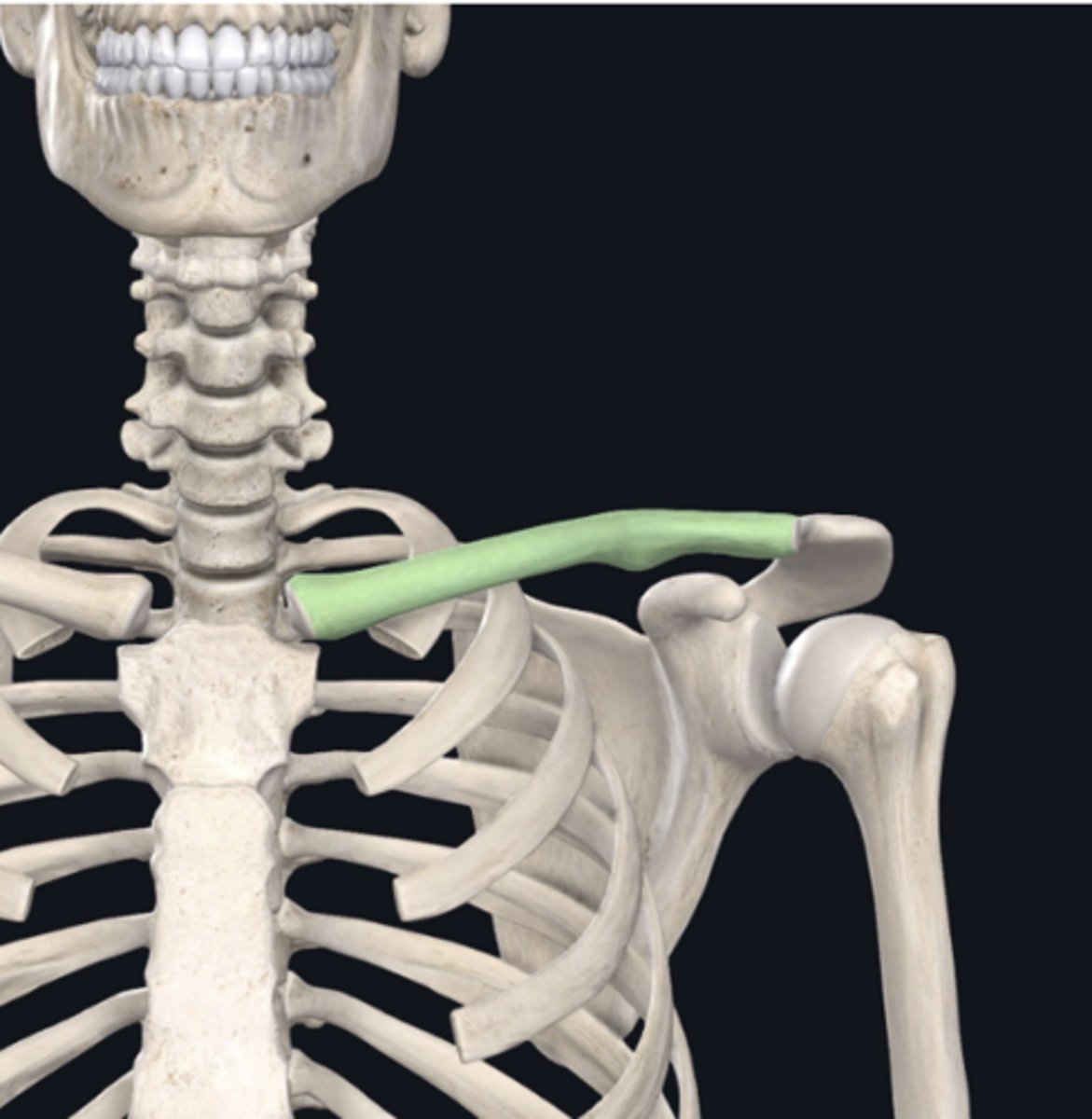
Clavicle
Identify the bone indicated in green:
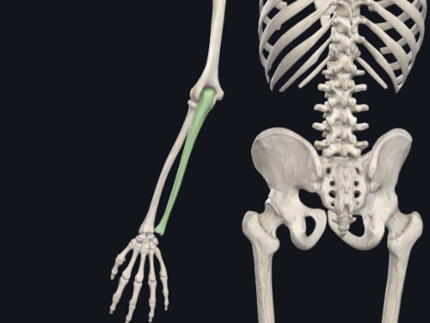
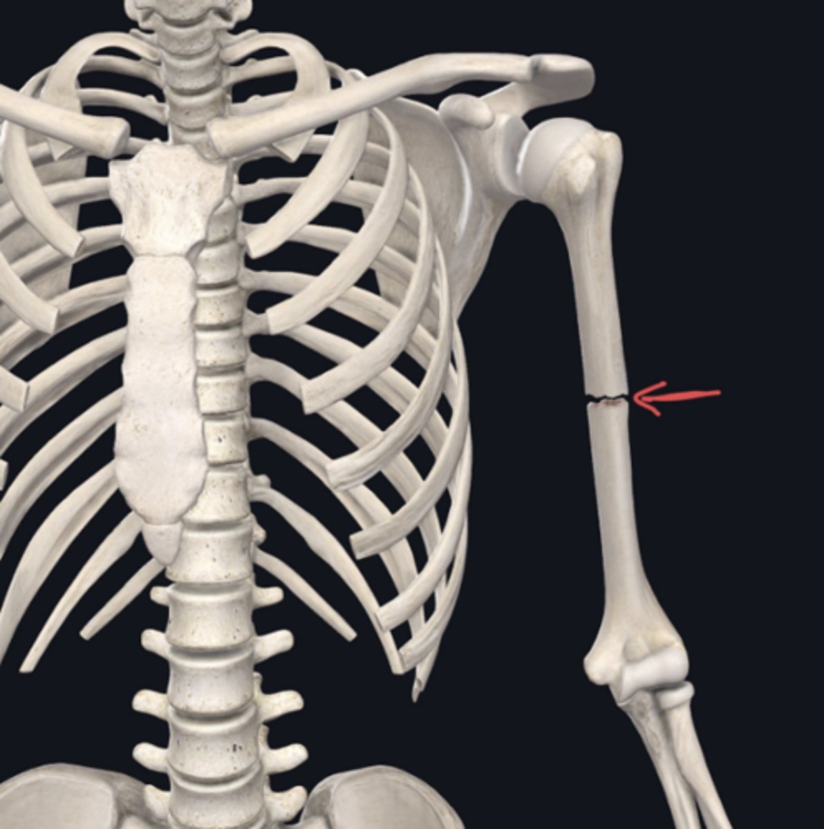
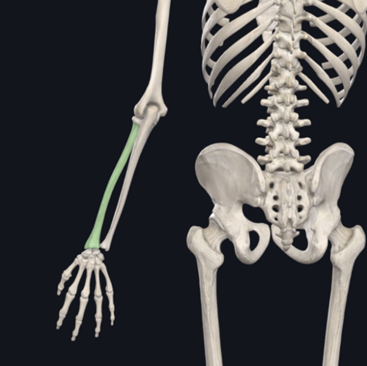
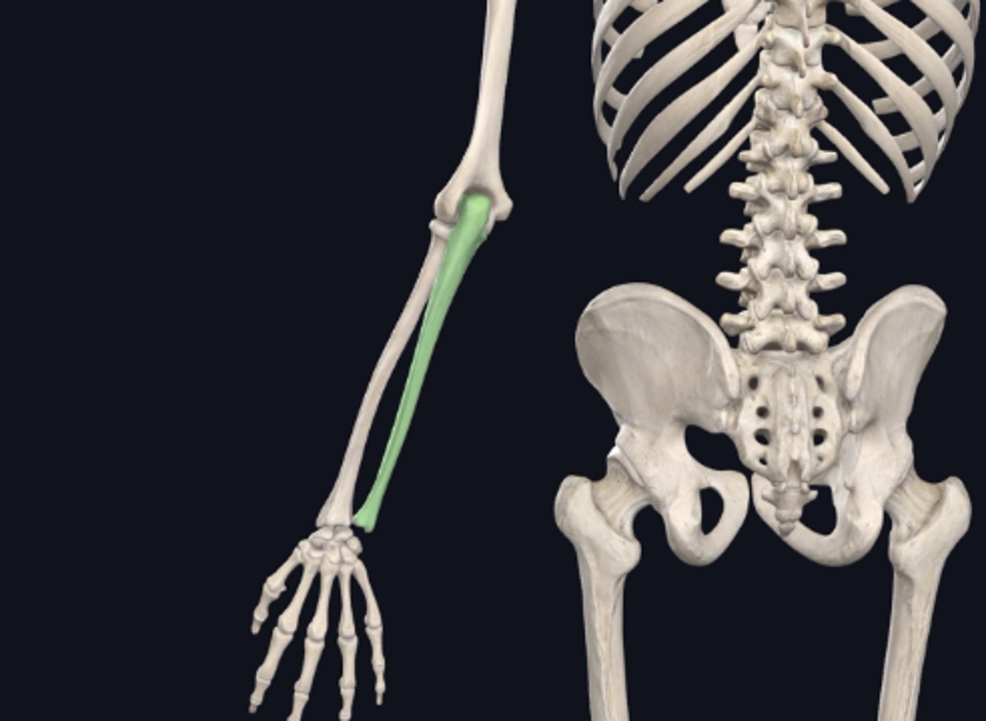
Ulna
Identify the bone indicated in green:
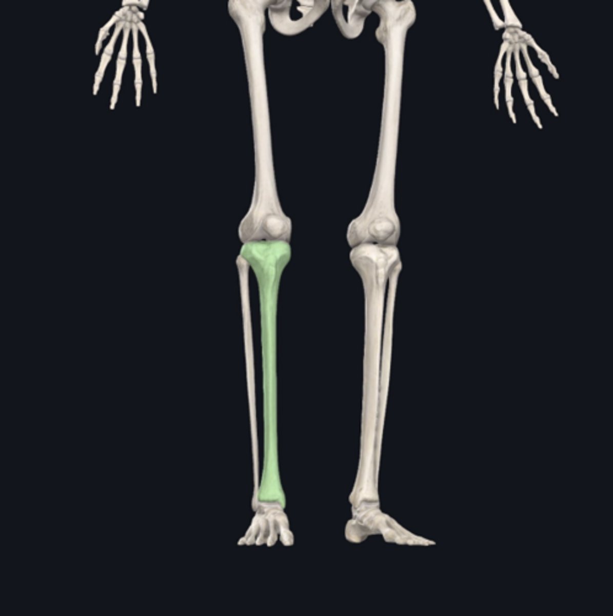
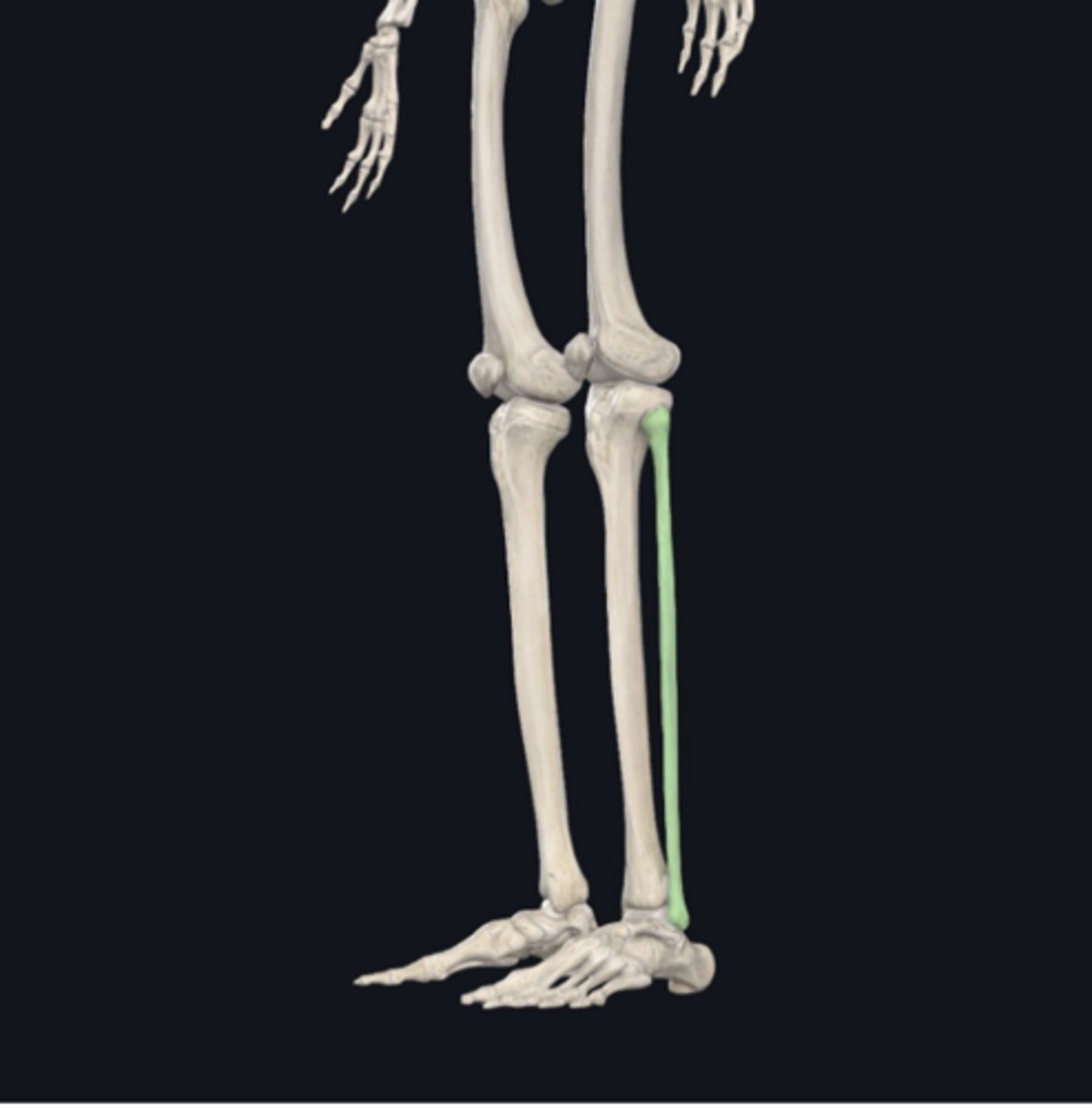
Tibia
Identify the bone indicated in green:
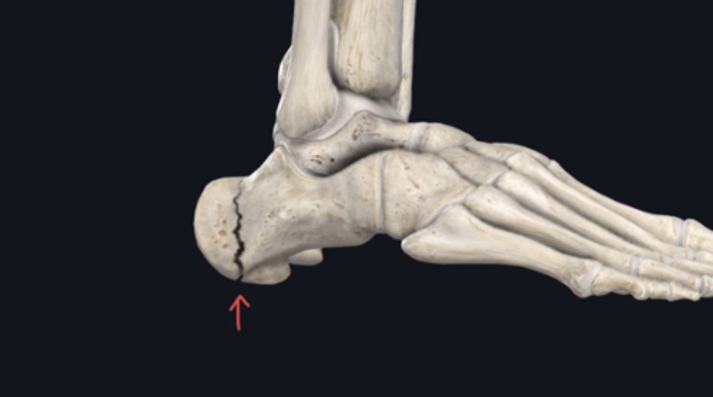
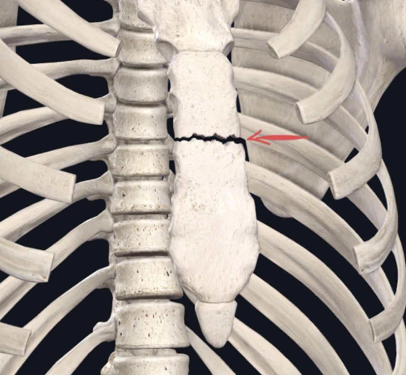
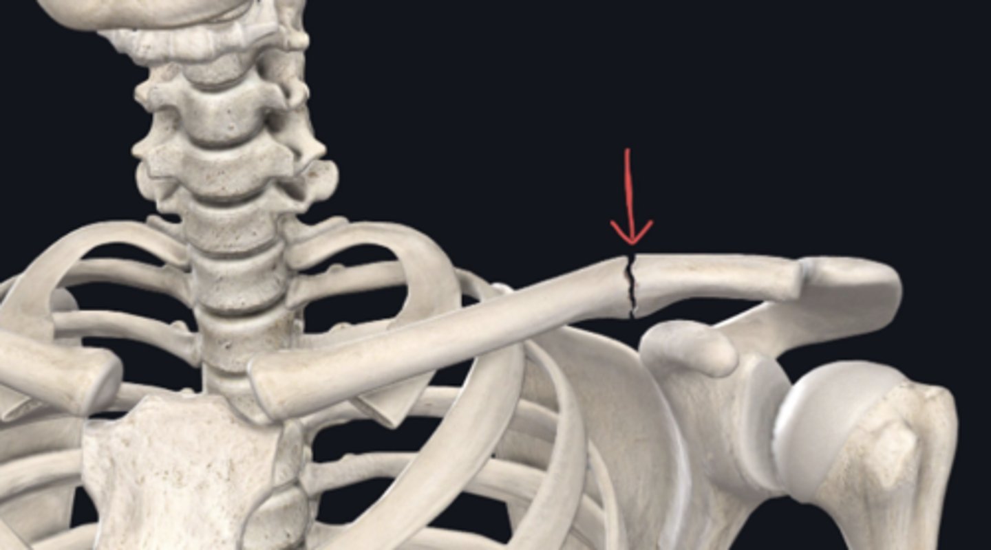
Coronal plane
Identify the plane of section in which the indicated fracture has occurred.
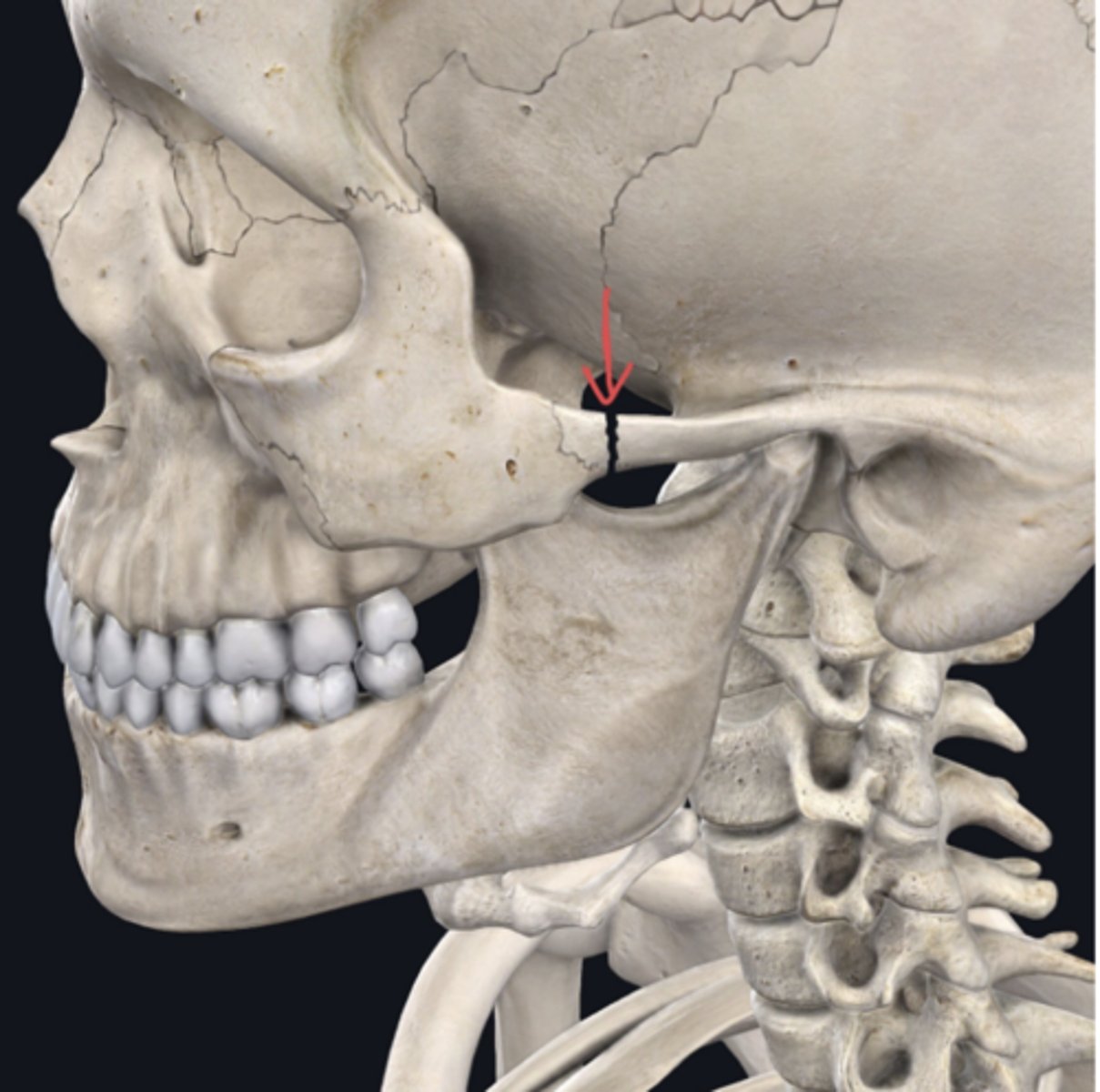
transverse plane (horizontal)
Identify the plane of section in which the indicated fracture has occurred.
longitudinal plane (Sagittal)
Identify the plane of section in which the indicated fracture has occurred.
Lower leg
Identify the region of the body containing the named structure below (for example, if the structure below is "Metatarsals," you would answer "Foot."
Tibia
Forearm
Identify the region of the body containing the named structure below (for example, if the structure below is "Metatarsals," you would answer "Foot."
Ulna
Thigh
Identify the region of the body containing the named structure below (for example, if the structure below is "Metatarsals," you would answer "Foot."
Femur
Thorax
Identify the region of the body containing the named structure below (for example, if the structure below is "Metatarsals," you would answer "Foot."
Manubrium
Hand
Identify the region of the body containing the named structure below (for example, if the structure below is "Metatarsals," you would answer "Foot."
Metacarpals
Distal
The tarsals are ________ to the patella:
Medial
Superficial
Deep
Proximal
Lateral
Distal
Lateral
Ribs 2-5 are ______ to the sternum:
Inferior
Superior
Deep
Superficial
Medial
Lateral
Cervical vertebrae
Which bone is most superior:
Humerus
Lumbar vertebrae
Clavicle
Cervical vertebrae
Fibula
Anterior
The sternum is ________ to the thoracic vertebrae:
Proximal
Posterior
Anterior
Distal
Axial
The sacrum is part of the _____ skeleton:
Amazing
Appendicular
Alien
Axial
Appendicular
The pelvic bone is part of the ____ skeleton:
Artificial
Appendicular
Astounding
Axial
Appendicular
The radius is part of the ____ skeleton:
Artificial
Appendicular
Astounding
Axial
Fibula
Identify the bone indicated in green. (Free response)
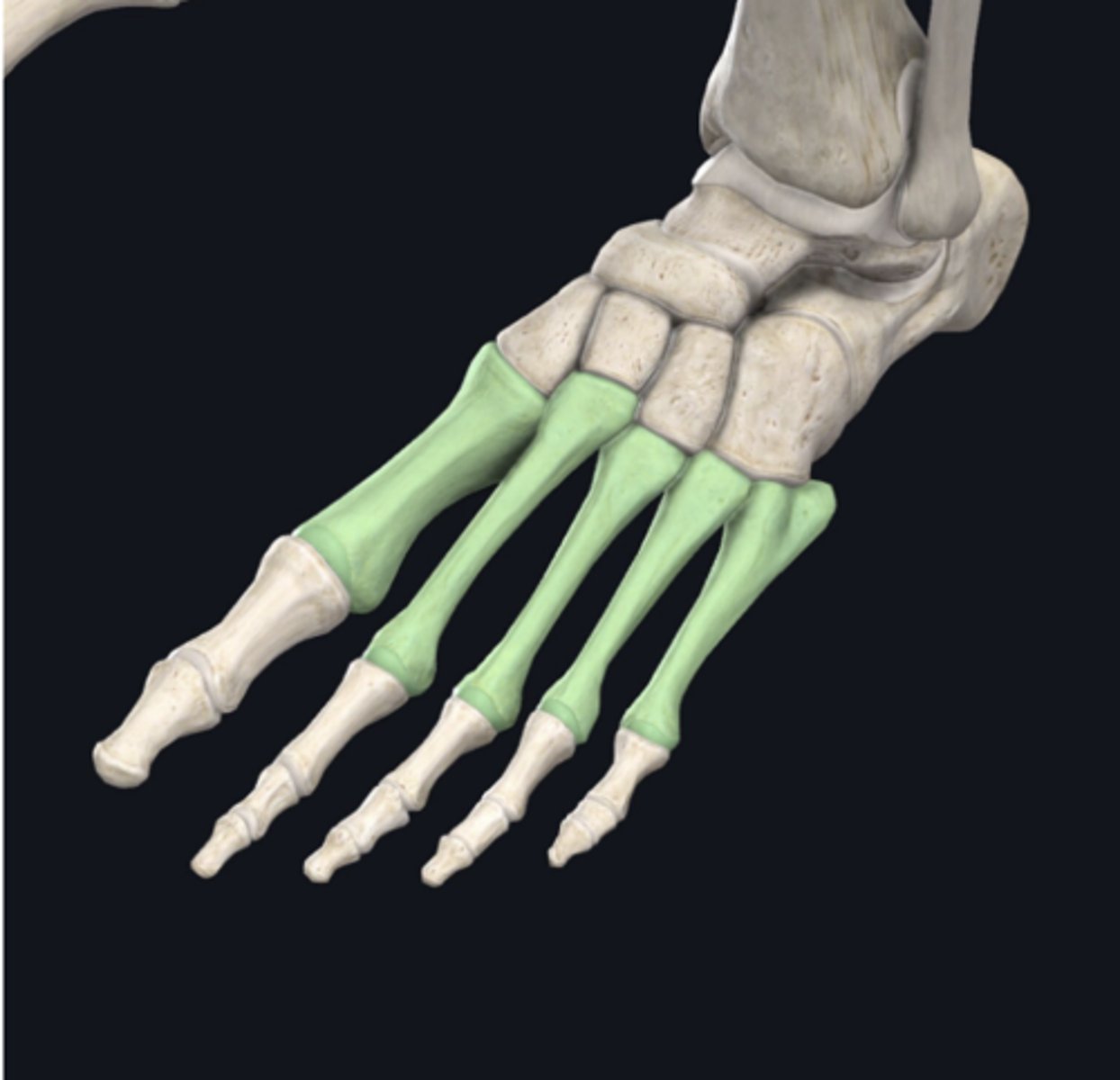
Metatarsals
Identify the bone indicated in green. (Free response)
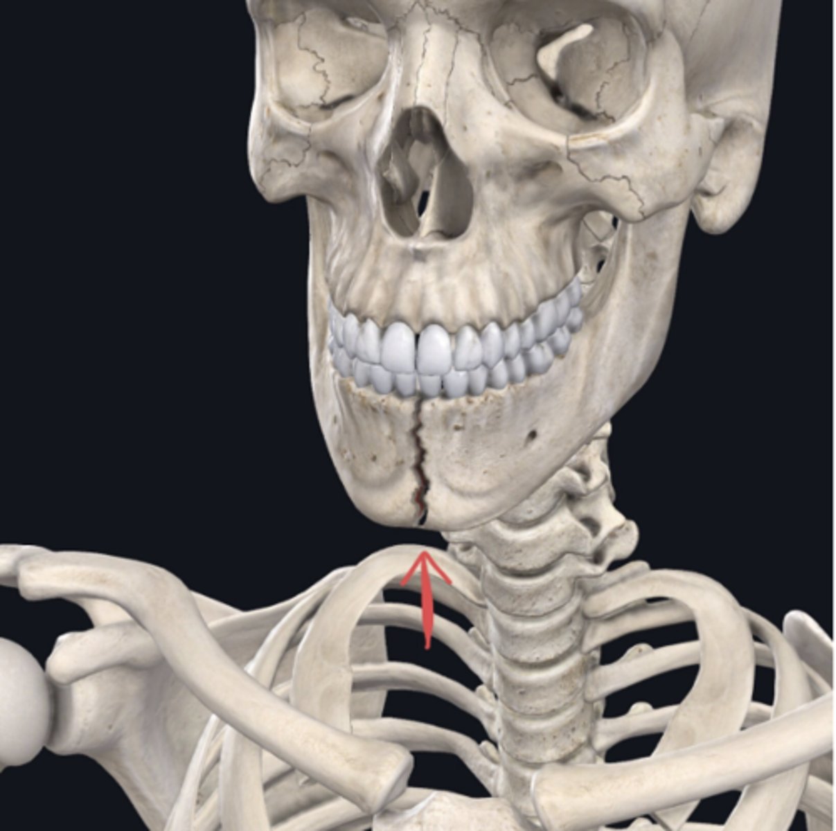
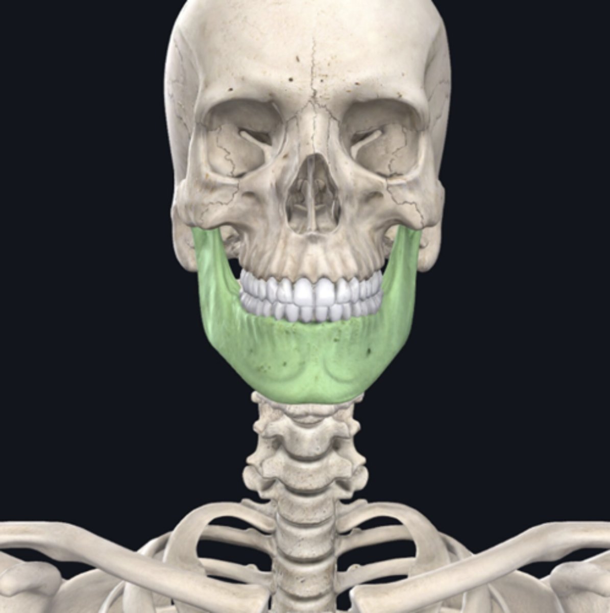
Mandible
Identify the bone indicated in green. (Free response)
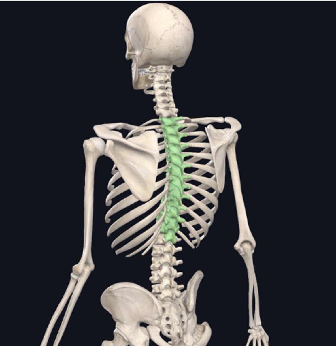
Thoracic vertebrae
Identify the bone indicated in green. (Free response)

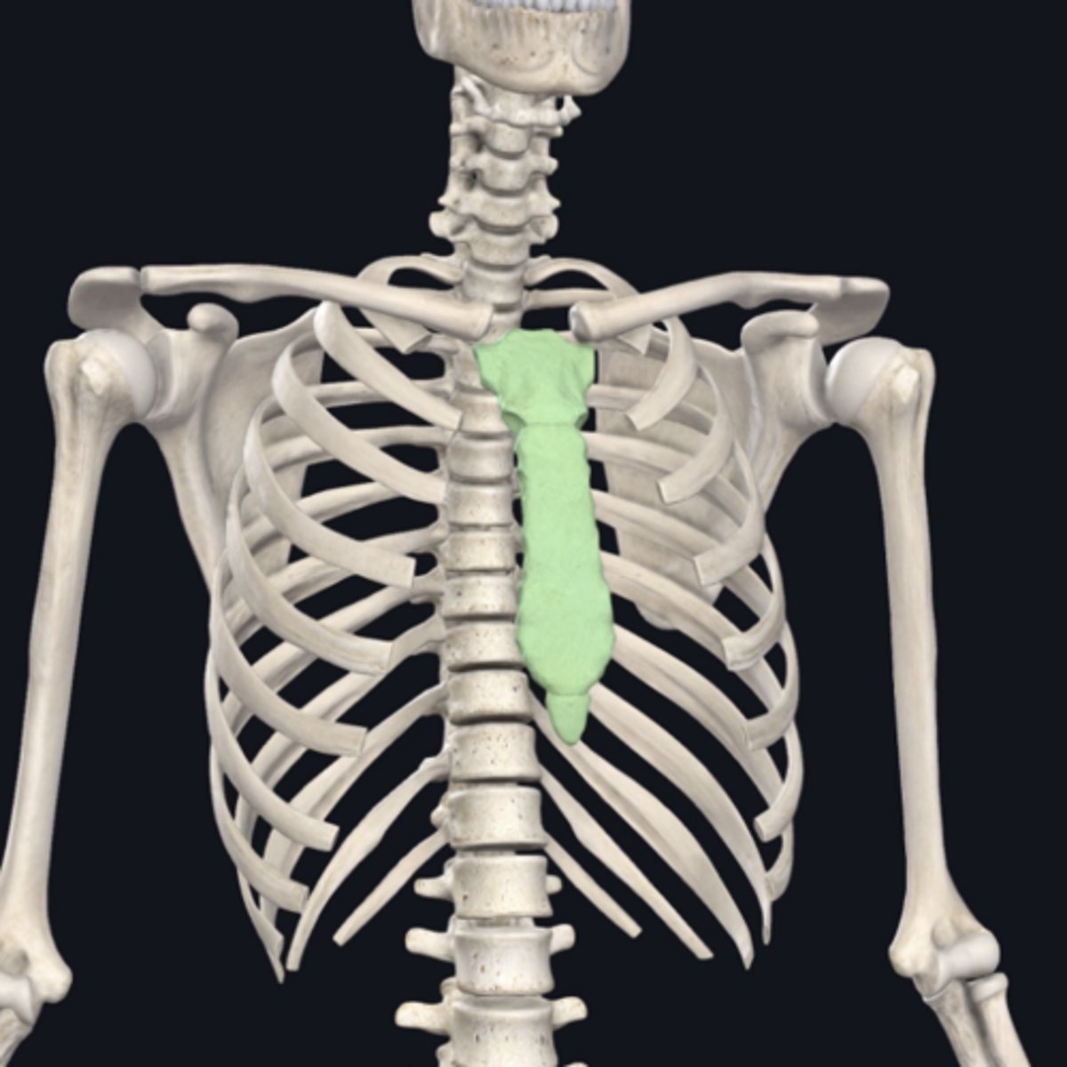
Sternum
Identify the bone indicated in green. (Free response)
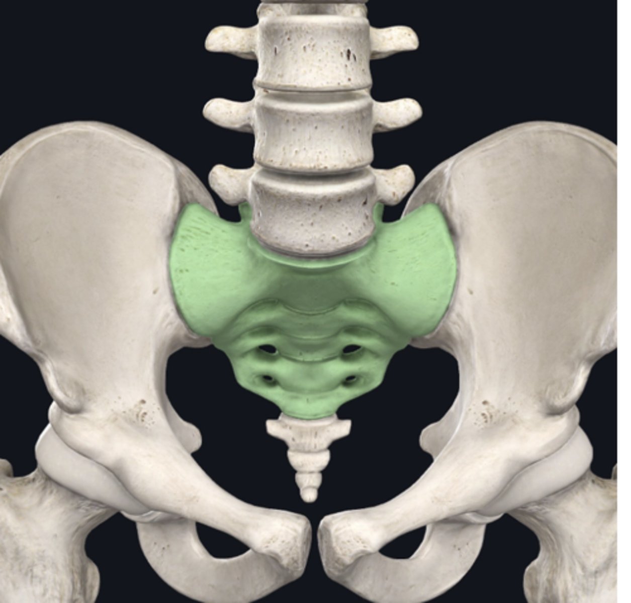
Sacrum
Identify the bone indicated in green. (Free response)
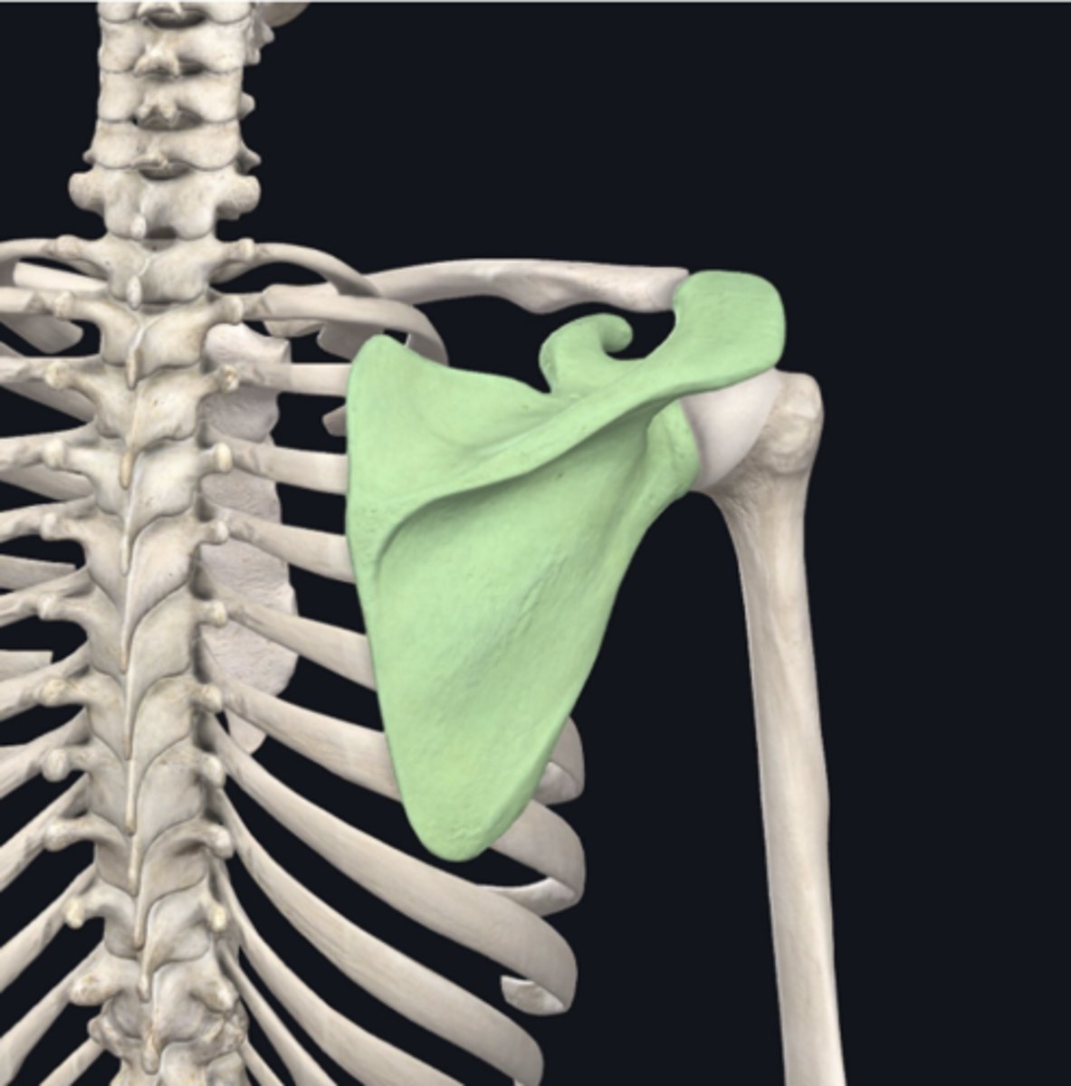
Scapula
Identify the bone indicated in green. (Free response)
Radius
Identify the bone indicated in green. (Free response)
Femur
Identify the bone indicated in green. (Free response)
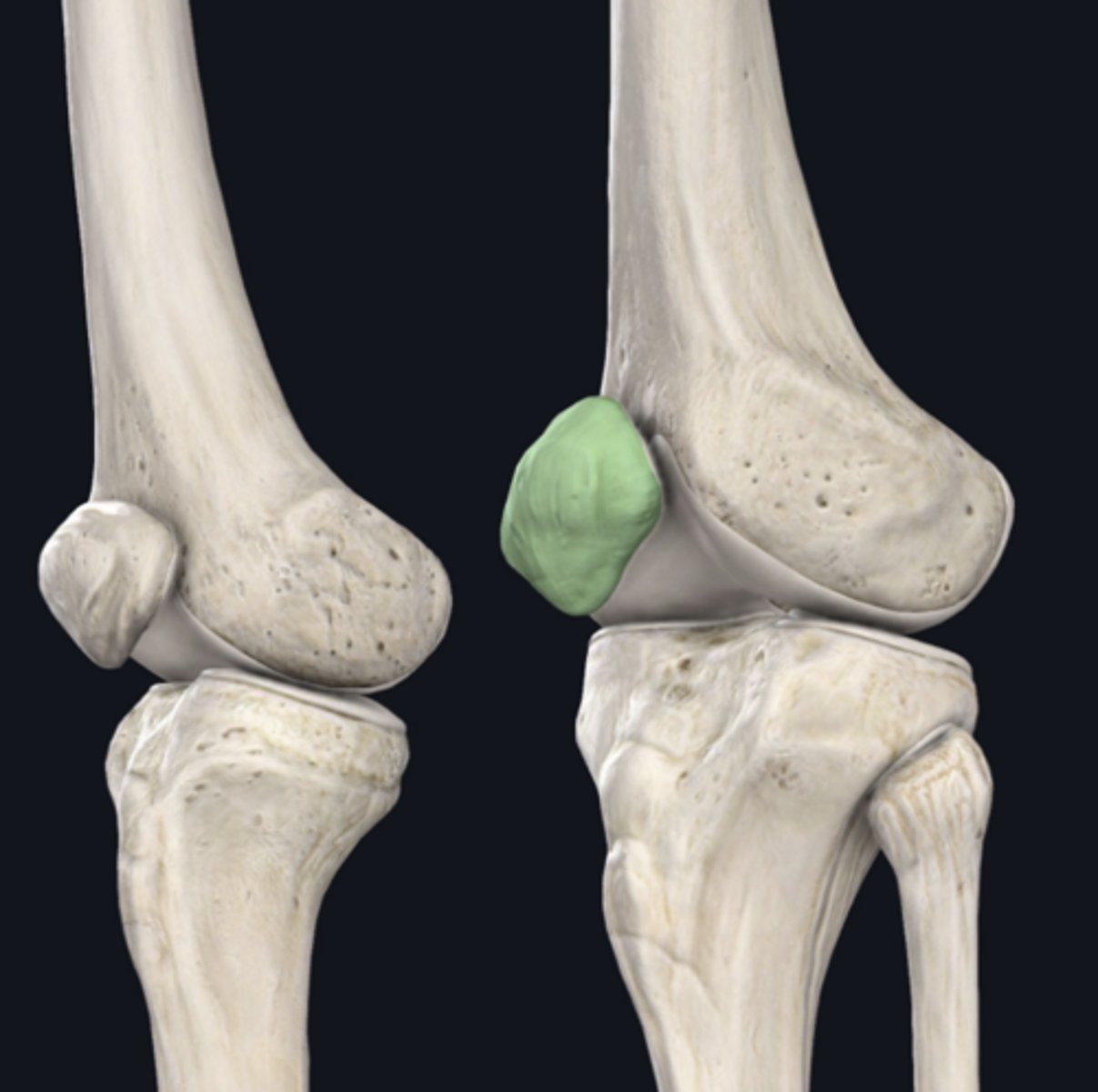
Patella
Identify the bone indicated in green. (Free response)
Transverse
(Note: Horizontal or axial would also be correct. Exam soft used in School ofMedicine only recognizes one correct answer, so if you answered either of these, the program may initially mark you wrong; your score would have to be corrected after the fact. For this reason, we encourage you to use the preferred term, listed first on the list of bold terms in your manual, and to spell it correctly!)
Identify the plane of section in which the indicated fracture has occurred. (Free response)
Sagittal (longitudinal also correct)
Identify the plane of section in which the indicated fracture has occurred. (Free response)
Frontal (coronal also correct)
Identify the plane of section in which the indicated fracture has occurred. (Free response)
Foot
Identify the region of the body containing the named structure. (Free response)
Metatartsals
Leg
Identify the region of the body containing the named structure. (Free response)
Fibula
Head
Identify the region of the body containing the named structure. (Free response)
Skull
Neck
Identify the region of the body containing the named structure. (Free response)
Cervical vertebrae
Arm
Identify the region of the body containing the named structure. (Free response)
Humerus
Proximal
The femur is ________ to the patella:
Distal
Proximal
Deep
Superficial
Lateral
Medial
Inferior
The ribs are ____ to the skull
Superior
Inferior
Lateral
Medial
Fibula
Which bone is most inferior
Humerus
Clavicle
Lumbar Vertebrae
Cervical Vertebrae
Fibula
Posterior
The thoracic vertebrae are ____ to the manubrium
Anterior
Posterior
Proximal
Distal
Appendicular
The scapula is part of the _____ skeleton
Axial
Appendicular
Appendicular
The clavicle is part of the ____ skeleton
Axial Appendicular
Axial
The skull is part of the ____ skeleton
Axial
Appendicular
connective tissue
Which of the four major tissue types is presented in the following image of the mesentery? (Hint: just answer one of the four major tissue types here, we don't need subtypes)
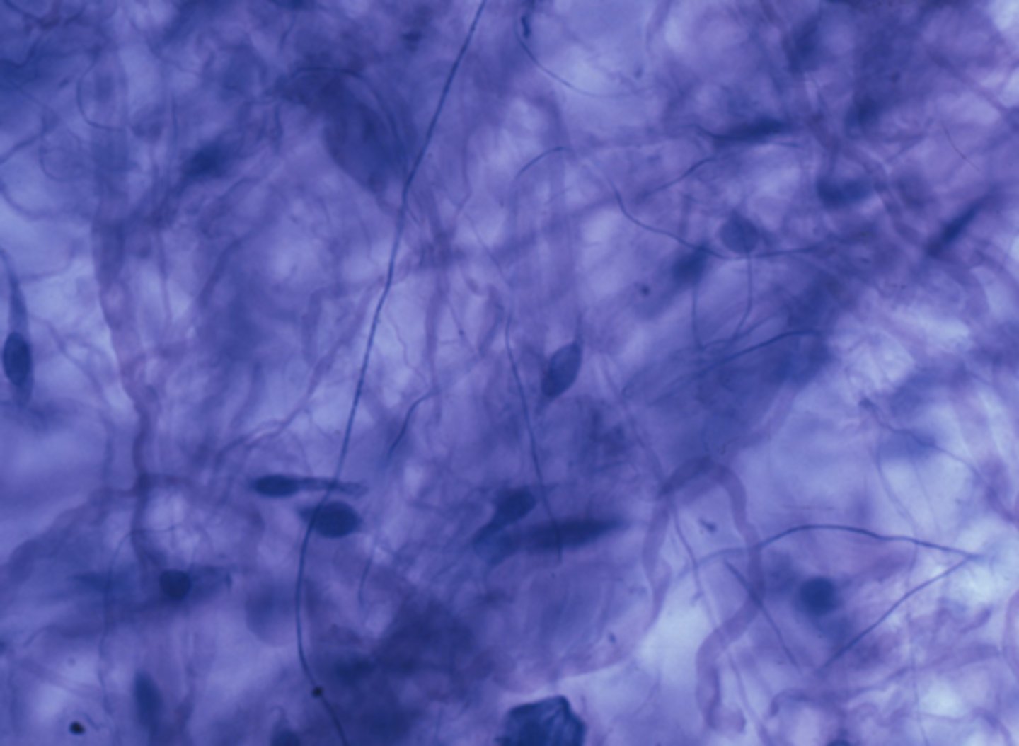
Cells produce an extracellular matrix
Which is a characteristic of the tissue type in the previous image: Connective Tissue
Cells produce an extracellular matrix
Cells are contractile
Cells have an apical and basolateral surface
Cells conduct electrochemical impulses
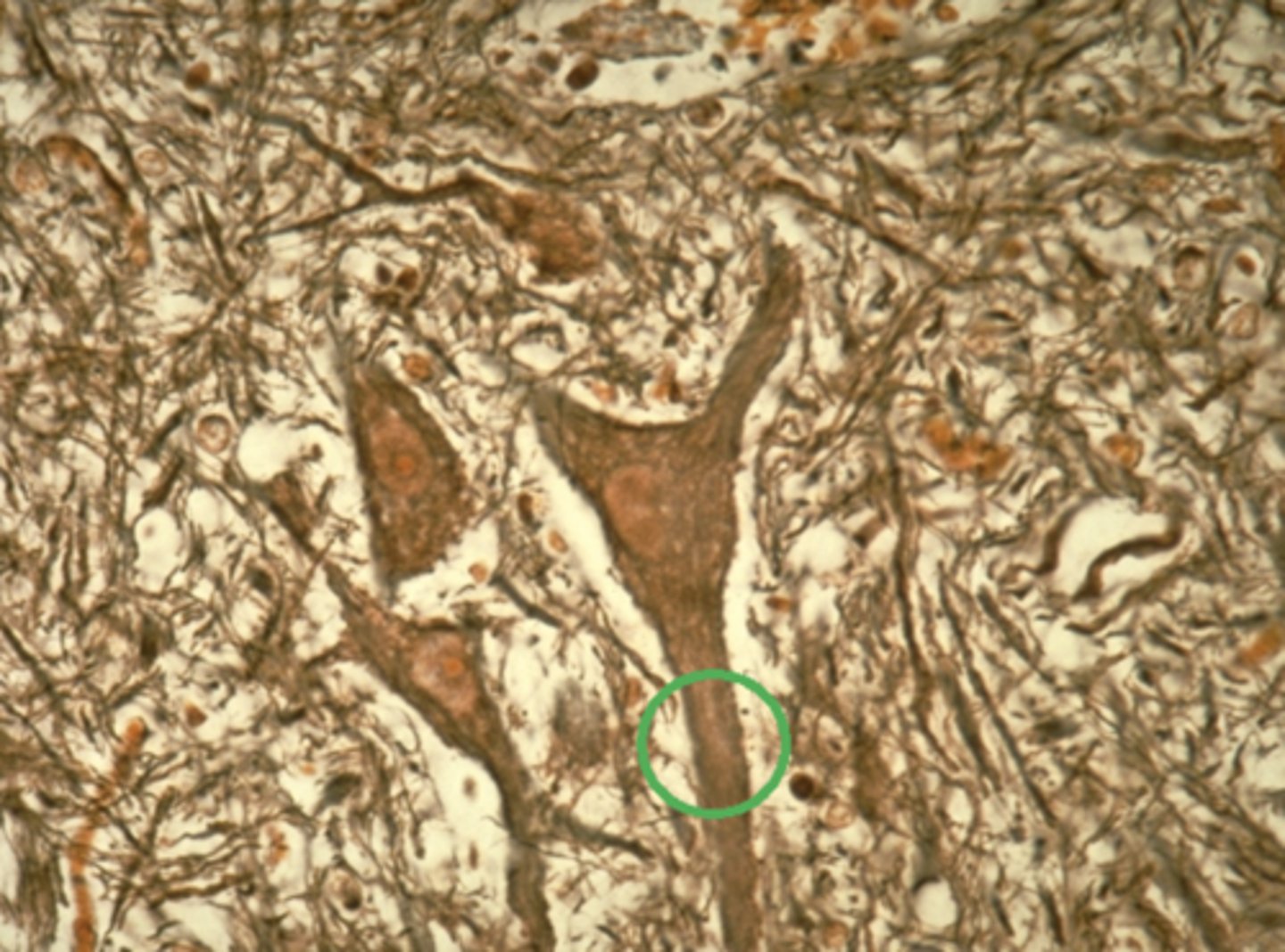
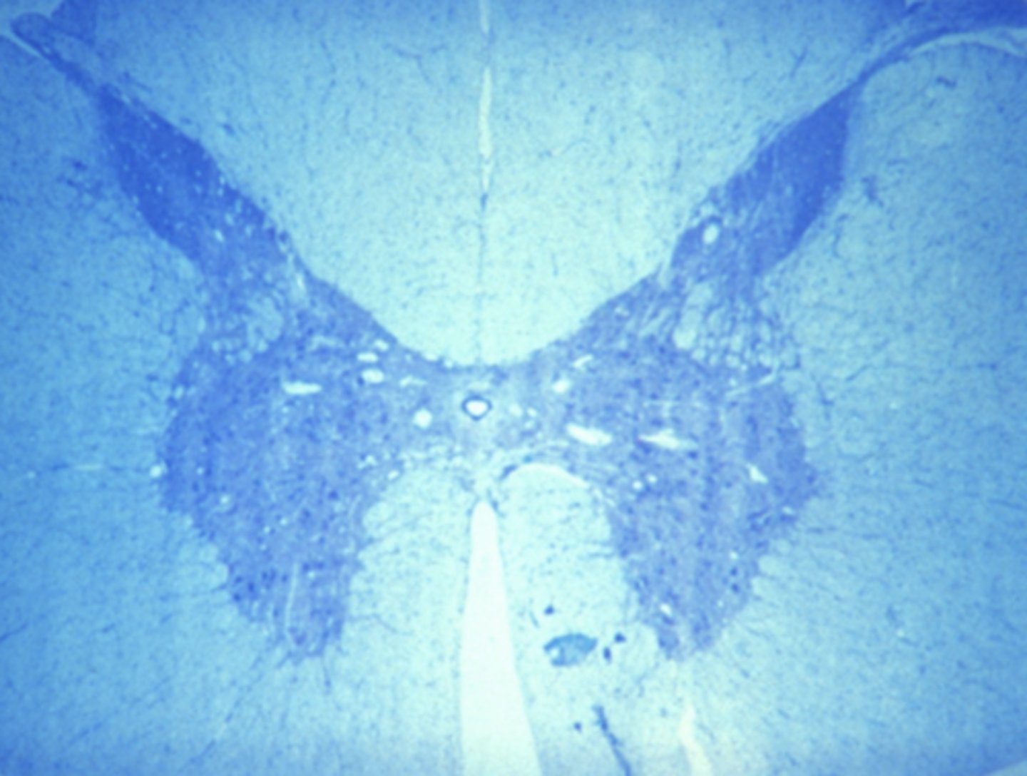
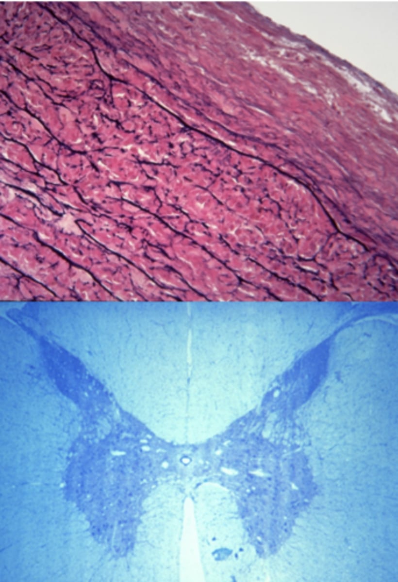
nervous tissue
Which of the four major tissue types is presented in the following image of the spinal cord:
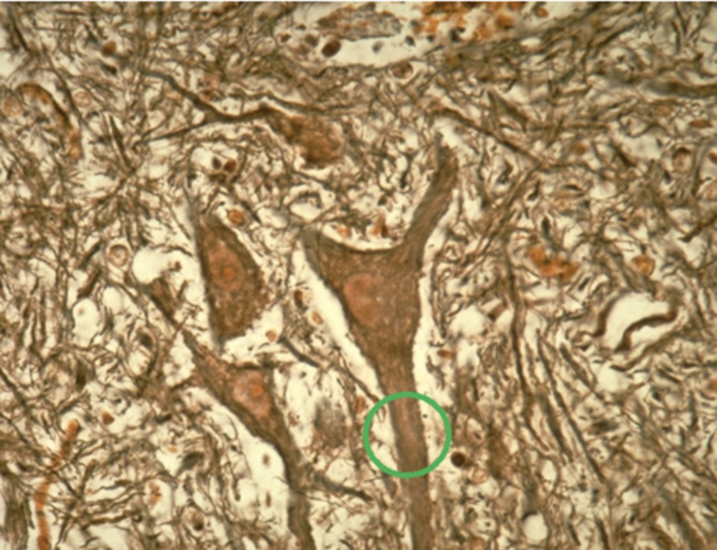
Axon
Which feature, circled in green in the previous image, is responsible for transmitting impulses away from the cell body? Nervous tissue
Cell body
Dendrite
Glial cell
Axon
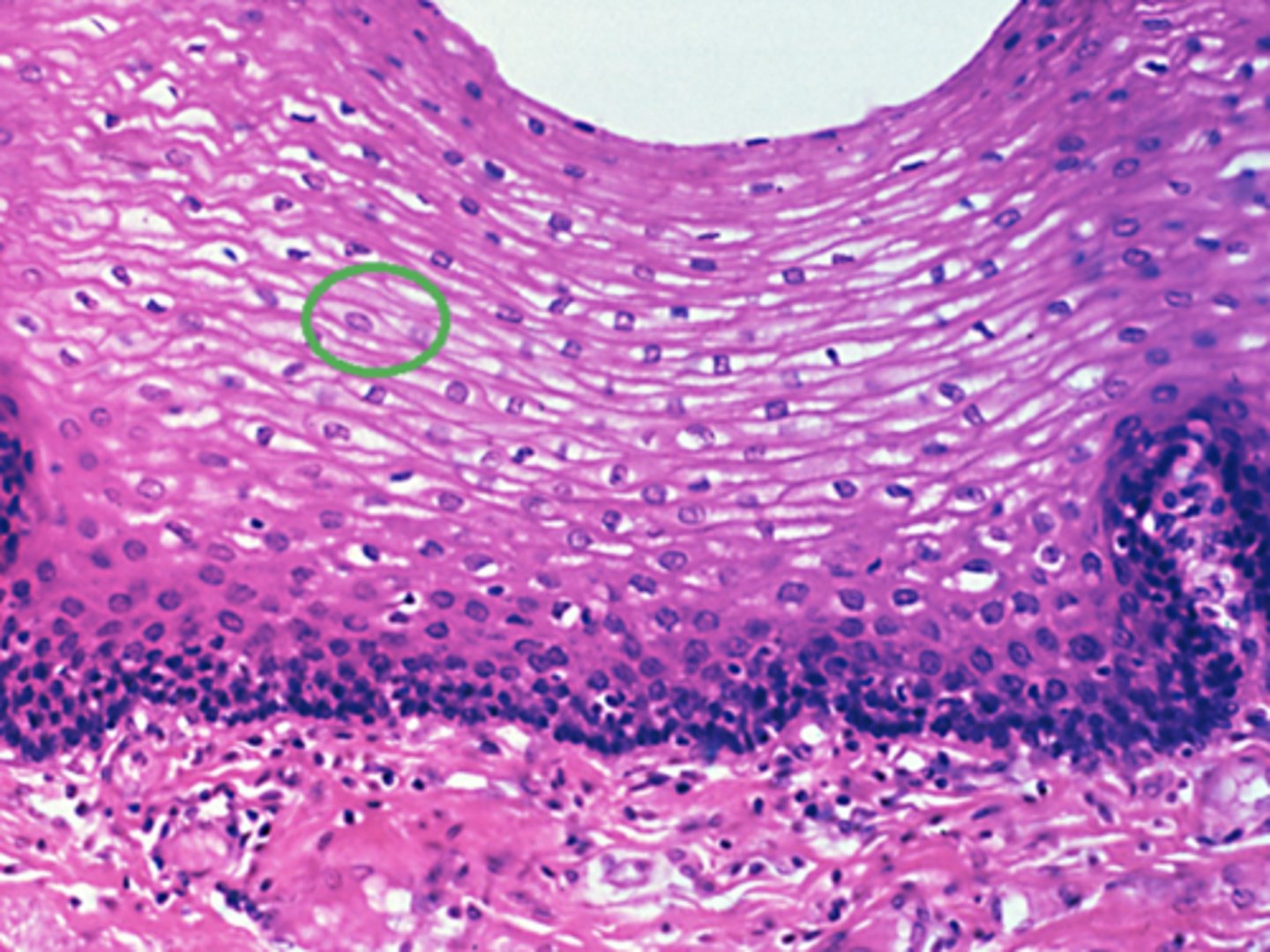
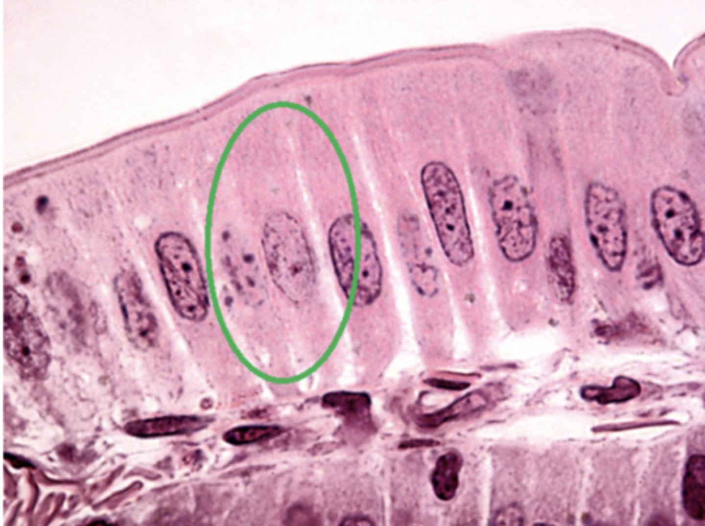
Epithelial tissue
Which of the four major tissue types is presented in the following image of the esophagus: (again, just name one of the four major tissue types, we don't need subtypes)
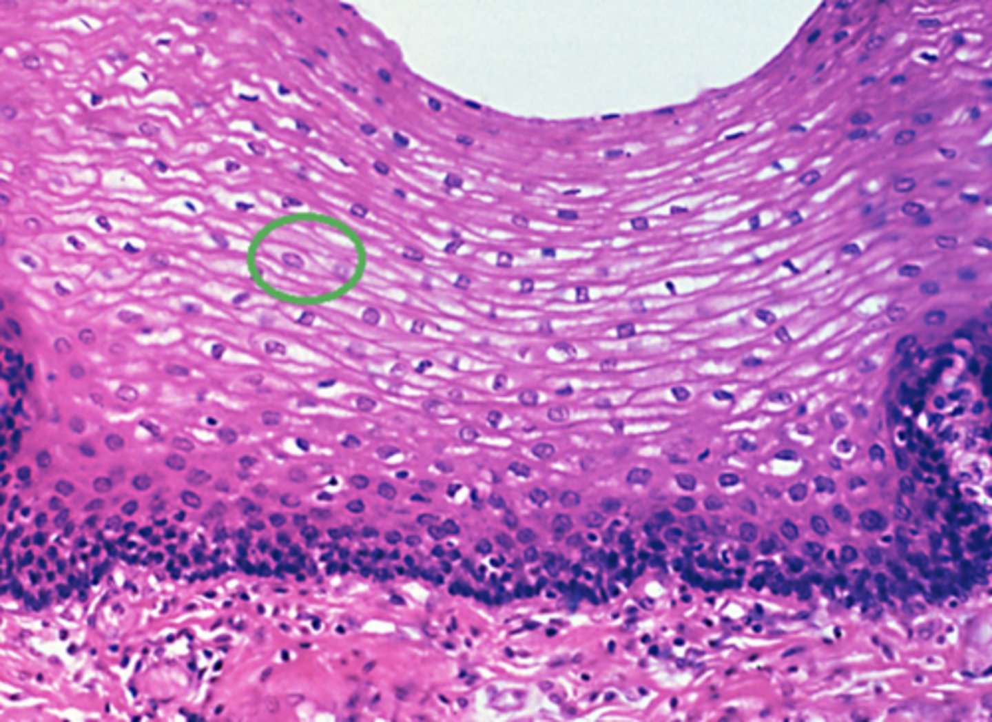
Squamous
Which cell type is circled in green in the this image of epithelial tissue?
Columnar
Squamous
Cuboidal
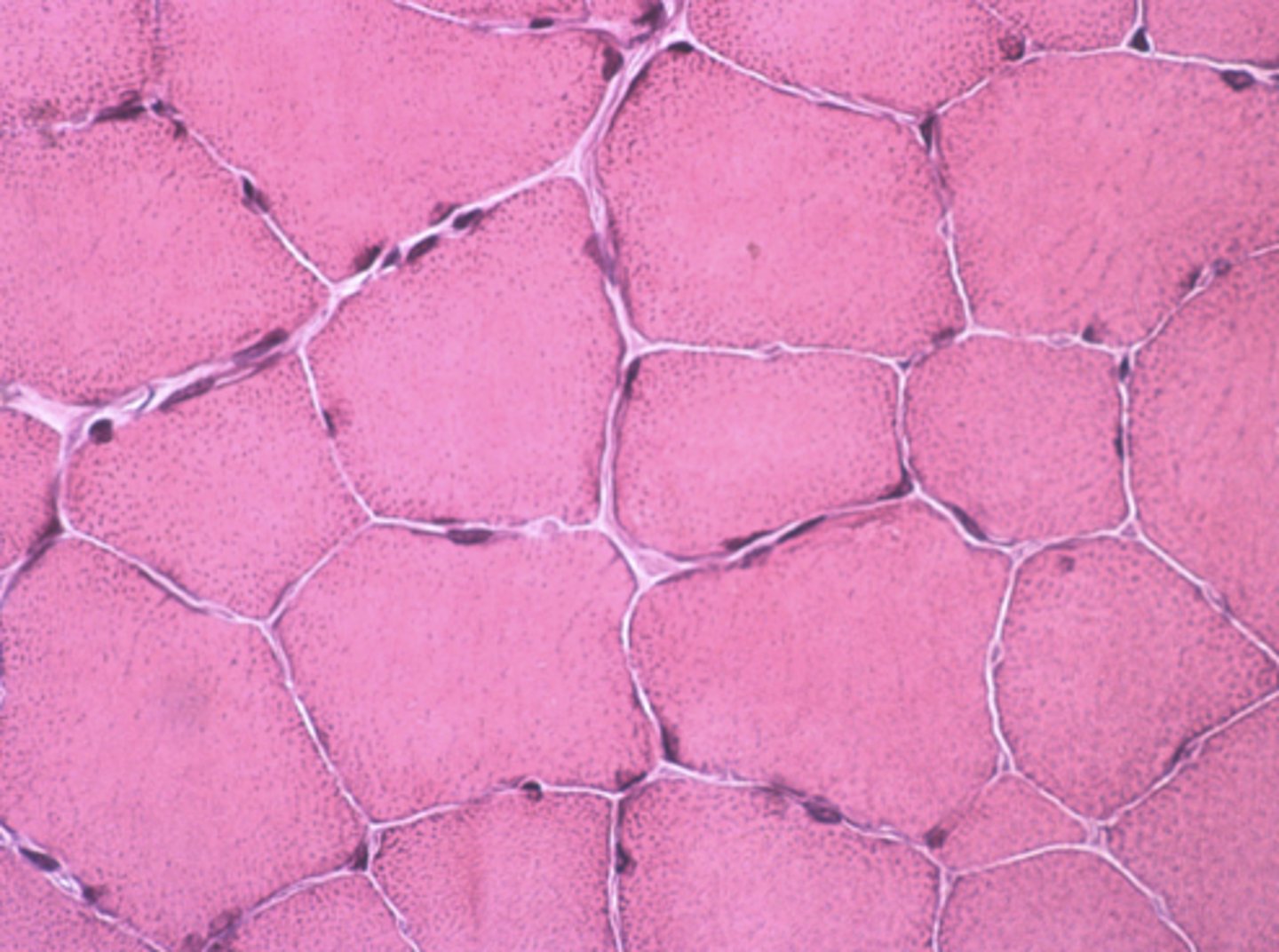
Muscle tissue
Which of the four major tissue types is presented in the following image of the small intestine:
Contractile cells that squeeze the contents of hollow organs
Which is a characteristic of the muscle tissue in this image:
Cells have an apical and basolateral surface
Contractile cells that move the extremities
Contractile cells that squeeze the contents of hollow organs
Cells produce an extracellular matrix
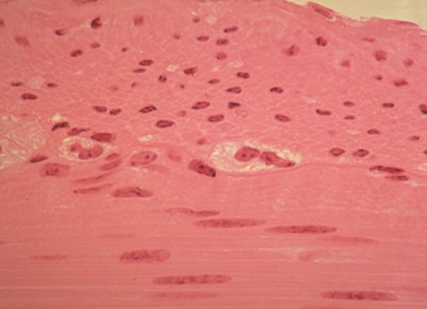
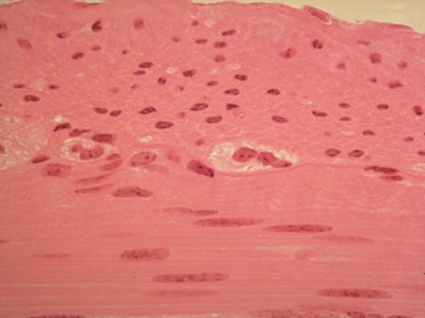
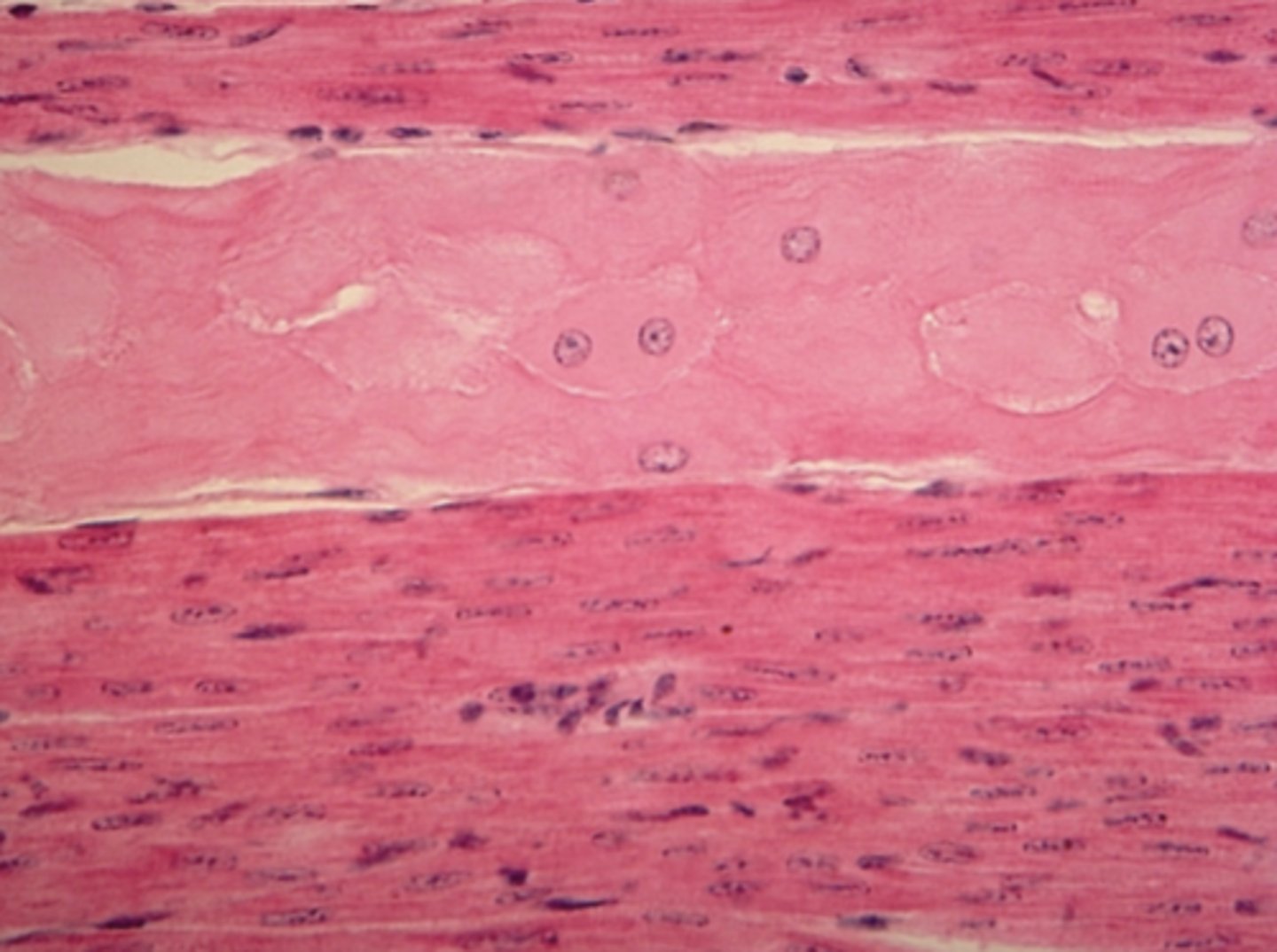
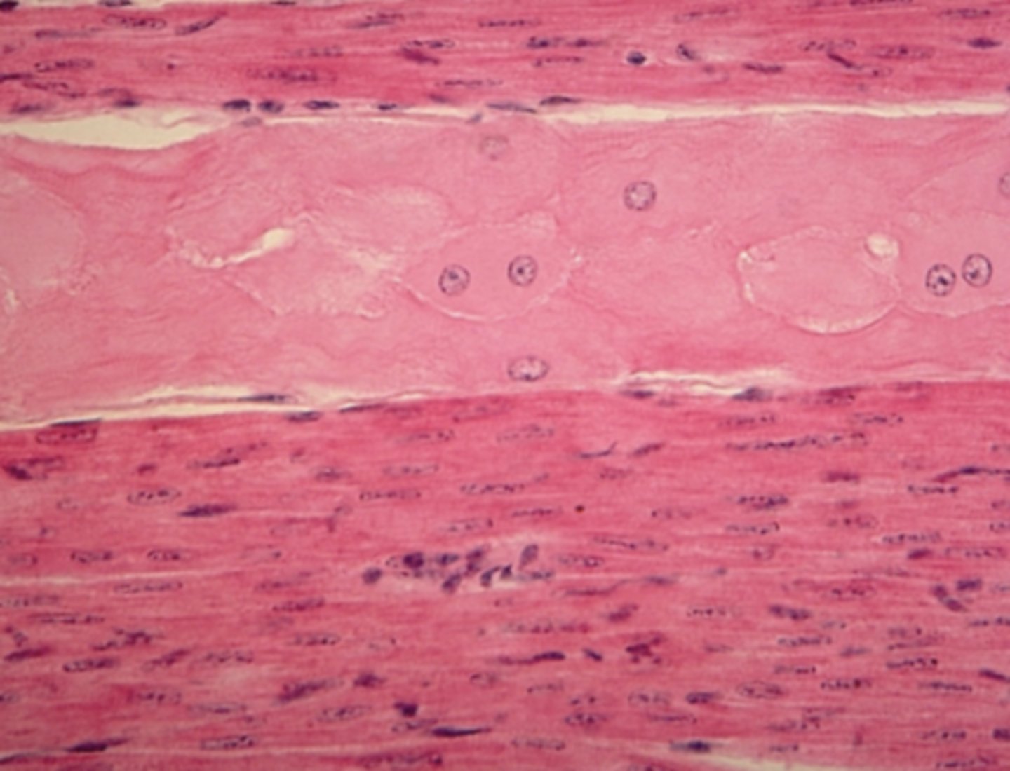
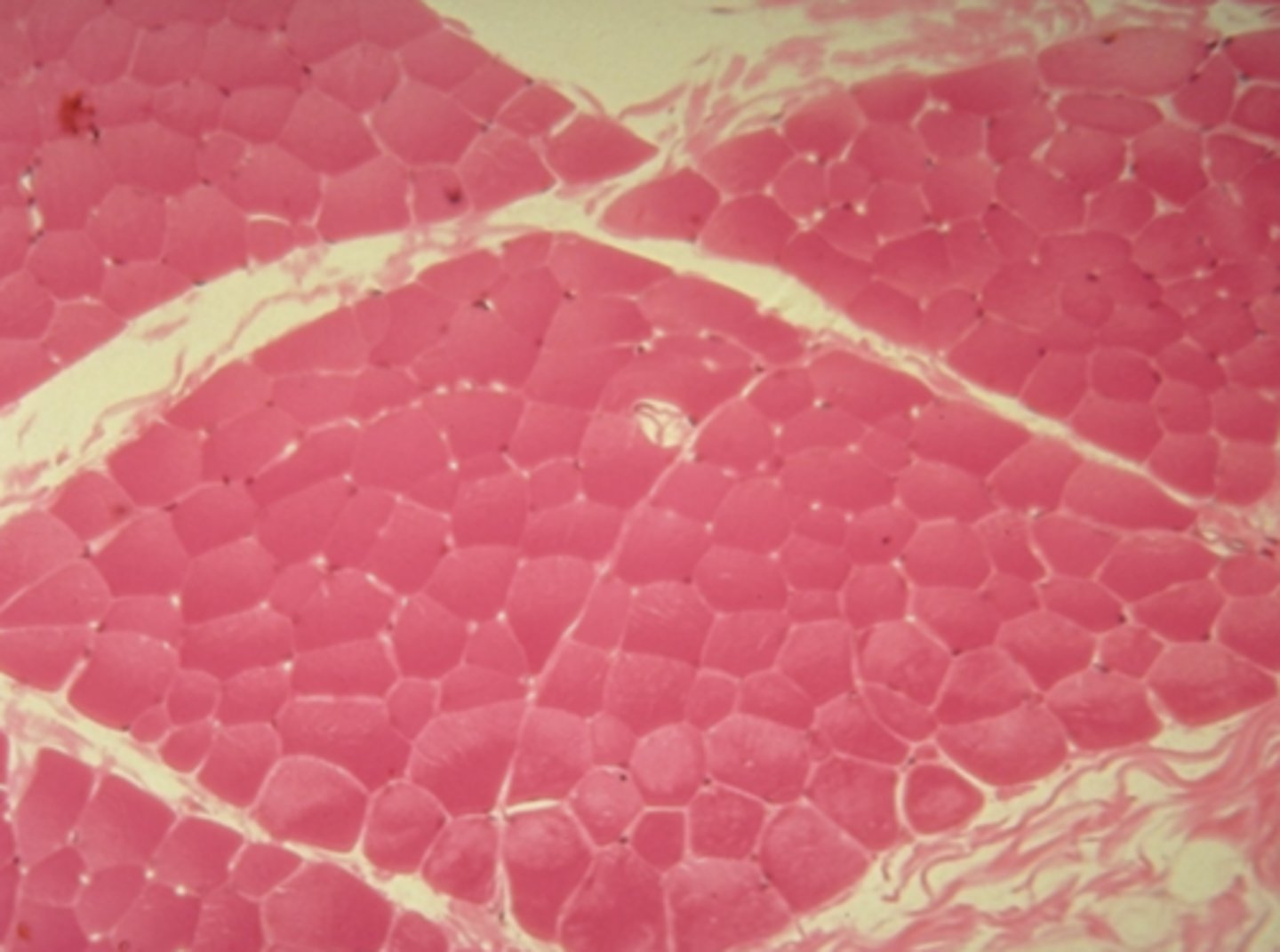
skeletal tissue
Which type of striated muscle tissue is presented in the following image:
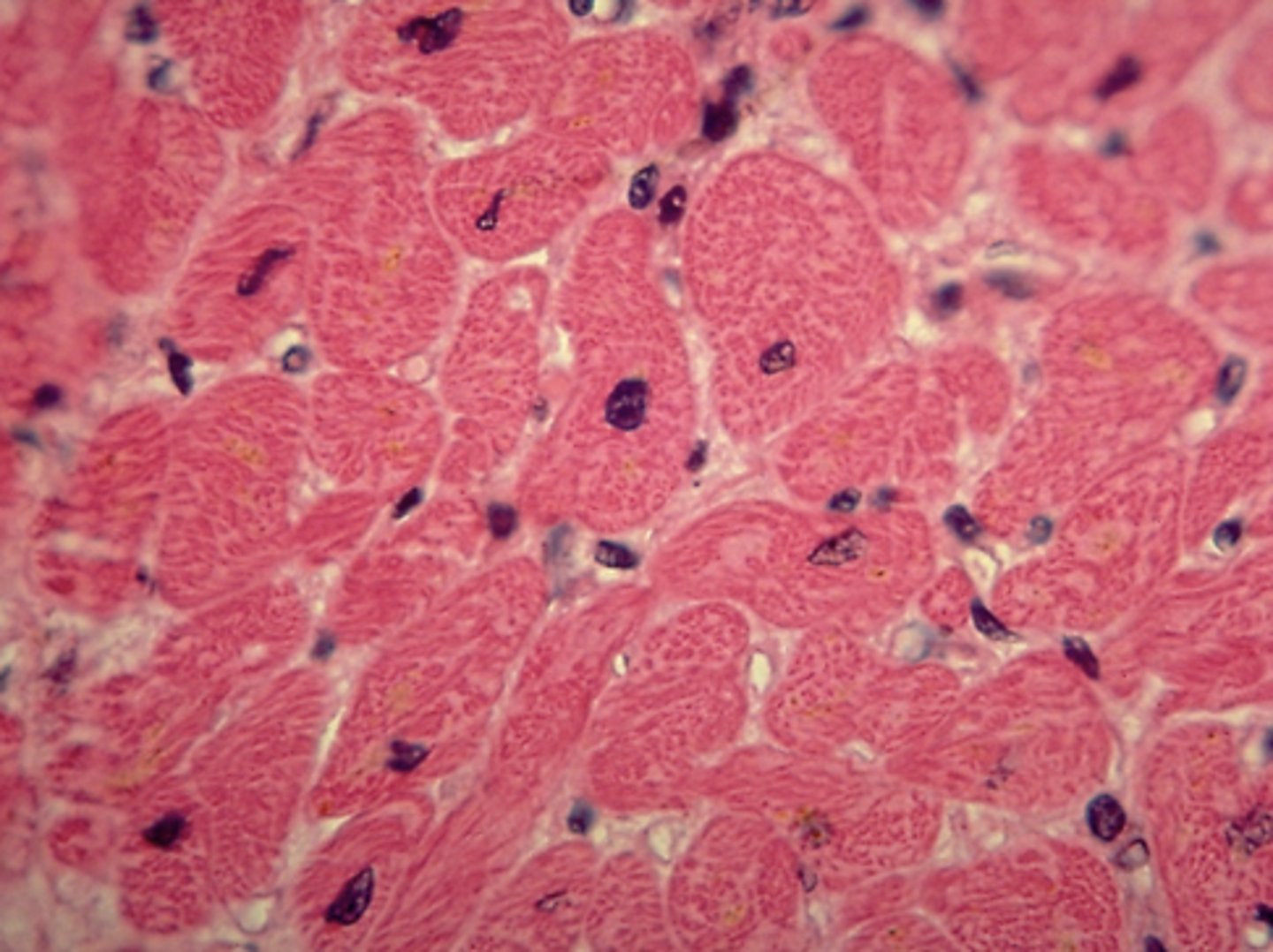
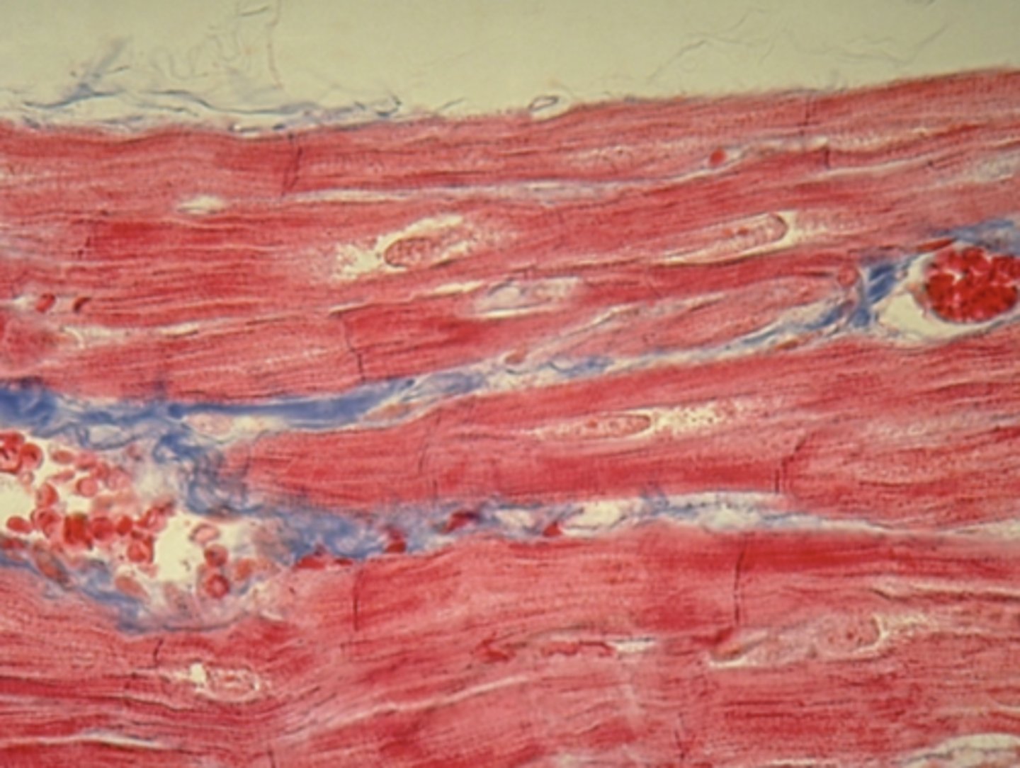
cardiac muscle tissue
Which type of striated muscle tissue is presented in the following image:
A
Which of the following tissues is likely responsible for secretion or absorption
Image A
Image B
Neither
Both
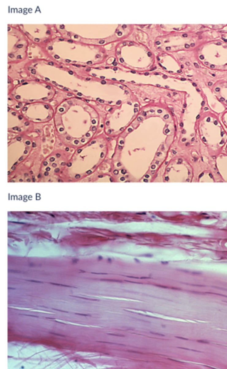
B
Which of the following tissues is contractile?
A
B
Both
Neither
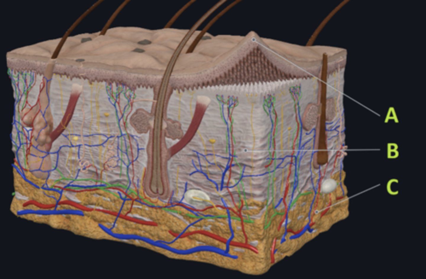
C - hypodermis
Answer the following questions based on the features indicated in the following image. Which letter indicates the administration site for subcutaneous injections?
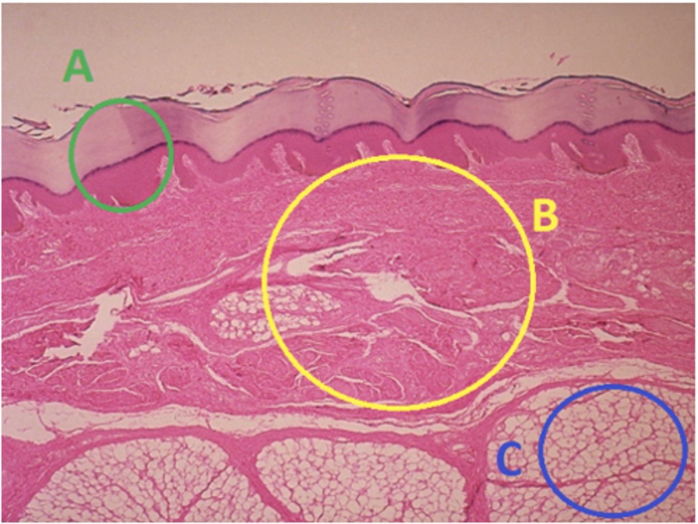
A - epidermis
Answer the following questions based on the features indicated in the following image. Which letter indicates the administration site for topical creams and ointments?
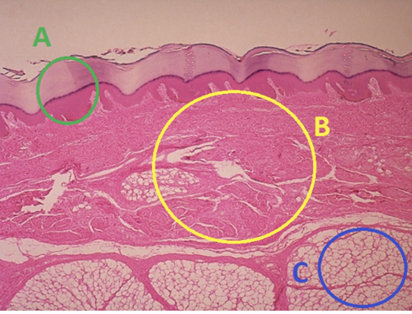
B - Dermis
Answer the following questions based on the features indicated in the following image. Which letter indicates the administration site for the tuberculin skin test?

A - epidermis
Answer the following questions based on the features indicated in the following image. Which letter indicates a region of the skin with no nerves or blood vessels?

B - Dermis
Answer the following questions based on the features indicated in the following image. Which letter indicates the region where skin appendages (sweat glands, sebaceous glands, and hair follicles) have their roots?
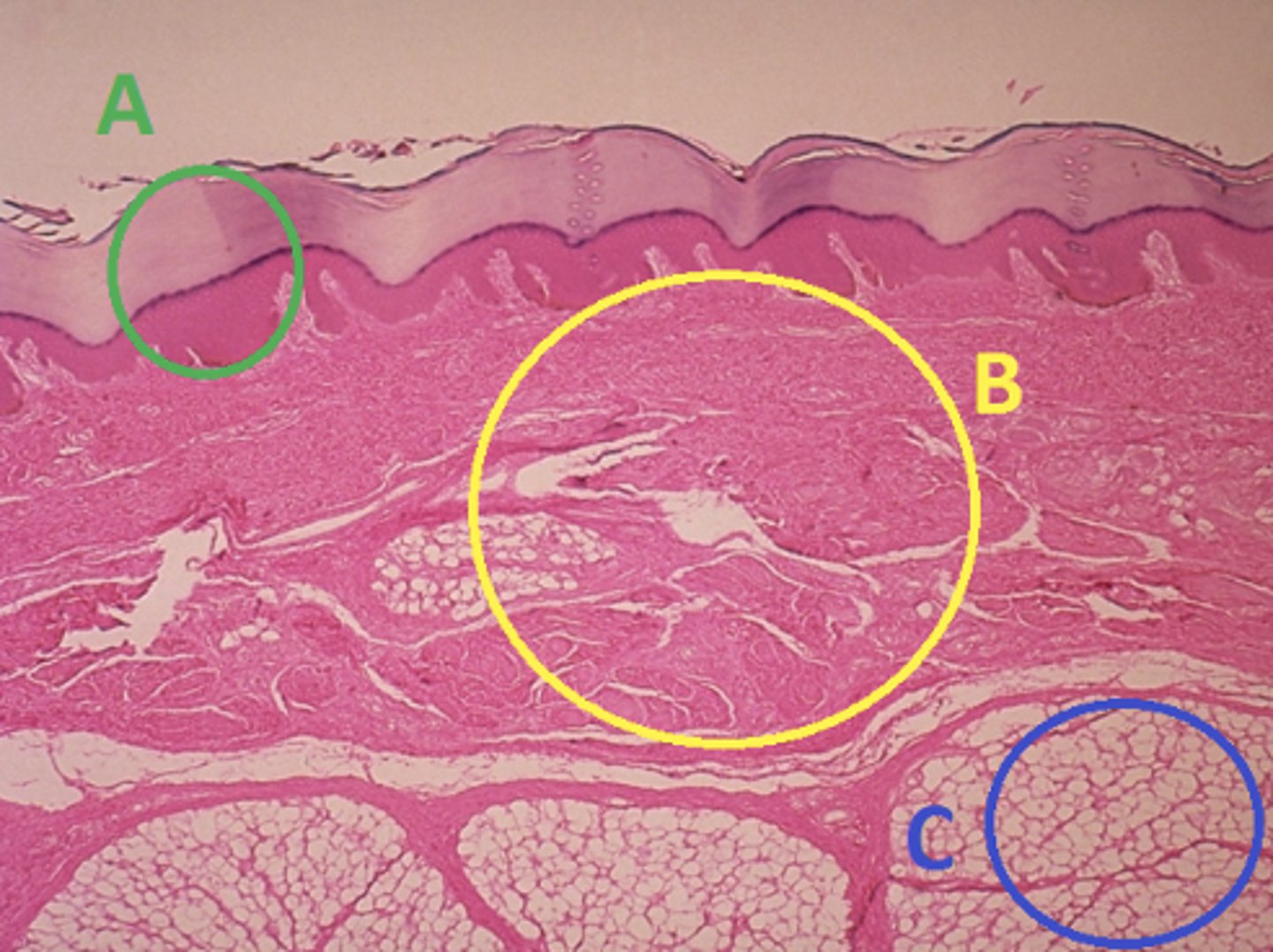
Epithelial tissue
Answer the following questions based on the features indicated in the following image. Which of the four major tissue types is indicated by letter A?
Skin of the soles of the feet
Where would hair follicles be absent?
Skin in the axilla (armpit)
Skin on the back of the hand
Skin of the dorsal surface of the feet
Skin of the soles of the feet
Cyanosis
What change in skin color might be a sign of poor circulation?
Jaundice
Erythema
Cyanosis
Pimples
Apocrine sweat glands
Which structure is responsible for most body odor?
Apocrine sweat glands
Broken shower head
Sebaceous glands
Eccrine sweat glands
Ulna
Name the bone indicated in green in the following image:
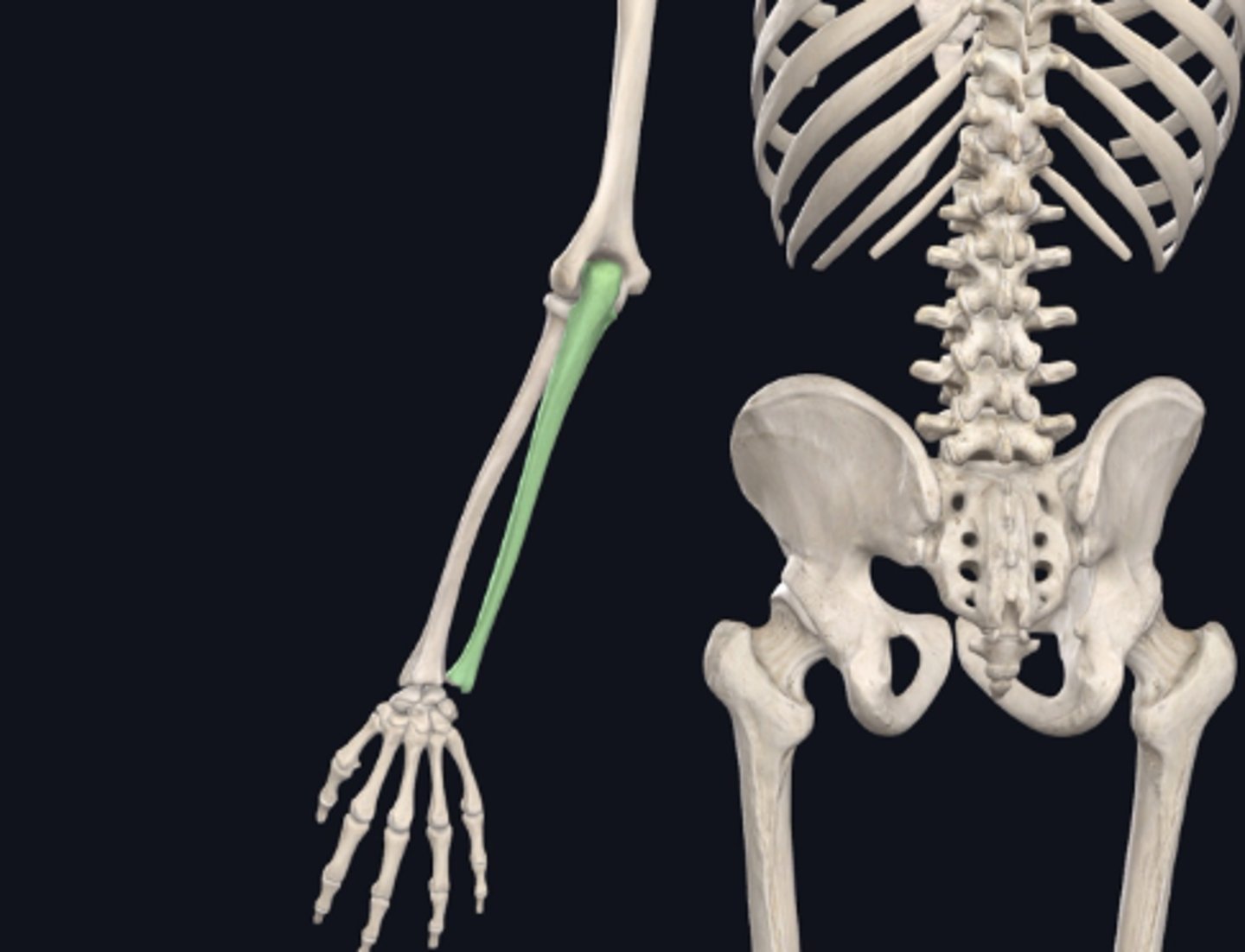
Humerus
Name the bone immediately proximal to the bone indicated in green in the previous image.
Thoracic vertebrae
Name the bones indicated in green in the following image:
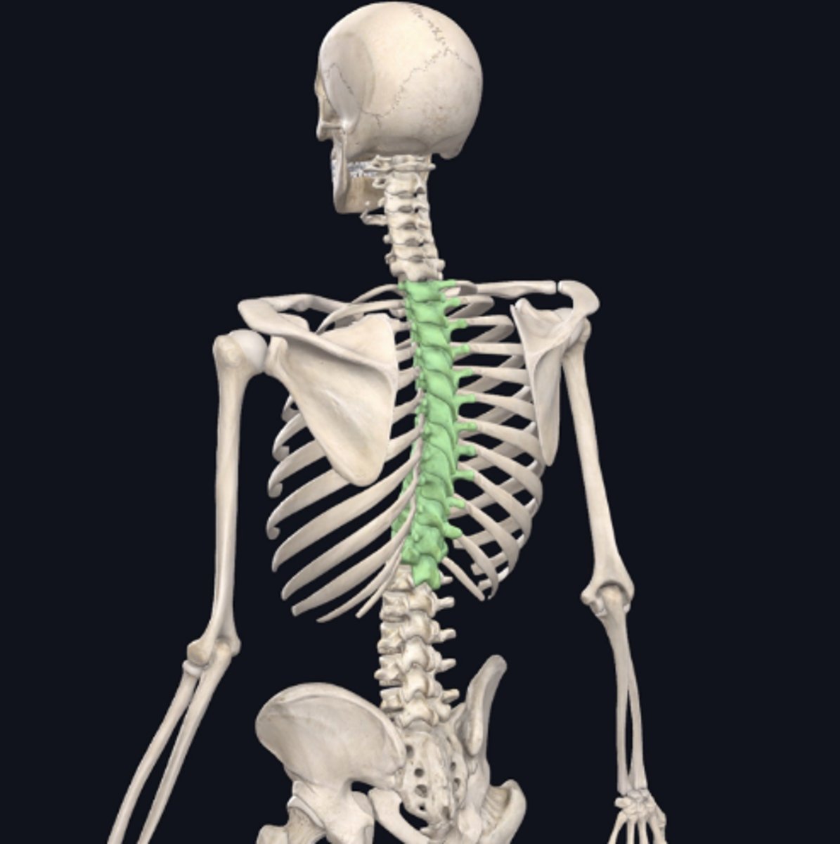
axial
In the previous image, the bones indicated in green are:
Axial
Appendicular
Nervous
Which of the four major tissue types is presented in the following image of the spinal cord:
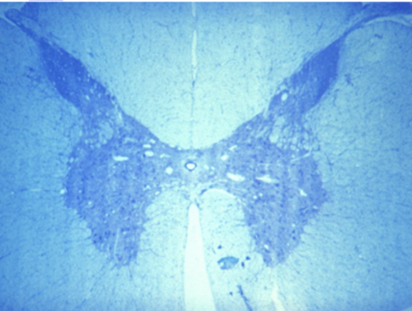
Cells conduct electrochemical impulses
Which is a characteristic of the tissue type in the previous image (nervous tissue):
Cells have an apical and basolateral surface
Cells conduct electrochemical impulses
Cells are contractile
Cells produce an extracellular matrix
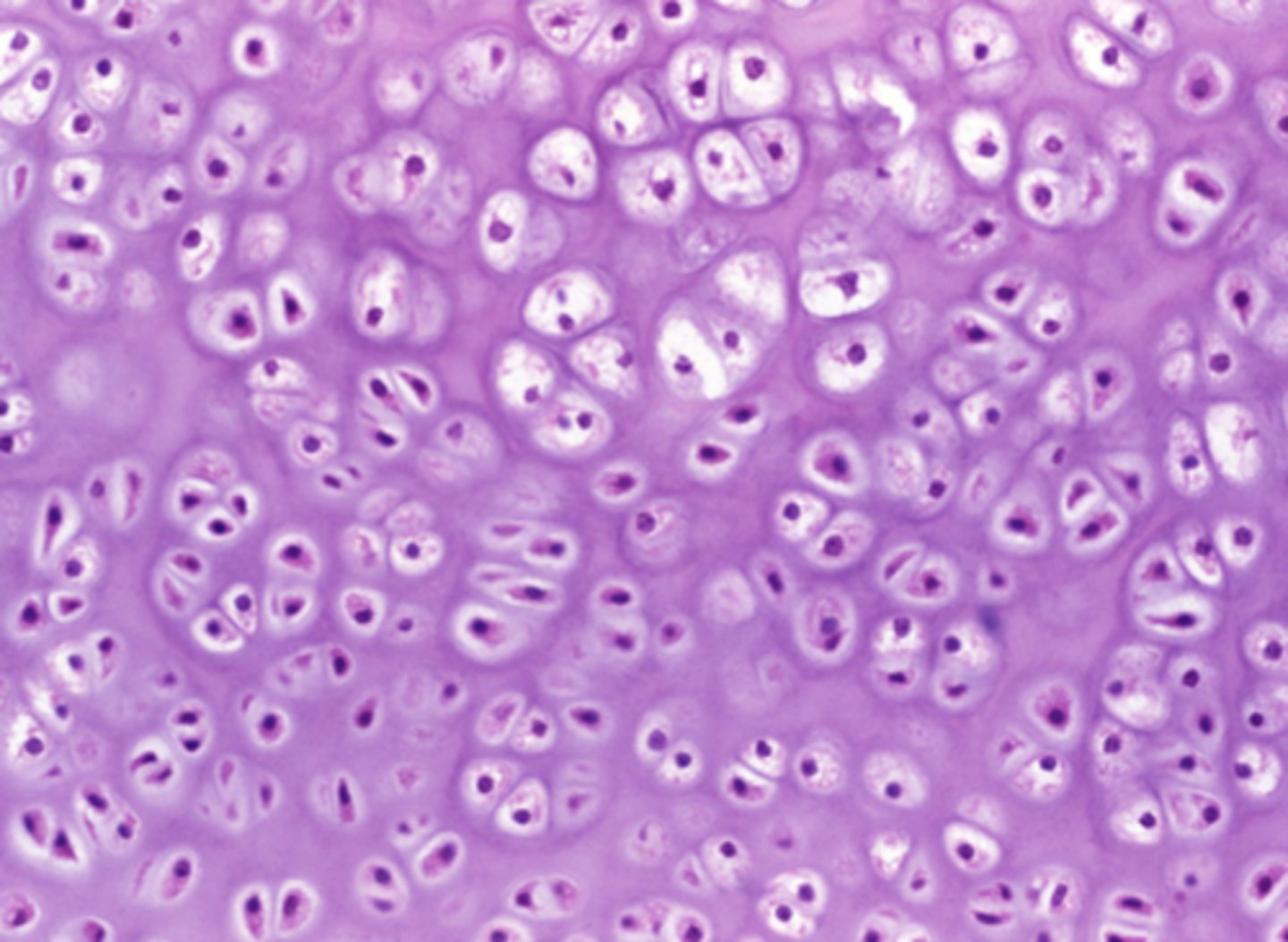
Connective tissue
Which of the four major tissue types is presented in the following image of the distal femur:
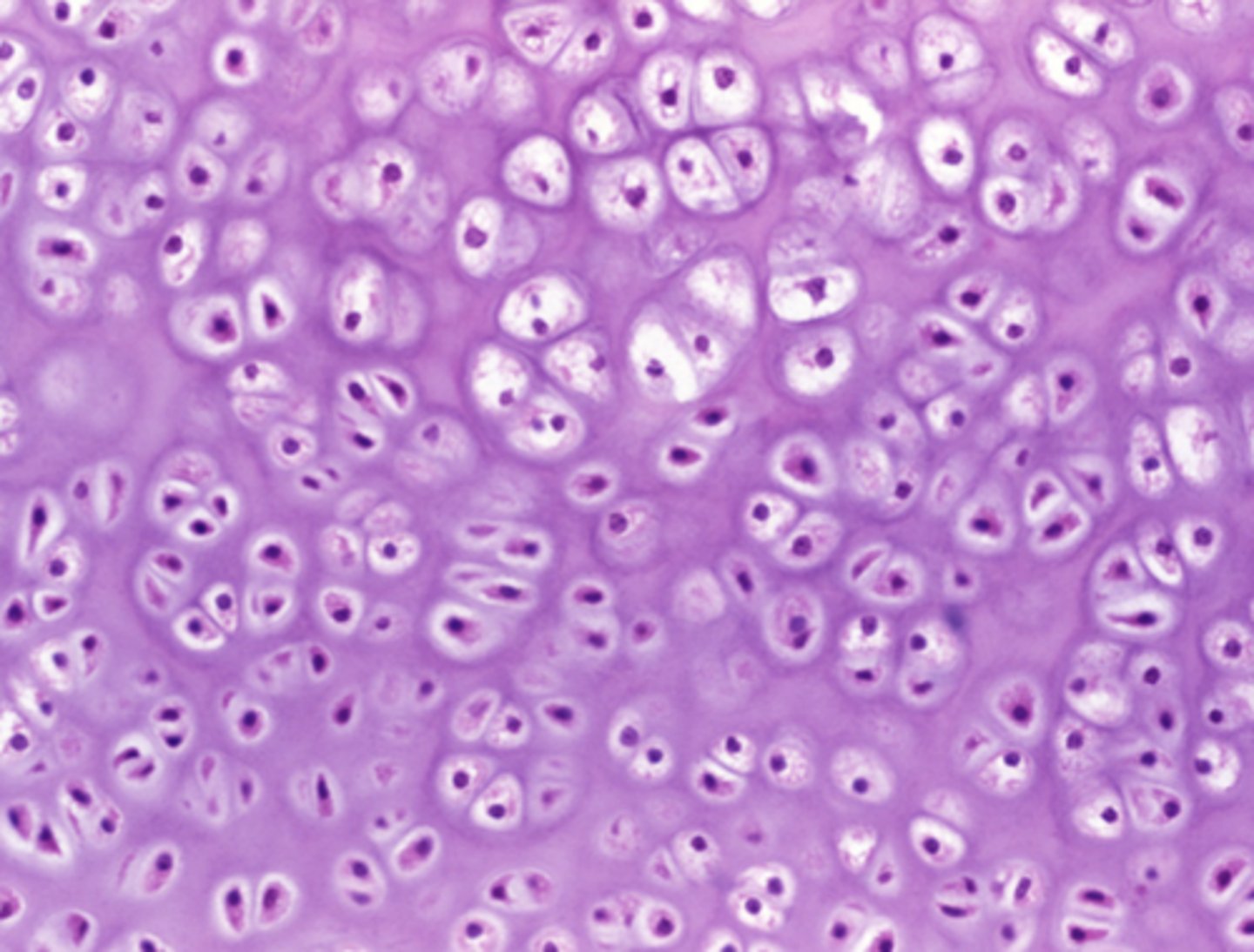
Flexible extracellular matrix with no blood or nerve supply
Which feature in the previous image is responsible for cushioning the body from mechanical impact?
Flexible extracellular matrix with no blood or nerve supply
Basement membrane supporting the cells' basolateral surface
Rigid extracellular matrix rich in calcium
Densely packed actin and myosin filaments
Muscle tissue
Which of the four major tissue types is presented in the following image of the heart:
Branched, striated cells with a central nucleus
Which is a feature of the tissue in the previous image of the heart (muscle tissue):
Tapered, non-striated cells with a central nucleusb. Branched, striated cells with a central nucleus
Branched, striated cells with a central nucleus
Long, striated cells with multiple peripheral nuclei
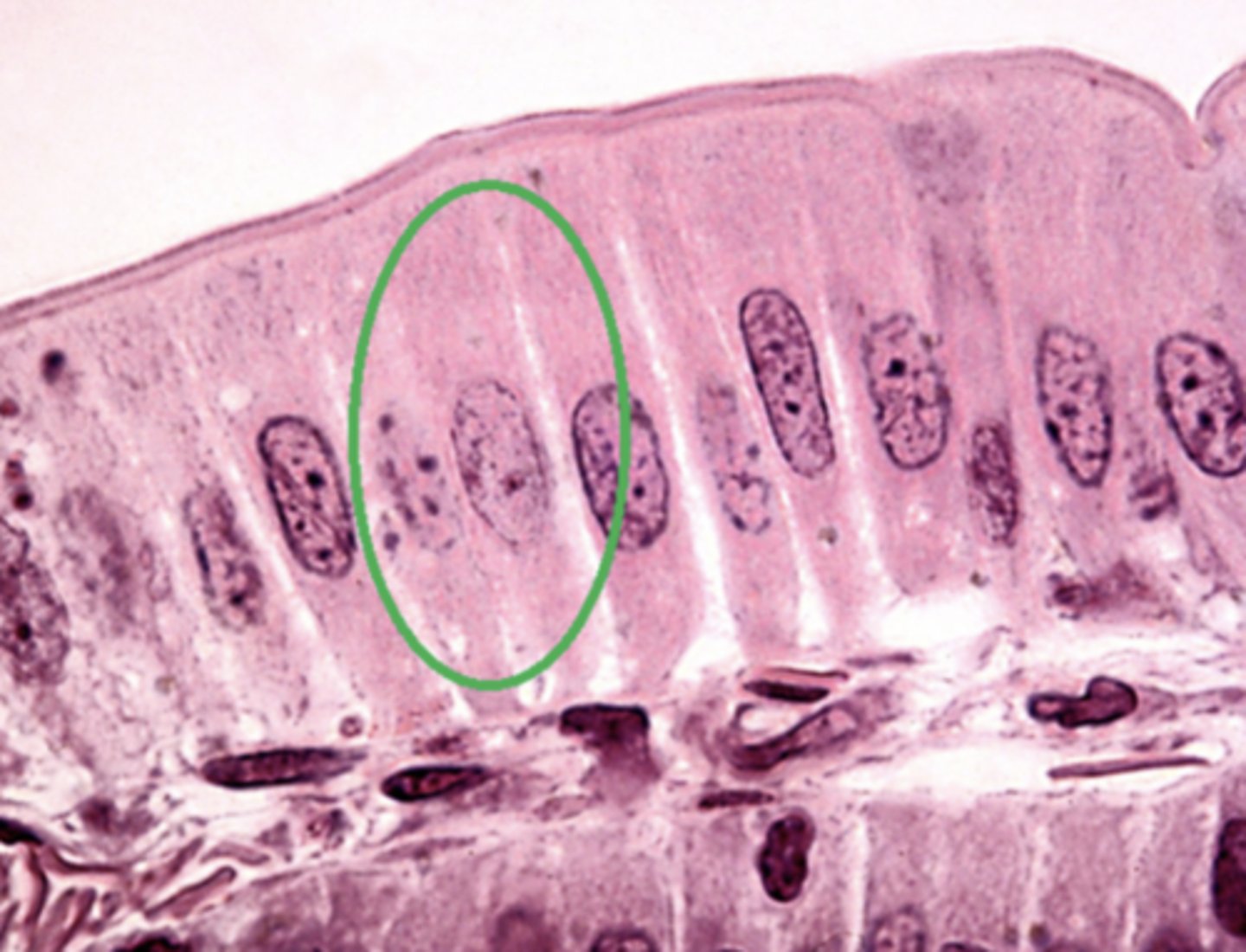
epithelial tissue
Which of the four major tissue types is presented in the following image of the small intestine:
Columnar
Which type of cell is circled in green in the previous image (epithelial tissue):
Squamous
Cuboidal
Columnar
skeletal muscle
Which of the three muscle tissue types is presented in the following image:
cardiac
Which of the three muscle tissue types is presented in the following image:
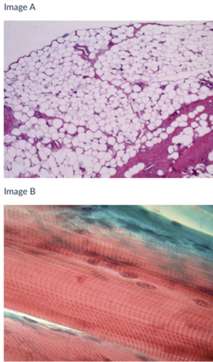
A
Which of the following tissues is likely responsible for protecting the surface of the body from friction
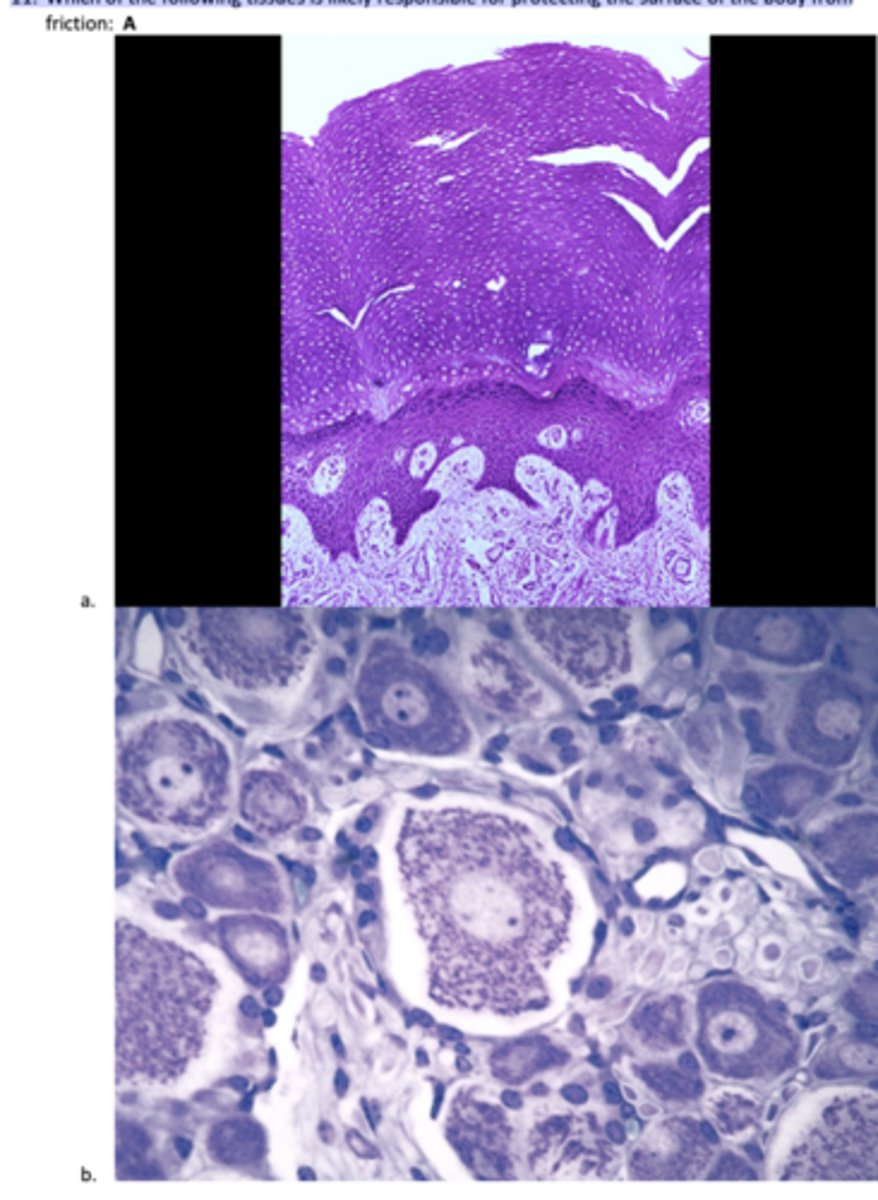
A
Which of the following tissues stretches to accommodate increases in pressure:
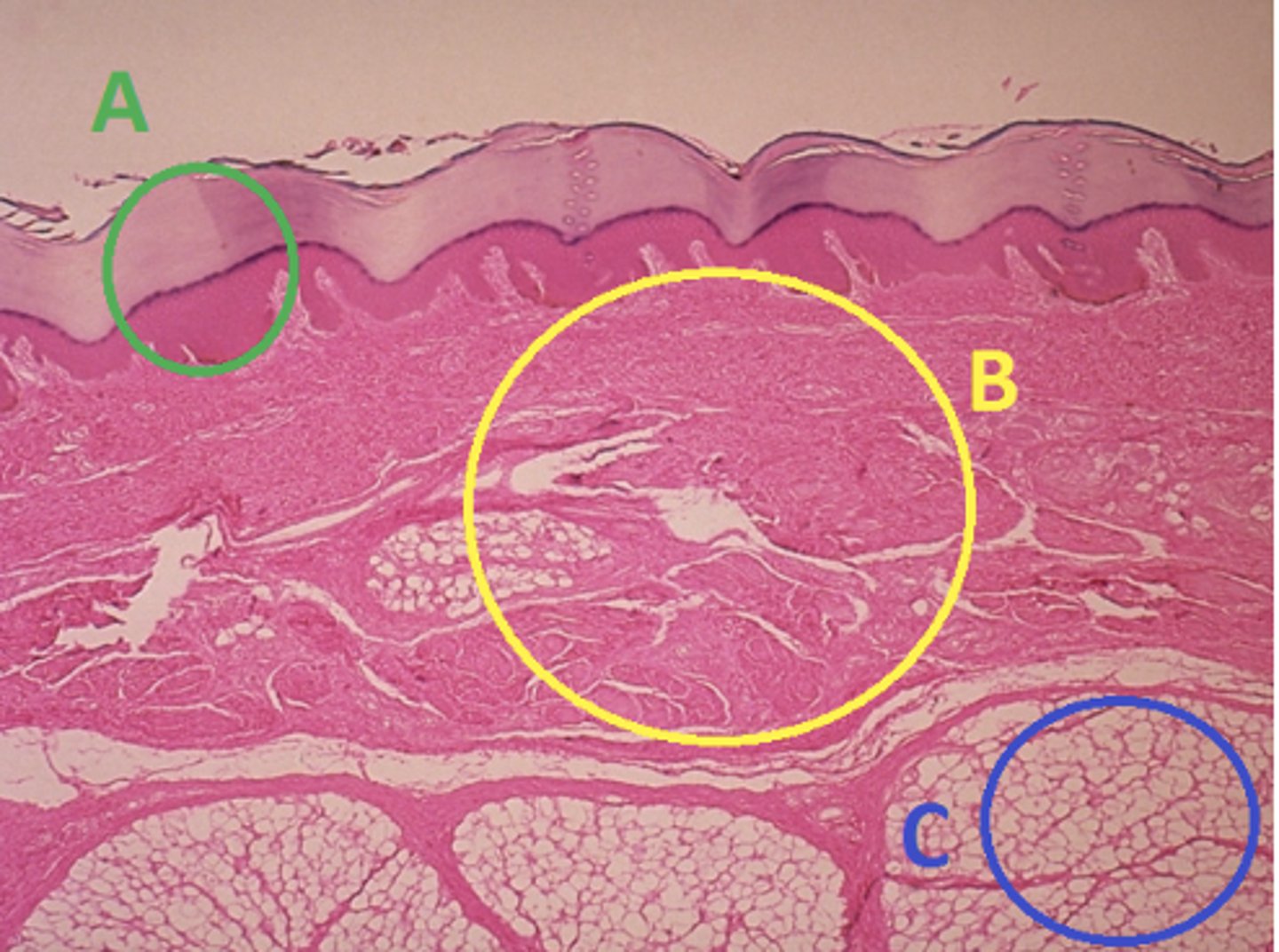
C
Which letter indicates the administration site for insulin injections?
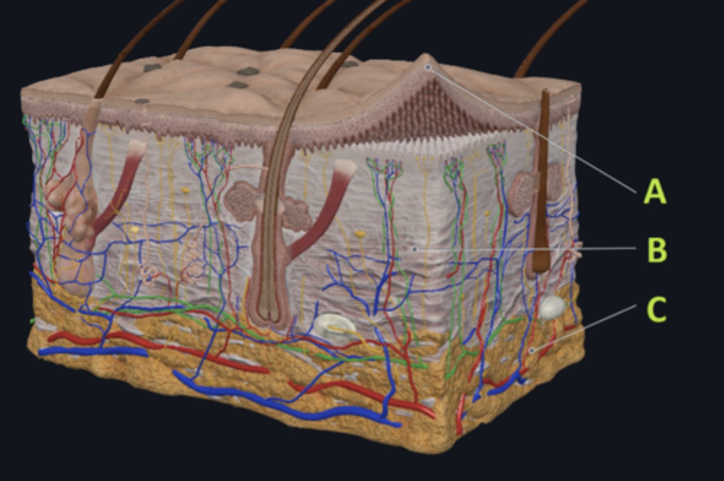
A
Which letter indicates the administration site for a lidocaine patch?
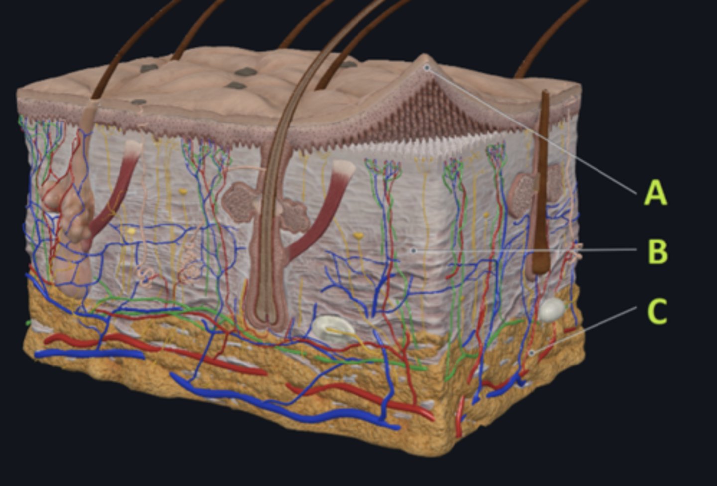
B
Which letter indicates the administration site for an allergy test?
B
Which letter indicates a region of the skin rich in dense, irregular connective tissue?

C
Which letter indicates the region most important for energy storage and thermal insulation?
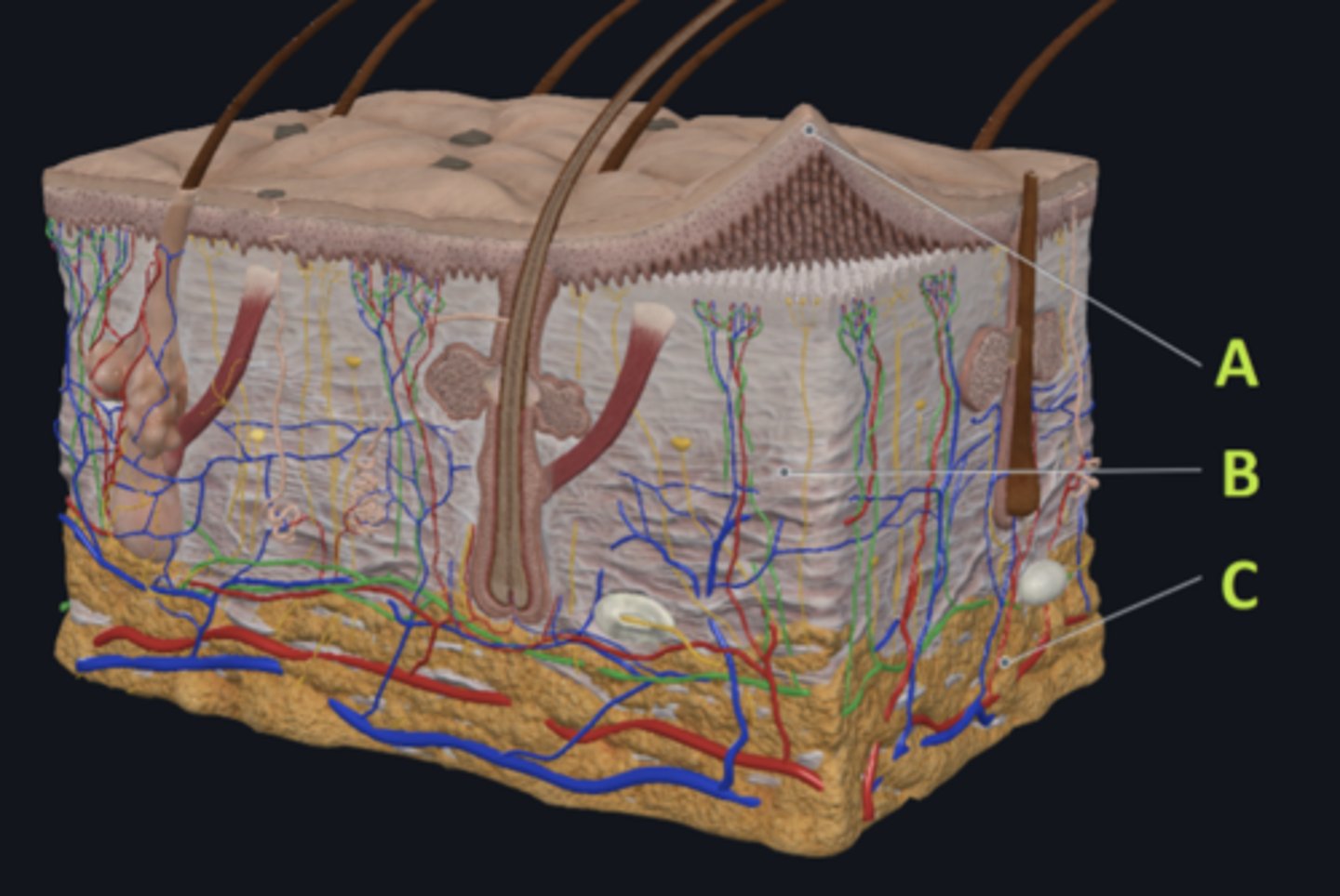
Connective
Which of the four major tissue types is indicated by letter C?
Skin of the dorsal surface (back) of the hand
Which part of the body could the previous image represent?
Skin of the ventral surface (palm) of the hand
Skin of the dorsal surface (back) of the hand
Jaundice
What change in skin color might be a sign of liver failure?
Cyanosis
Jaundice
Erythema
Sebaceous glands
Which structure is most responsible for acne?
Sebaceous glands
Eccrine sweat glands
Apocrine sweat glands
Scapula
Name the bone indicated in green in the following image:
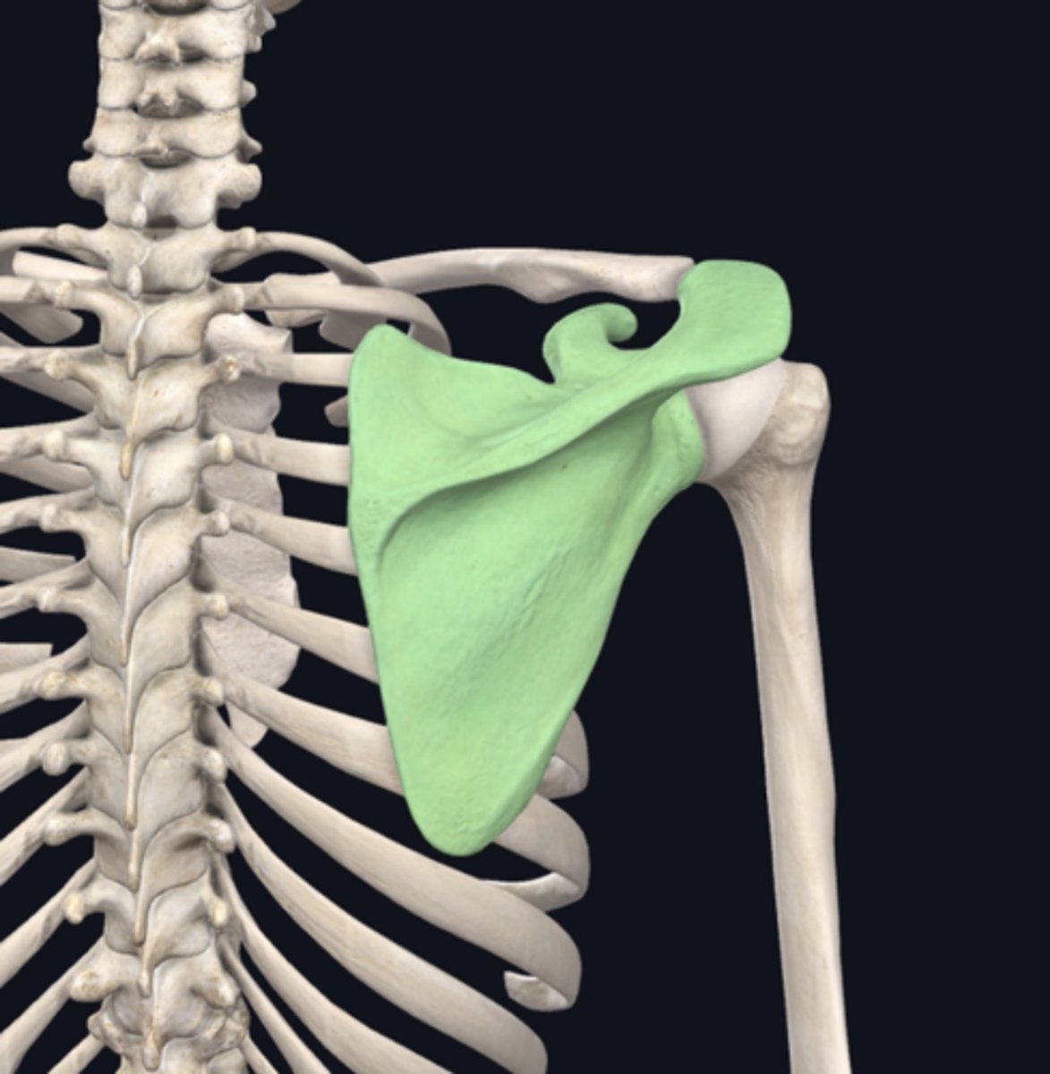
Humerus
Name the bone immediately lateral to the bone indicated in green in the previous image.

Metatarsals
24. Name the bones indicated in green in the following image:
Appendicular
In the previous image, the bones indicated in green are:
Axial
Appendicular