Small animal interactive cardio cases notes
 Call Kai
 Learn
Learn Practice Test
 Spaced Repetition
 Match
Match Flashcards
 Knowt Play
1/23

There's no tags or description
Looks like no tags are added yet.
Name | Mastery | Learn | Test | Matching | Spaced | Call with Kai | Chat |
|---|
No analytics yet
Send a link to your students to track their progress
24 Terms
What is paradoxical breathing?
What could it be suggestive of?
Abdomen sucking inwards when insp. and pushing outwards when exp. (should be opposite)
Could be suggestive of pleural dx
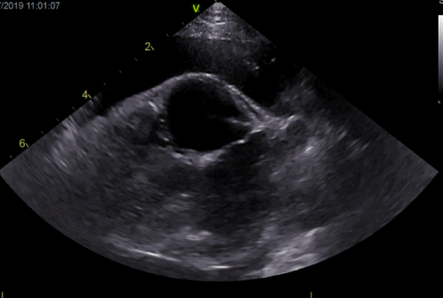
What view is this echo and label?
What abnormalities is it showing?
PS long axis view
Showing pleural effusion + bi-atrial dilation

What is the normal ratio of LA:Ao?
Should be <1.5x (short axis view)
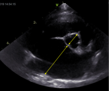
LA should be <16.5x on long axis view
What are these rads showing?

Unclear cardiac sil
DV worse cardiac sil visibility
R lat = loss of cardiac sil ventrally —> pleural effusion causing effacement
Lung fields = hazy = incr. sofft tissue opacity + air bronchogram
mix interstitial + alveolar pattern in cd.dorsal lung fields
Should improve after thoracocentesis
What drug is used for pleural effusion and how much?
Furosemide 2mg/kg IV (better than oral —> works quicker)

What is this echo showing?
What is the most likely diagnosis?
LV concentric hypertrophy + pericardial effusion (+ small amount of pericardial effusion @ bottom?)
Diag = stage C HCM —> thickened wall —> congested i.e. lungs @ risk of failure
Is jugular venous distension seen in R or L sided CHF?
R sided
Which type of AV block is this?

3rd degree
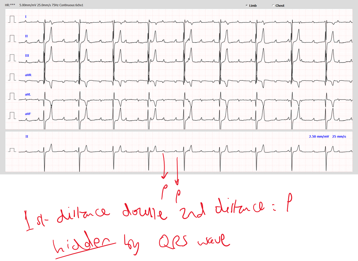
No ass. between atria & ventricle i.e. complete block
How does the ECG present when AP travels from R→L vs L→R in lead II?
R→L = +ve (above baseline)
L→R = -ve (below baseline)
What is the tx option for 3rd AV block?
Pacemaker implantation (R jugular vein implantation)
What should you NOT do after placing a R jugular vein implantation?
DO NOT take bloods from here —> take bloods from legs
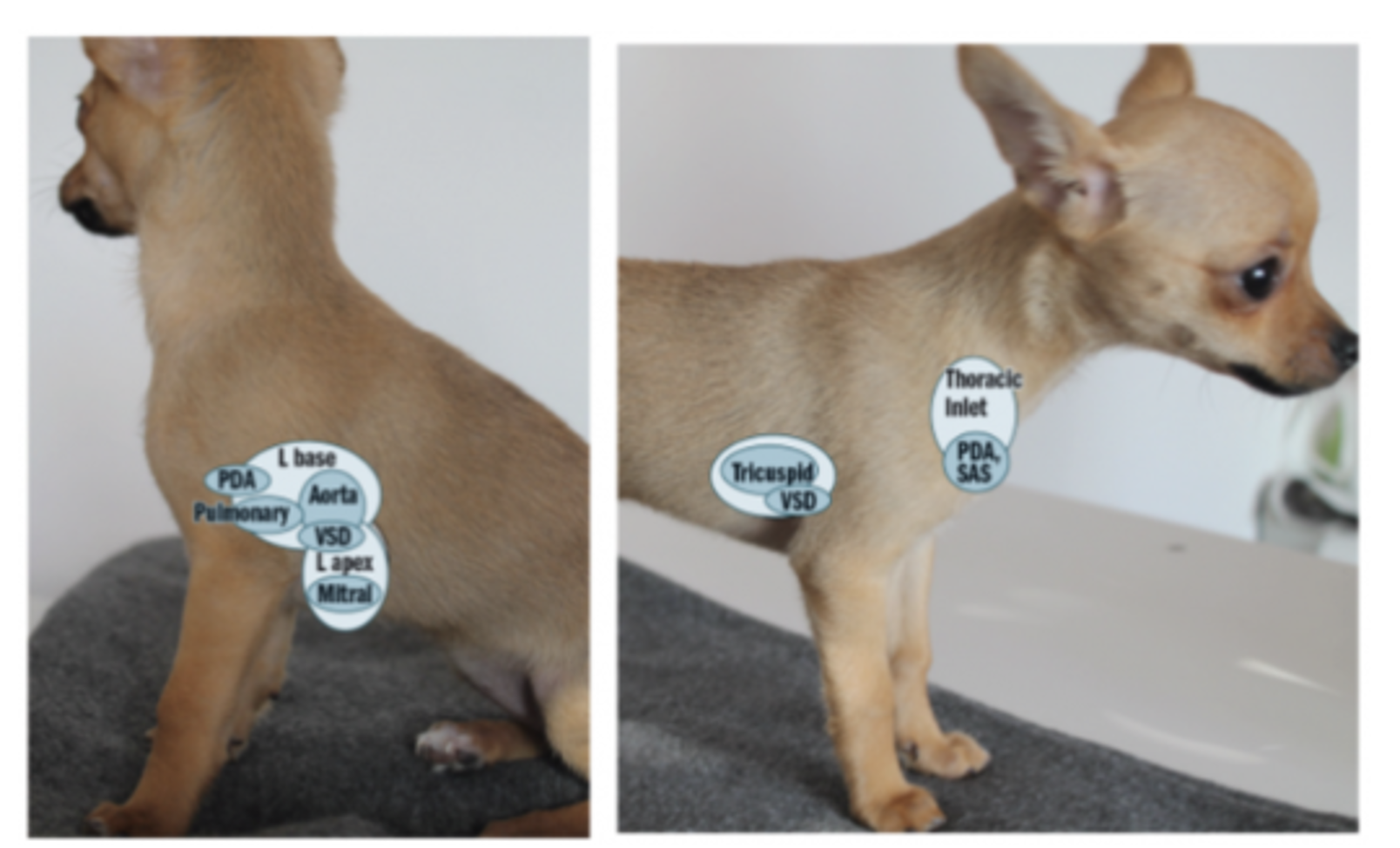
Where are the different heart sounds & abnormalities heard on auscultation?
What is this ECG showing?

Sinus arrhythmia — regularly irregular
Not wide
P for every QRST
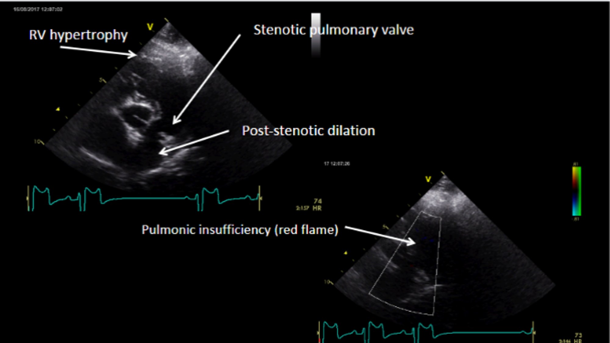
What are these ECGs showing?


RV hypertrophy + post-stenotic dilation
What does this ECG show?

R bundle branch block
P waves + wide QRS
L→R = -ve inflection
What does this colour dopple echo show?
Green = turbulence = regurg
Poor systolic function
Hyperdynamic —> vol overload, decr. afterload
(BART colour system)
When is LA considered dilated when compared with aorta?
When La:Ao >1.6
Describe the abnormalities

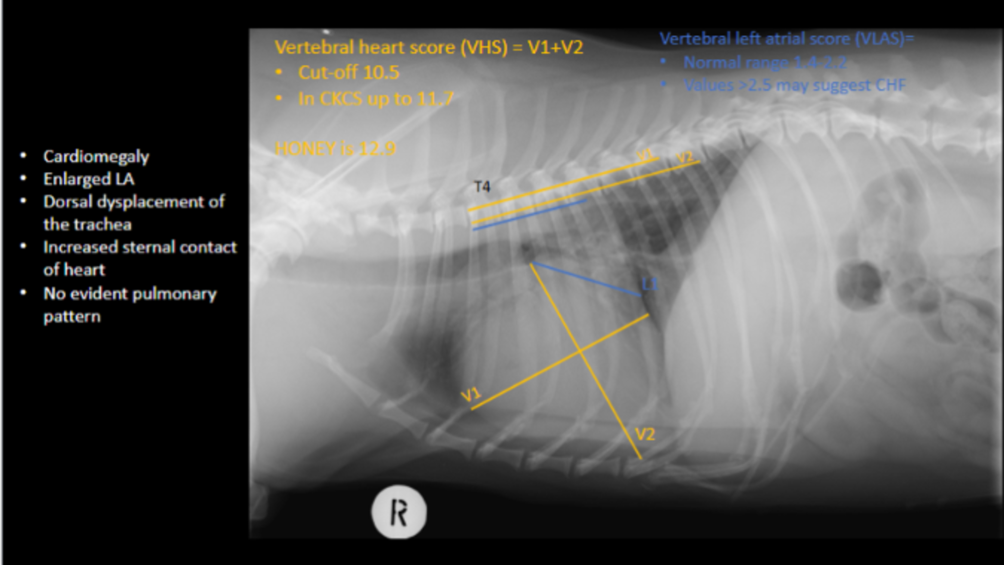
What stage of MMVD does this show?

What is the tx for this stage?
B2 —> mitral regurg + atrial and ventricular remodelling (enlargement)
Pimobendan —> improve contractility —> decr. time to progression to CHF
What needs to be done next after discovering the MMVD is stage B2?
Repeat rads —> to see pulmon oedema? → L-CHF
Repeat echo —> enlarged heart, systolic function?
BP measurement —> incr. = stress added to valves → worsens MMVD
Describe the abnormalities
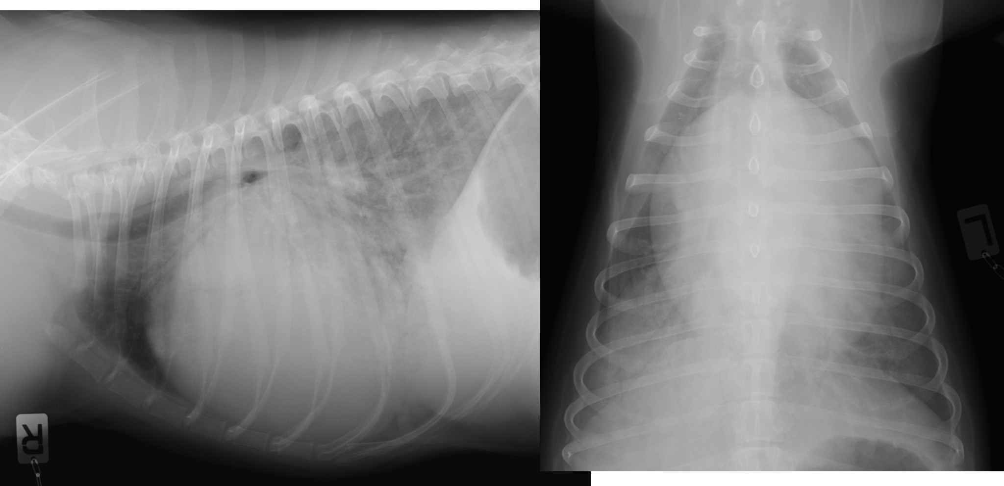
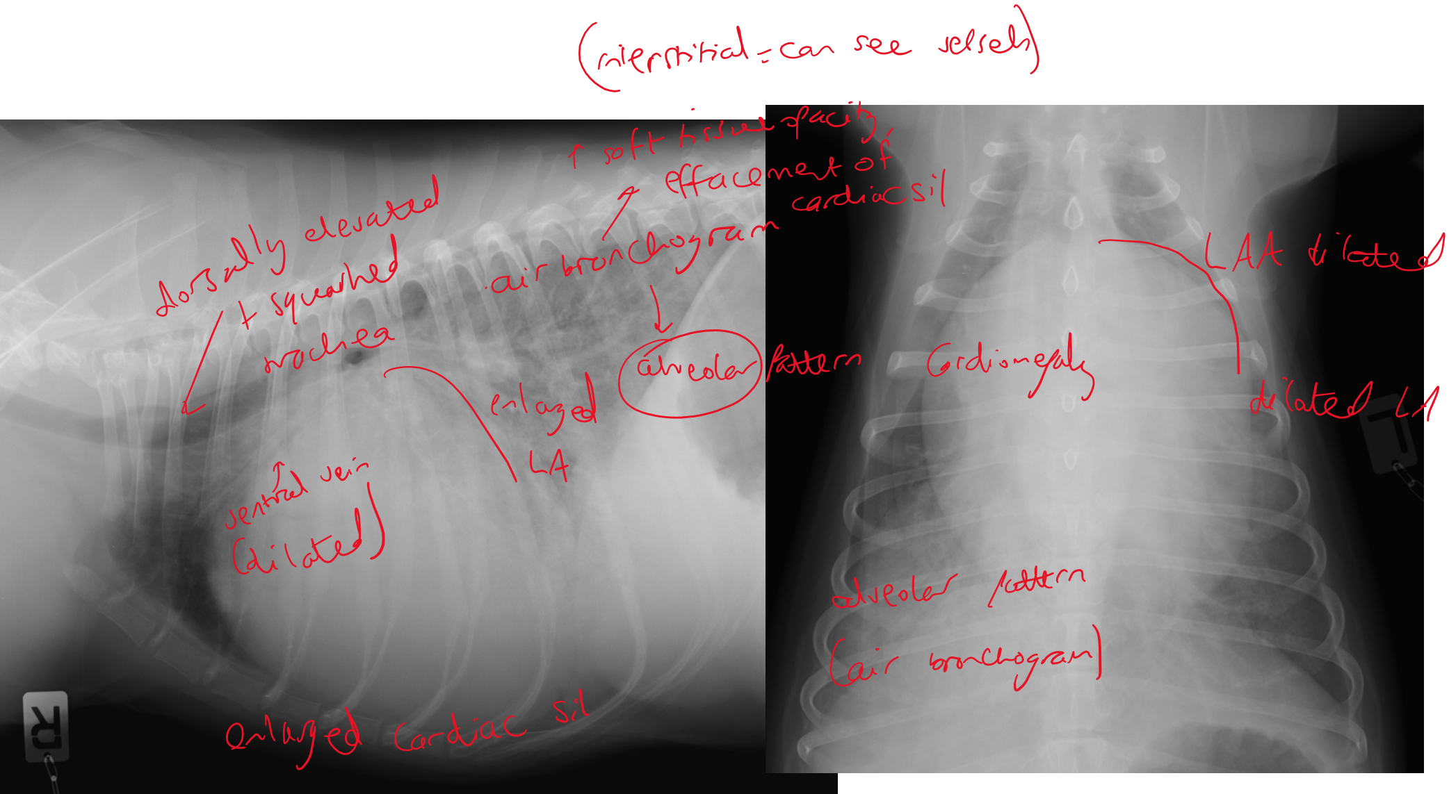
Progression to CHF = stage C
What medication can stage C be treated with if BP & renal function are normal?
Cont. pimobendan, start furosemide, benzaepril & spironolactone
Pimo = lifelong
Furosemide = affects kidneys so check K levels
Monitor BP regularly
What should be monitored regularly @ home once MMVD has progressed to CHF?
Sleeping resp rate —> should be <30 consistently otherwise suggestive of pulmon oedema?