DAT Biology: Circulatory System
 Call Kai
 Learn
Learn Practice Test
 Spaced Repetition
 Match
Match Flashcards
 Knowt Play
1/82

There's no tags or description
Looks like no tags are added yet.
Name | Mastery | Learn | Test | Matching | Spaced | Call with Kai | Chat |
|---|
No analytics yet
Send a link to your students to track their progress
83 Terms
Heart
Pump at the center of the circulatory system, responsible for making sure that the body tissues have access to required nutrients and gases are carried in blood
Right Atrium
Receives deoxygenated blood from the superior vena cava and inferior vena cava
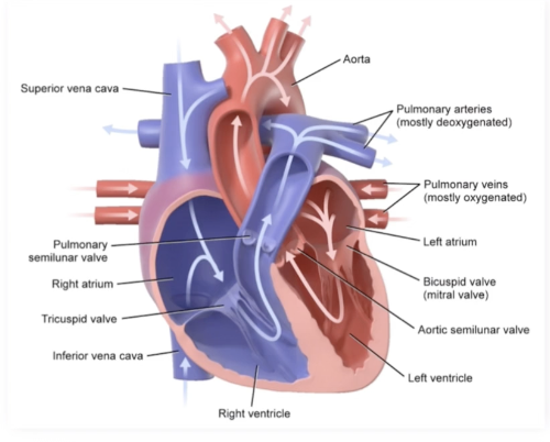
Superior Vena Cava
Collects deoxygenated blood from the chest, arms, head, and neck
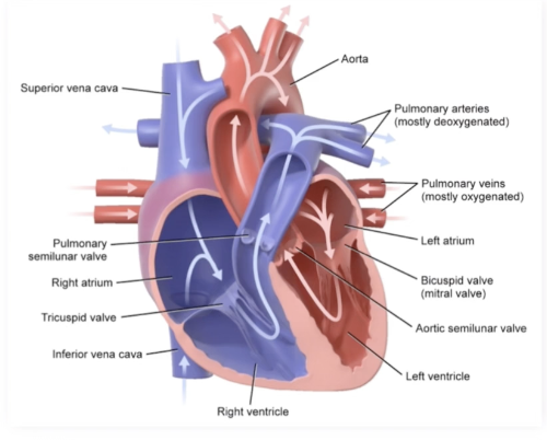
Inferior Vena Cava
Collects deoxygenated blood from the abdomen, pelvis, and lower limbs
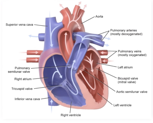
Right Atrioventricular (AV) Valve (Tricupsid Valve)
Allows blood to pass from the right atrium to the right ventricle
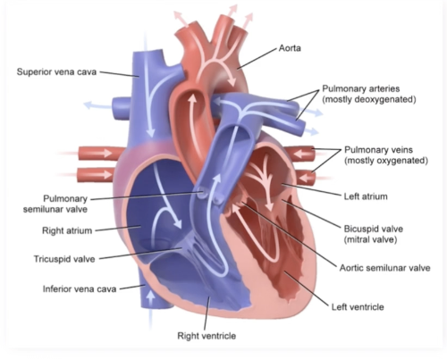
Right Ventricle
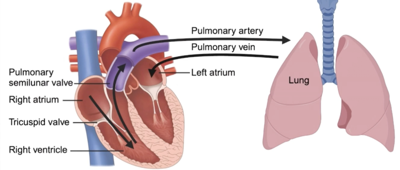
Pumps deoxygenated blood into the pulmonary artery
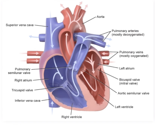
Pulmonary Artery
Carries deoxygenated blood to the lungs
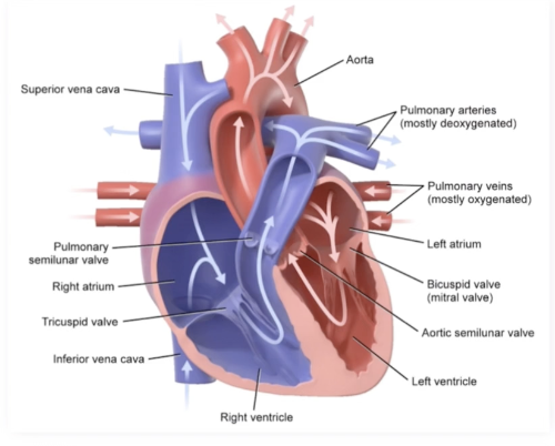
Pulmonary Semilunar Valve
Allows blood to pass from the right ventricle into the pulmonary artery
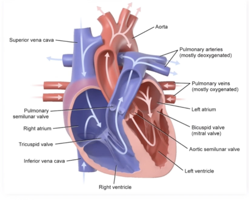
Right Ventricular Contraction
Tricuspid valve closes and the pulmonary semilunar valve opens, allowing for ventricular emptying without backflow into the atrium
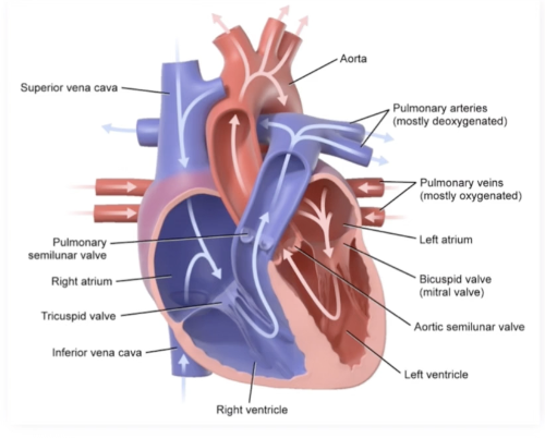
Right Ventricular Relaxation
Tricuspid valve opens and the pulmonary semilunar valve closes, allowing for ventricular filling from the atrium only without backflow from the pulmonary artery
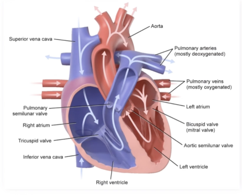
Left Atrium
Receives oxygenated blood from the pulmonary vein
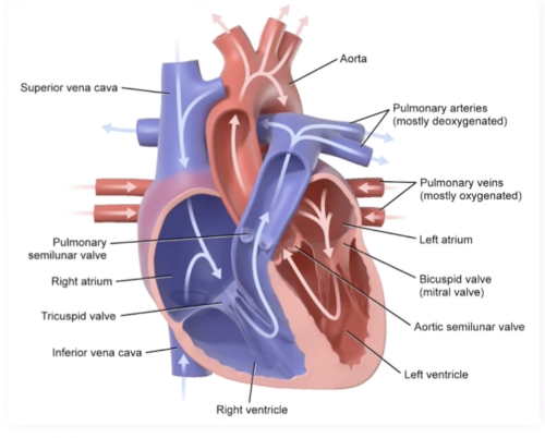
Pulmonary Vein
Carries oxygenated blood from the lungs to the heart
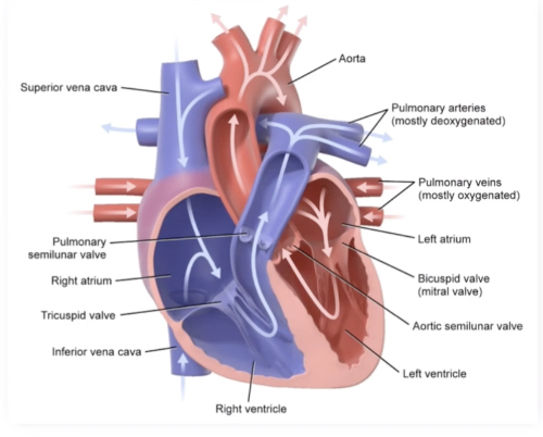
Left Atrioventricular (AV) Valve
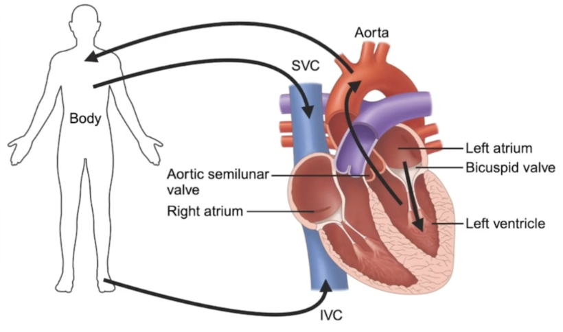
Bicuspid or mitral valve, allowing oxygenated blood to flow from the left atrium to the left ventricle
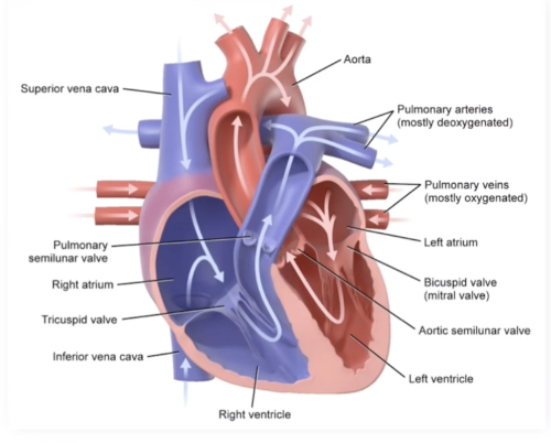
Left Ventricle
Pumps oxygenated blood into the aorta, it is the most muscular chamber of the heart, and the contraction will generate more pressure than that of the right ventricle
Aorta
Directs oxygenated blood from the heart into the systemic circulation
Pulmonary Circulation
Transports deoxygenated blood from the heart to the lungs via the pulmonary artery and oxygenated blood from the lungs back to the heart via the pulmonary veins
Systemic Circulation
Transports oxygenated blood from the heart to the body via the aorta and deoxygenated blood from the body to the heart via the superior and inferior vena cava
Cardiac Rhythm
Heart contracting and relaxing in a rhythmic pattern to ensure the most efficient transfer of blood
Proper relaxation of a heart chamber allows for maximal filling
Proper contraction after filling allows for maximal ejection of blood
Cardiomyocytes
Heart Muscle Cells
Automaticity
Ability of cardiac cells to generate electrical impulses spontaneously without external stimulation
Sinoatrial (SA) Node
Pacemaker of the heart
Located in the upper wall of the right atrium, has greatest automaticity (most likely to reach threshold and initiate an action potential), simultaneously stimulates contraction of the right and left atra, and transmits the electrical signal to the AV node
Atrioventricular (AV) Node
Briefly delays the electrical current coming from the SA node
Delay allowing the ventricles time to fill before the signal to contract is sent to the ventricles
Located in the lower wall of the right atrium
Passes electrical signal to the Bundle of His
Bundle of His
Passes the signal from the AV node to the bundle left and right bundle branches
Purkinje Fibers
Receive electrical signal from the bundle branches and stimulates ventricular contraction
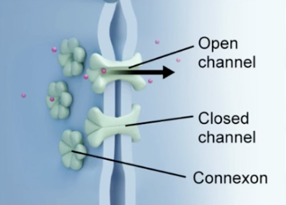
Intercalated Disks
Cell junctions between cardiomycotes, providing physical/electrical connection (gap junctions), allowing the contraction signal to be propagated in a coordinated, rhythmic fashion
Systole
Ventricular Ejection Phase
Ventricles contract, increasing the pressure within the chambers until it is enough to push open the semilunar valves, blood enters the pulmonary and systemic arteries, and arteriol blood pressure is at its highest during systole, the artery with the highest pressure during systole is aorta
Diastole
Ventricular Filling Phase
Ventricular myocardium relaxes, allowing a maximal amount of blood to fill each chamber from the atria, and arterial blood pressure is at its lowest point
Cardiac Output
Volume of blood being pumped by the heart per minute
Heart Rate X Stroke Volume
Heart Rate
How fast the heart beats (beats/minute)
Stroke Volume
Volume ejected per single beat, calculated by subtracting end systolic volume from end diastolic volume
Blood Vessels
Transport blodo to and from the heart, helping to form a closed circulatory system with the heart
Arteries
Transport blood away from the heart
Pressure highest in the arteries due to the hydrostatic pressure of the heart
Carry oxygenated blood in the systemic circulation
Carry deoxygenated blood in the pulmonary circulation
Arteriole
Branches off of arteries with smaller diameters than their parent arteries
Greatest drop in bp is when going from arteries to arterioles
Capillaries
One-cell-thick branches off of arterioles, connecting the arterial and venous circulations
Exchange gases and nutrients by diffusion
Body Tissues: Collect waste and CO2, deposit O2 and nutrients
Lungs: deposit CO2 and collect O2
Venules
Intermittent vessel that transports blood from the capillaries to the veins
Veins
Transport blood towards the heart
Venous circulation holds more blood tha the arterial circulation and veins have lower pressure than arteries
Veins have valves (pocket or venous valves) to prevent back flow in the low pressure system
Contraction of the surrounding skeletal muscles is used to generate the pressure necessary to push blood through the veins
Blood Plasma
Fluid component of blood
Composed of waterm proteins, nutrients,hormones, and other water soluble substances
Makes up most of the blood by volume (55%)
White Blood Cells (Leukocytes)
Cells of the immune system that defend against infection
Travel to site of infectino through blodo and enter the infected tissue to mount a response against the invading pathogens
Neutrophil
Most common white blood cell
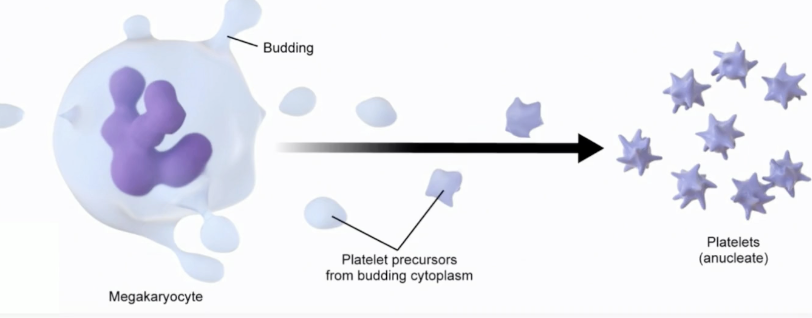
Platelets
Aka thrombocytes, immune cells, anucleate, cytoplasmic cell fragments responsible for clotting
Release clotting factors helping to turn fibrinogen into fibrin, creating a net to stop bleeding
Megakaryocytes
Large, platelet precursor/parent cells that are bone marrow residents
Vitamin K
Necessary cofactor for several clotting factors; deficiency can lead to a bleeding disorder
Red Blood Cells (Erthrocytes)
Responsible for transporting gases via hemoglobin proteins, making up 45% of blood volume
Transports O2 from the lungs to the tissues and CO2 from the tissues to the lungs
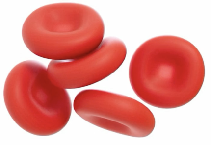
Bioncave Disk Shape
Bioncave Disk Shape
Looks like donut without the hole, just the indentation
Blood Clotting
A protective mechanism meant to repair damage to blood vessels and prevent extensive bleeding
Tears in blood vessels initiate blood clotting cascade helping to plug the tear, sealing it off to prevent further leakage
Blood Clotting Cascade
Positive Feedback Mechanism
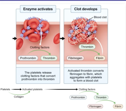
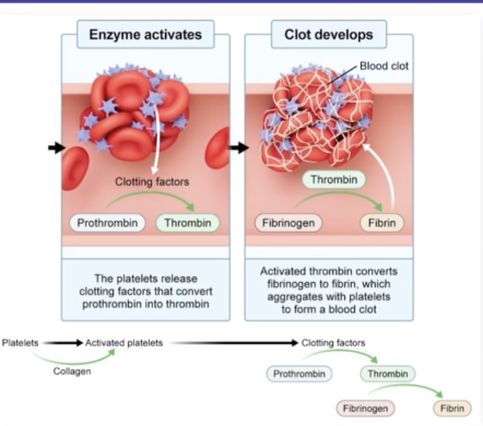
Damaged Tissues signal for platelet accumulation, each platelet then signals for the attraction and activation of other platelets
Tissue Damage
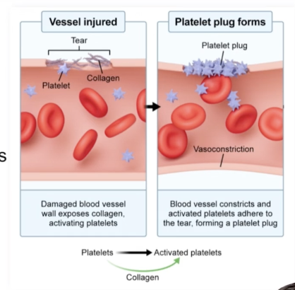
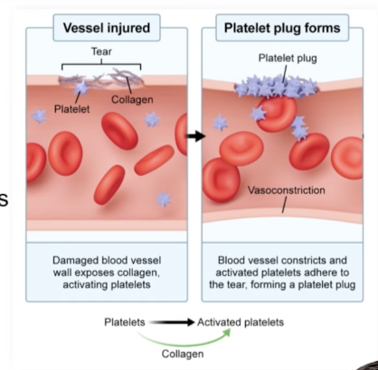
A tear occurs in the blood vessel wall, exposing structural collagen
Platelet Activation
Exposed collagen initiates platelet adhesion and activation
Platelet Plug
Accumulation of aggregated platelets that initially plug the tear
Activated Platelets
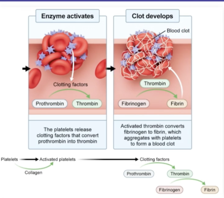
Release clotting factors converting prothrombin (inactive form) into thrombin (active form)
Clot Formation
Activated thrombin converts fibrinogen (inactive form) into fibrin (active form)
Activated fibrin strands polymerize together, forming the hemostatic plug
Hemostatic Plug
Blood clot made of fibrin meshwork and attached platelets
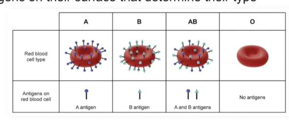
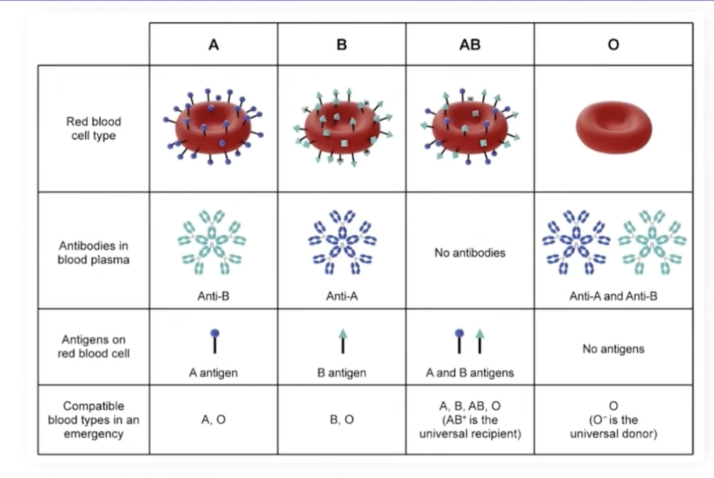
RBC Antigens
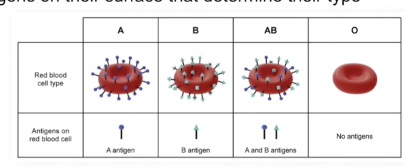
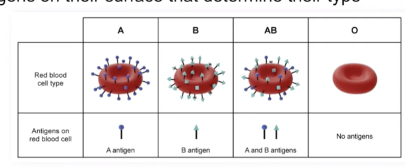
Sugar and protein identity markign molecules on cells
Type A
RBCs have the A antigen
Type B
RBCs have the B antigen
Type AB
RBCs have both the A and B antigens
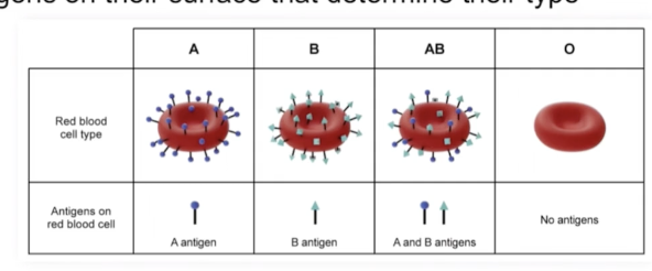
Type O
RBCs have neither the A or B antigens
Agglutination
Clumping together of red blood cells and red blood cell antibodies, caused by receiving an incorrect blood type
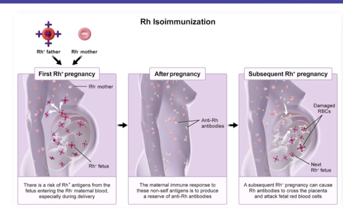
Rhesus Factor
Additional red blood cell surface marker with two phenotypes: Rh + and Rh -
Rh + donors cannot give blood to Rh - because the foreign Rh antigens will promote an immune response in the receiving, attacking it, rendering it ineffective
Universal Donors
Blood donors who can donate blood to anyone, type O (-) blood
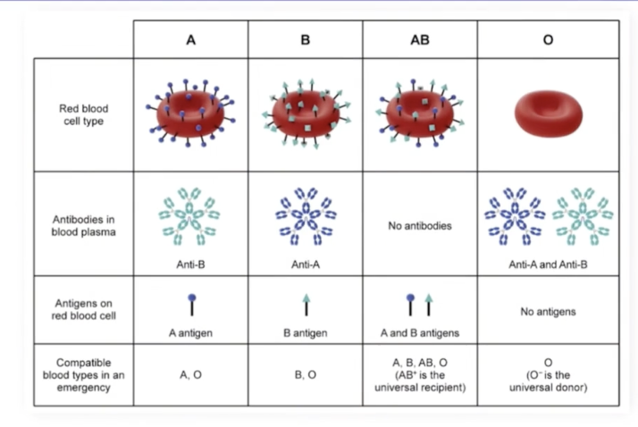
Type O (-)
Universal Donor: red blood cells that do not have A, B, or Rh antigens
No red blood cell antigens are present to stimulate an immune response in the receiving patient
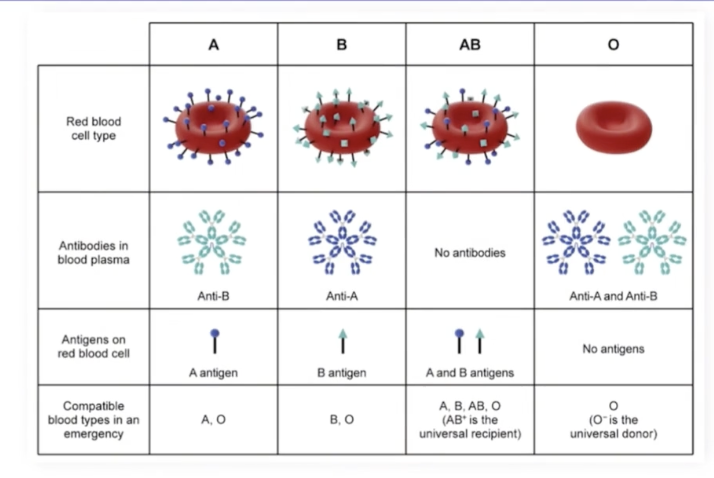
Universial Acceptor
Patients who can receive blood from anyone (can receive any blood type)
Type AB (+)
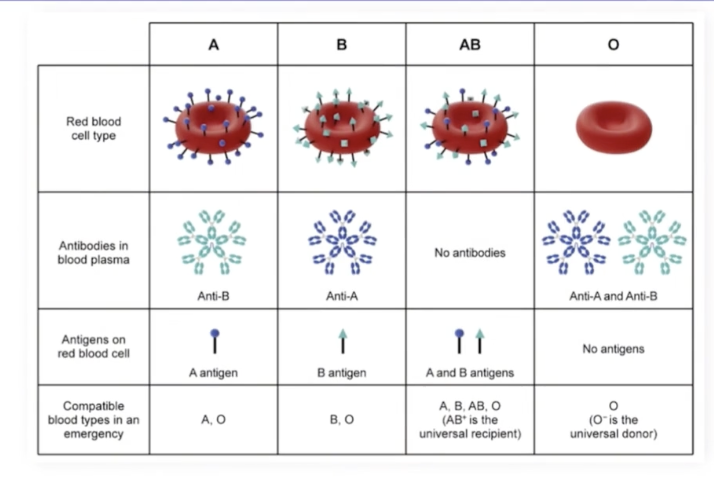
Type AB (+)
AB (+) red blood cell have the A, B, and Rh antigens, and the host immune system will be familiar with all three, so no immune response will be stimulated by any of the three antigens
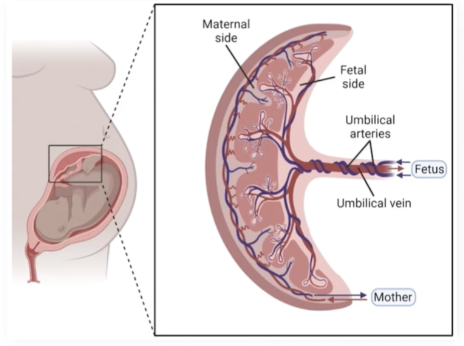
Placenta
Source of fetal nutrients from the mother, delivered via the umbilical cord: barrier for gas/nutrient exchange
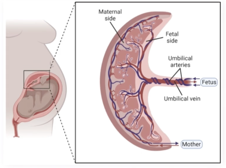
Umbilical Cord
Houses the umbilical vein and umbilical artery
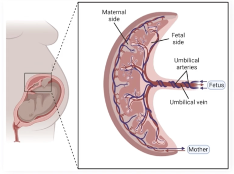
Umbilical Vein
Carries oxygenated blood from the placenta to the fetal heart
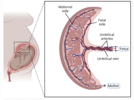
Umbilical Artery
Carries deoxygenated blood from the fetal heart back to the placenta
Removes wastes and CO2 from the fetal circulation
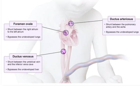
Fetal Circulatory Shunts
Fetal lungs and liver are nonfunctional/liquid-filled, so they help to ensure the fetus gets oxygen to its brain without wasting energy trying to pump blood into fluid-filled, collapsed lungs or liver that palcenta is alrfeady taking care of; overall just more efficient
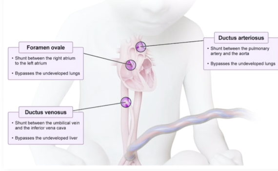
Ductus Venosus
A duct connecting the umbilical vein to the inferior vena cava
Blood passing through this duct will bypass the nonfunctional liver
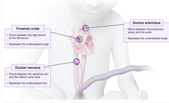
Foramen Ovale
A hole connecting the right and left atria, blood is allowed to flow from the right to the left atrium, the right ventricle wants to pump blood to the lungs but the fetal lungs are nonfunctional, so blood passing through this duct bypasses the pulmonary circulation
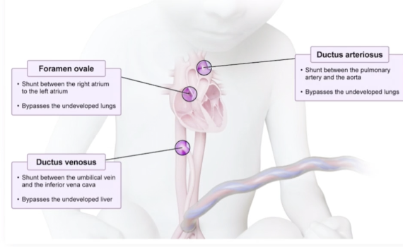
Ductus Arteriosus
A duct that connects the pulmonary artery to the aorta
Some blood is inevitably pumped by the right ventricle into the pulmonary artery, so it allows blodo to still bypass the pulmonary circulation
Erythroblastosis Fetallis
Disorder initiated by a Rh (-) mother carrying an Rh (+) fetus, becomes problematic upon a second pregnancy with another Rh (+) fetus
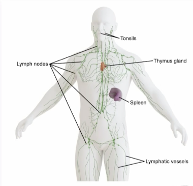
Lymphatic System
Subsystem of the circulatory system that regulates fluid levels and produces immune cells
Thymus
Major immune center within the lympathic system
Location of T cell lymphocyte maturation
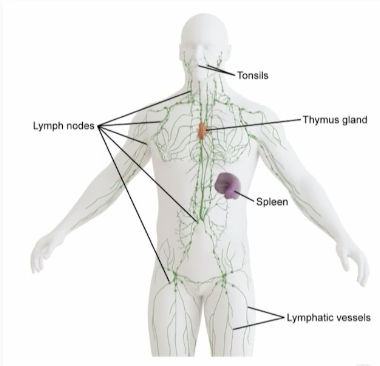
Spleen
Site of blood filtration within lymphatic system, filtering blood for pathogens and recycles old red blood cells
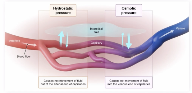
Hydrostatic Pressure
Pushes fluid out of the blood vessels (arterial end) and into the interstitial space, pressure exerted by vessel fluid
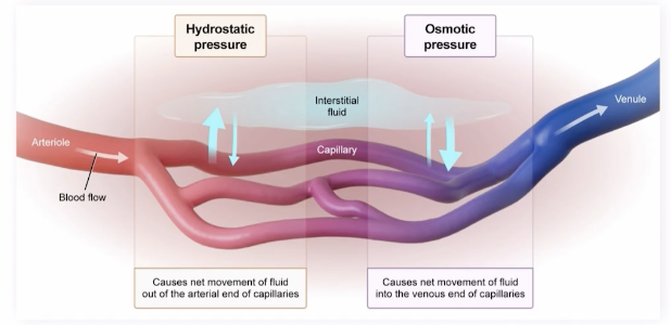
Oncotic Pressure
Brings fluid back into the capillary (venous end), type of osmotic pressure
Proteins on one side of a membrane pull water across to that side, protein concentrations higher in the blood than in the interstitium, blood protein oncotic pressure pulls fluid back into capillaries, not all fluid is reabsorbed from the interstitial space
Lymph
Consists of remaining fluid in the interstitial space, bacteria, fats, and proteins
Lymphatic Capillaries
Colelcts the lymph out of the interstitial space, merging to form larger vessels that travel to the heart
Lymph Nodes
Centers for the immune system to eliminate infections, lymph traveling to the heart is filtered through lymph nodes
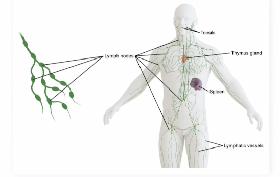
Lymphatic Vessels
No pressure, similar to veins, valves in them to prevent backflow
Lymph Propulsion
Relies on contraction of surrounding skeletal muscle and smooth muscle, achieved via peristalsis
Peristalsis
Involuintary, wave-like contraction and relaxation of smooth muscles lining the digestive tract propelling food, liquids, waste forward through the esophagus, stomach, and intestines