Humerus and Shoulder Anatomy and Positioning
 Call Kai
 Learn
Learn Practice Test
 Spaced Repetition
 Match
Match Flashcards
 Knowt Play
1/121

There's no tags or description
Looks like no tags are added yet.
Name | Mastery | Learn | Test | Matching | Spaced | Call with Kai | Chat |
|---|
No analytics yet
Send a link to your students to track their progress
122 Terms
Radius
lateral bone of the forearm, aligns with 1st digit
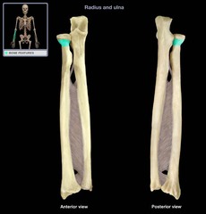
head of radius

radial notch
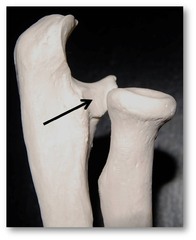
proximal radioulnar joint

neck of radius
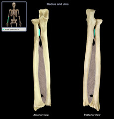
radial tuberosity
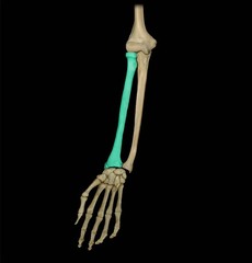
body or shaft of radius

Ulna
Medially bone of forearm, aligns with 5th digit
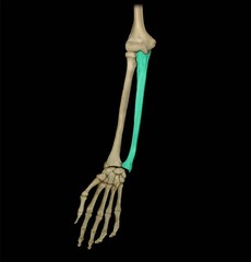
olecranon process of ulna
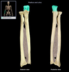
trochlear notch (semilunar notch) of ulna

coronoid process of ulna
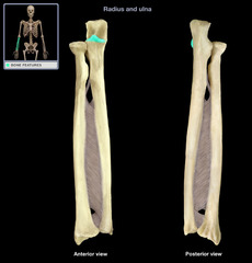
coronoid tubercle

Humerus
Largest & longest bone in the upper extremity About 1/5 of an adult's height Articulates with the scapula at the shoulder joint

Lateral epicondyle

Capitulum

Trochlea

Medial epicondyle

Humeral condyle
Expanded distal end of the humerus

Coronoid Fossa

Radial Fossa
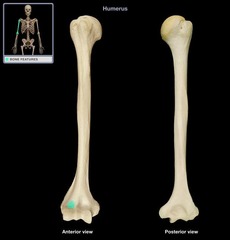
Olecranon Fossa

Concentric Arches
1st and smallest arc - Trochlear sulcus
2nd intermediate arc double lined -
smallest of the 2 - Capitulum
Largest of the 2 - Trochlea
3rd arc - Trochlear notch of the ulna

Elbow joint
hinge joint formed by humerus, ulna, and radius

body or shaft of humerus (diapysis)
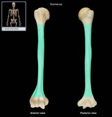
deltoid tuberosity of humerus
rough raised elevation along the anterolateral surface of the body (Deltoid muscle attaches here)
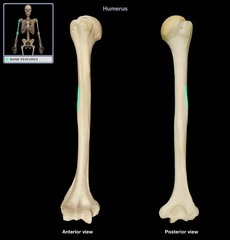
surgical neck of humerus

greater tubercle (tuberosity)
Larger lateral process, Pectoralis major and supraspinatus muscles attach
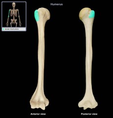
Intertubercular groove (bicipital groove)
Deep groove between the two tubercles

Lesser tubercle (tuberosity)
The process directly below the anatomic neck on the anterior surface
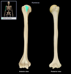
Anatomic neck

Head of humerus
articulates with glenoid cavity of scapula, Most proximal part of the humerus

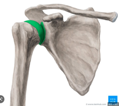
Scapulohumeral joint (glenohumeral joint)
The humeral head articulate with the glenoid cavity of the scapula
Neutral rotation of humerus
natural arm position without internal or external rotation, proximal humerus in an oblique position (looks like 2 eye balls).
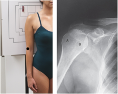
Internal rotation of humerus
dorsal aspect of hand on hip with entire humerus rolled inward. Humerus in true lateral position. Greater tubercle "A" and lesser tubercle is "B".

External rotation of humerus
Arm abducted and supinated with the greater tubercle "A" and lesser tubercle "B".

Internal
How is the humerus rotated? External, Internal, or Neutral?

Neutral
How is the humerus rotated? External, Internal, or Neutral?

External
How is the humerus rotated?
A=greater tubercle B=lesser tubercle

Head of humerus
A?

greater tubercle (tuberosity)
B?
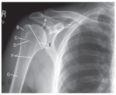
Intertubercular groove (bicipital groove)
C?
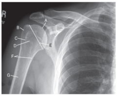
Lesser tubercle (tuberosity)
D?
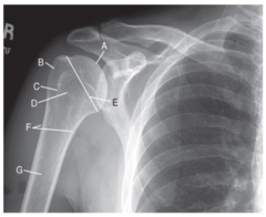
Anatomic neck
E?

surgical neck of humerus
F?

body or shaft of humerus (diapysis)
G?

Hill Sachs defect
Compression fracture of the articular surface of the humeral head

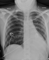
radiopaque objects, clothing removed placed in gown , long hair, oxygen tubing, and pacemaker leads to avoid reduced detail/scatter
During patient preparation, what must be removed?
1. Limit the number of exposures
2. Collimation
3. shielding (led apron)
4. Position correctly
What 4 things help protect you and the patient against radiation?
BEFORE the examination
When must the anatomical marker be placed?
kVp: 70-85
grid: use if greater than 4in (10cm)
mA: dependent
What technical factors are needed for a good Humerus (kVp, grid, mA)?
AP
Lateral (Lateromedial and mediolateral)
Routine projections of Humerus?
What is the evaluation criteria and procedure for a AP Humerus Xray?
SID: 40inches
Grid: yes if bigger than 4in
kVp: 70-85
CR: Midpoint of humerus
Patient position: Erect or supine, rotate body to affected side to bring shoulder and proximal humerus onto IR, extend hand and forearm, supinate hand pinky on hip
Breathing instructions: suspend respiration during exposure
Evaluation criteria: Entire humerus including shoulder and elbow, no motion, greater tubercle is seen laterally, humeral head is partially seen in profile medially, lateral and medial epicondlyes, Minimal superimposition of the glenoid cavity

What is the evaluation criteria and procedure for a Mediolateral Humerus (true lateral) Xray?
SID: 40inches
Grid: yes if bigger than 4in
kVp: 70-85
CR: Midpoint of humerus, rotate to humerus angle
Patient position: Face patient towards IR, oblique as needed to allow close contact of humerus to IR, flex elbow 90°
Breathing instructions: suspend respiration during exposure
Evaluation criteria: Includes both joints, True Lateral Projection, Epicondyles superimposed, Lesser tubercle in profile medially
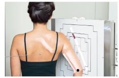
What is the evaluation criteria and procedure for a Lateromedial Humerus Xray?
SID: 40inches
Grid: yes if bigger than 4in
kVp: 70-85
CR: Midpoint of humerus
Patient position: Patients back to IR ,Elbow partially flexed, internally rotate arm so epicondyles are perpendicular to IR, abduct arm slightly
Breathing instructions: suspend respiration during exposure
Evaluation criteria: entire humerus including elbow and shoulder joints, epicondyles superimposed, lesser tubercle is shown in profile medially, partial superimposition by lower portion of glenoid cavity
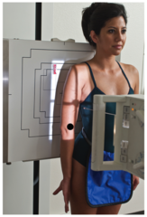
Horizontal beam lateral
Transthoracic Lateral
AP neutral rotation
Special projections of Humerus?
What is the evaluation criteria and procedure for a Horizontal beam Lateral Xray?
SID: 40inches
Grid: yes if bigger than 4in
kVp: 70-85
CR: distal 2/3 of humerus
Patient position: With patient recumbent, place support under patients' arm, Flex elbow to 90° if possible, Place IR between arm and thorax
Breathing instructions: suspend respiration during exposure
Evaluation criteria: elbow flexed to 90°, includes elbow joining and distal humerus

What is the evaluation criteria and procedure for a transthoracic lateral (trauma) projection Xray?
MUST INCLUDE AP NEUTRAL ROTATION
SID: 40inches
Grid: yes if bigger than 4in
kVp: 70-85
CR: mid diaphysis directed through thorax
Patient position: position affected arm in a neutral rotation, raise opposite arm (unaffected) above head, thorax in true lateral
Breathing instructions: Orthostatic breathing (gently and short)
Evaluation criteria: entire humerus and glenohumeral joint demonstrated

What is the evaluation criteria and procedure for a AP Neutral Xray?
SID: 40inches
Grid: yes if bigger than 4in
kVp: 70-85
CR: midpoint of humerus
Patient position: leave arm in a neutral position (No rotating) • "AS IS"
Breathing instructions: suspend respiration during exposure
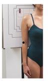
Shoulder girdle
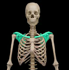
Scapula
Forms the posterior part of the shoulder girdle, Flat triangular bone

Superior border of scapula

Lateral (axillary) border of scapula

Medial (vertebral) border of scapula

superior angle
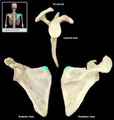
inferior angle
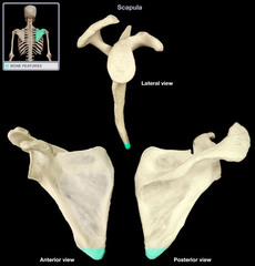
Lateral angle (head of scapula)
Thickest part of scapula, ends laterally in a shallow depression called the glenoid cavity (fossa)

Glenohumeral joint

Neck of scapula
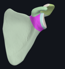
Body (blade) of scapula
Arched in shape for greater strength, Thin flat lower portion of the body is referred to as the Wing or ala

Costal or ventral surface of scapula (subscapular fossa)
Anterior surface of scapula, closest to ribs

Acromion
Long, curved process that extends laterally over the head of the humerus

Coracoid process
Thick, beaklike process that projects anteriorly beneath the clavicle

Subscapular notch
notch on the superior border that is partially formed by the base of the coracoid process

Dorsal surface of scapula

Crest of spine

Supraspinous fossa

infraspinous fossa
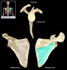
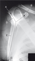
Acromion
Coracoid process
Body
Which structures from the "Y" on the scapula?

Acromion
8?
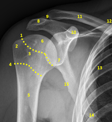
Coracoid process
10?
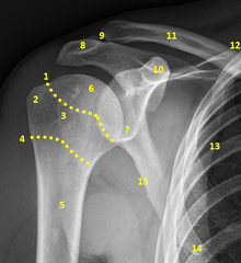
Glenohumeral joint
7?
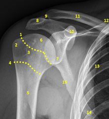
Lateral (axillary) border of scapula
15?
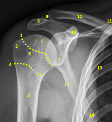
inferior angle
14?
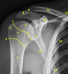
Medial (vertebral) border of scapula / Body (blade) of scapula
13?

Acromion
A?

Coracoid process
B?
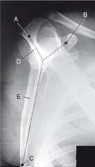
inferior angle
C?
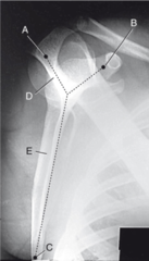
Medial (vertebral) border of scapula / Body (blade) of scapula AND Lateral (axillary) border of scapula
E?
AP External rotation
AP Internal rotation
Routine projections of the (NONTRAUMA) shoulder?
What is the evaluation criteria and procedure for a AP external rotation Xray?
SID: 40inches
Grid: depends
kVp: 70-85
CR: 1 inch inferior to coracoid process
Patient position: abduct and externally rotate arm, erect or supine
Breathing instructions: suspend respiration during exposure
Evaluation criteria: proximal humerus and 2/3 clavicle and upper scapula and glenoid cavity included, Greater tubercle profiled laterally

What is the evaluation criteria and procedure for a AP Internal rotation Xray?
SID: 40inches
Grid: depends
kVp: 70-85
CR: 1 inch inferior to coracoid process
Patient position: abduct and internally rotate arm
Breathing instructions: suspend respiration during exposure
Evaluation criteria: proximal humerus and 2/3 clavicle and upper scapula , Scapulohumeral joint centered, Lesser tubercle profiled medially
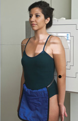
Inferosuperior axial projection (Lawrence method)
AP oblique projection (Grashey Method)
Special Projections of (NONTRAUMA) shoulder?
What is the evaluation criteria and procedure for a Inferosuperior axial projection (Lawrence method) Xray?
SID: 40inches
Grid: depends
kVp: 70-85
CR: medially 25-30° horizontal to axilla/humeral head for 90° abducted arm, less than 90° CR decreased to 15-20° (more arm abduction greater CR angle)
Patient position: supine, shoulder raised 2in to line up to IR,
Breathing instructions: suspend respiration during exposure
Evaluation criteria: Lesser tubercle profiled anteriorly, Humeral head and glenoid fossa profiled, Optimal exposure factors

What is the evaluation criteria and procedure for a AP oblique projection (Grashey Method) Xray?
SID: 40inches
Grid: depends
kVp: 70-85
CR: scapulohumeral joint (2in inferior and 2in medial from superolateral border of shoulder)
Patient position: erect or supine, rotate body 35-40° toward affected side, arm slightly flexed in neutral rotation, LPO/RPO
Breathing instructions: suspend respiration during exposure
Evaluation criteria: Glenoid Cavity should be visualized, Scapulohumeral joint centered, Optimal exposure factors
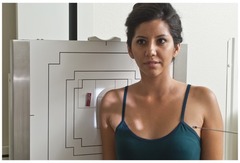
AP Neutral Rotation
PA Oblique (scapular Y lateral)
Routine TRAUMA projections of shoulder?
What is the evaluation criteria and procedure for a AP Neutral Rotation Xray?
SID: 40inches
Grid: depends
kVp: 70-85
CR: midscapulohumeral joint (3/4in inferior and 2in slightly lateral to corcoid process)
Patient position: erect or supine, rotate body if necessary to place body on IR
Breathing instructions: suspend respiration during exposure
Evaluation criteria: Greater tubercle superimposed ,Scapulohumeral joint centered, Optimal exposure factors

What is the evaluation criteria and procedure for a PA Oblique (scapular Y lateral) Xray?
SID: 40inches
Grid: depends
kVp: 70-85
CR: scapulohumeral joint (palpate superior angle of scapula and AC joint)
Patient position: erect or recumbent, rotate patient until imaginary line between AC and superior angle is perpendicular to IR, angle may be 45-60° LAO/RAO
Breathing instructions: suspend respiration during exposure
Evaluation criteria: Body of scapula superimposed on end, Acromion and coracoid processes in profile, Humeral head and glenoid cavity superimposed, Optimal exposure factors
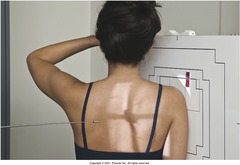
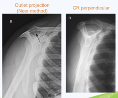
Supraspinatous outlet (Neer Method)
Special TRAUMA projections of shoulder?
What is the evaluation criteria and procedure for a Supraspinatous outlet (Neer Method) Xray?
SID: 40inches
Grid: depends
kVp: 70-85
CR: 10-15° caudal angle, centered posteriorly to pass through humeral head (1in above midscapula)
Patient position: erect or recumbent, rotate patient until imaginary line between AC and superior angle is perpendicular to IR, angle may be 45-60° LAO/RAO
Breathing instructions: suspend respiration during exposure
Evaluation criteria: Supraspinatus outlet open and in profile ,Demonstrates coracoacromial arch, Optimal exposure factors
AP
Lateral
Routine Scapula Projections?