Lec 12 - Anatomy
 Call Kai
 Learn
Learn Practice Test
 Spaced Repetition
 Match
Match Flashcards
 Knowt Play
1/35

There's no tags or description
Looks like no tags are added yet.
Name | Mastery | Learn | Test | Matching | Spaced | Call with Kai | Chat |
|---|
No analytics yet
Send a link to your students to track their progress
36 Terms
Neural Tissue (Nervous Tissue) Definition & Function
One of the 4 types of human tissue.
Function: Controls the body through electrical signaling (Sensory & Motor control).
Consists of CSF fluid, Neurons, and Glial cells.
Electrical signals in Dendrites
Graded potential (input signal)
Electrical signals in Axons
Action potential (output signal)
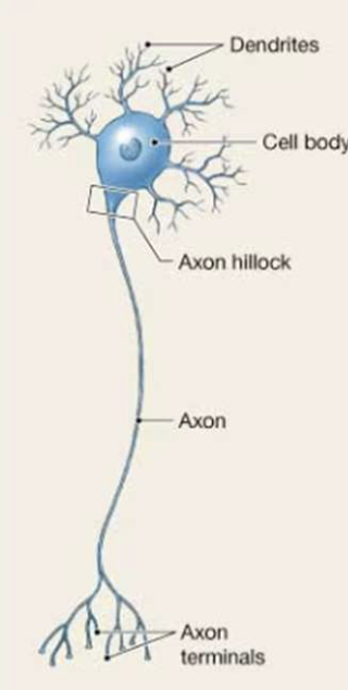
Multipolar Neuron
Most common neuron shape; has multiple dendrites and one single axon.
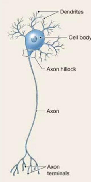
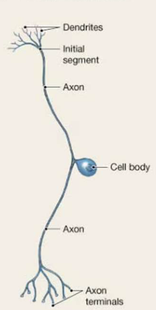
Pseudounipolar Neuron
Axons leave on each side of cell body; e.g., sensory neurons of PNS (dorsal root ganglion).
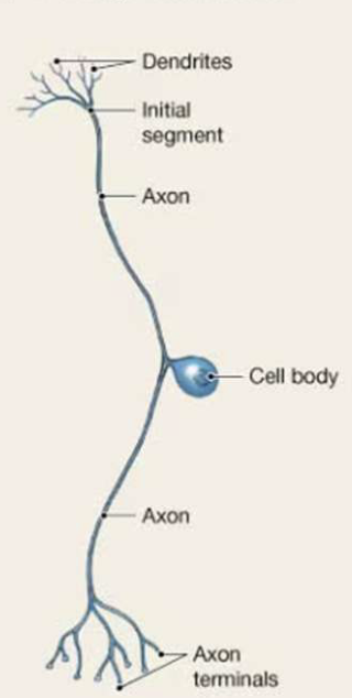
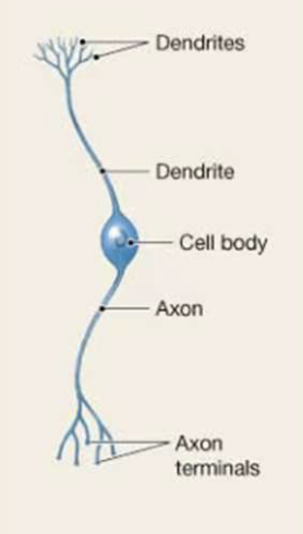
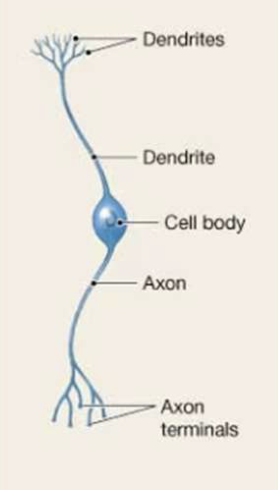
Bipolar Neuron
Dendrites are long and extend from one side of the cell body; found in ear, eyes, nose.
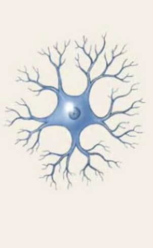
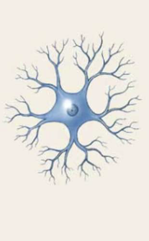
Anaxonic Neuron
Axons and dendrites cannot be distinguished; rare shaped, found in retina and brain.
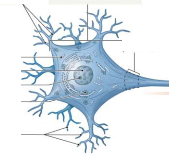
where you will fine the nucleus and the other typical cellular organelles you are familiarwith (such as mitochondria, endoplasmic reticulum, ribosomes)
Cell body
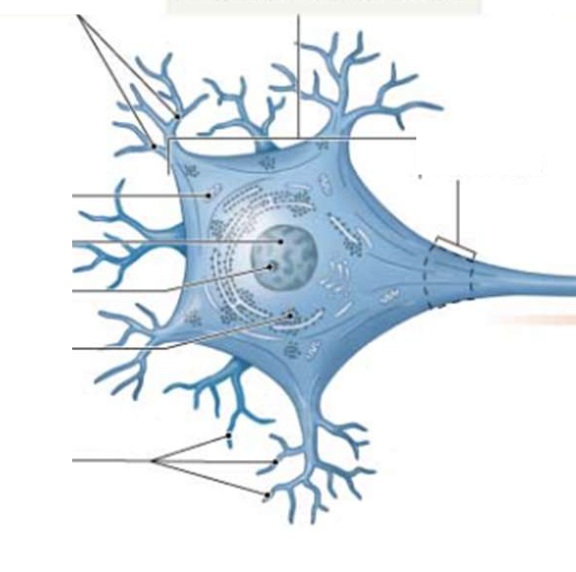
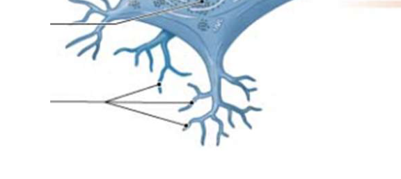
Contain receptors to receive signals from surrounding
environment and/or other neurons.
Denrites
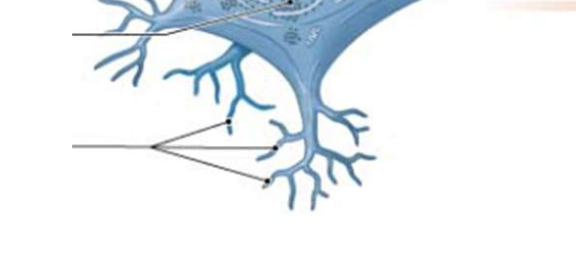
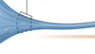
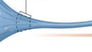
The initiating segment of axon (single long process), region of axon where action potential (nerve impulse) begins
Axon Hillock
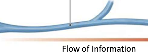
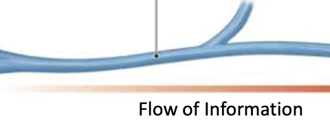
transmits (sends) nerve impulse (action potential) towards axon terminals to “signal” other cells.
Axon
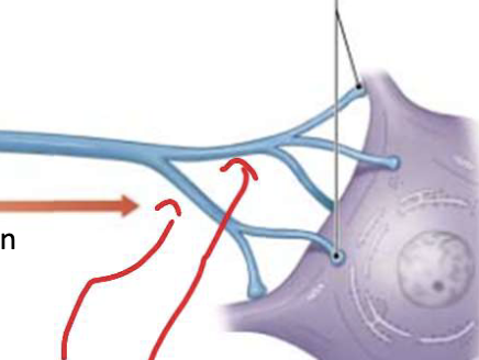
small axon branches sending action potential (nerve impulse) to terminal boutons.
Terminal arborization or Telodendria
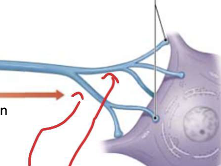
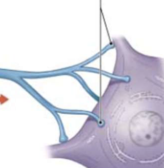
region found at the end of the axon where neurotransmitters release from vesicles. Action potentials end here too
Terminal Bouton
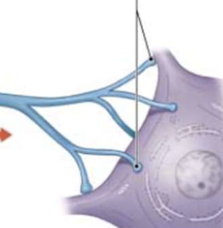
Condition for Action Potential
Only occurs if the threshold at the axon hillock is met.
Action Potential Characteristics
Output signal found in the plasma membrane of an axon;
Involves a rapid change in membrane potential/voltage.
Anatomical Location of Synapse
Occurs at the terminal bouton of one end and the dendrites at another end.
Cells a neuron can communicate with
Another neuron
Steps at a Synapse (Summary)
Action potential reaches terminal bouton.
2. Exocytosis releases neurotransmitter vesicles.
3. Neurotransmitters bind to postsynaptic receptors.
4. Postsynaptic cell fires or does not fire based on excitability.
Bob with spinal meningitis, recommends high doses of antibiotics…and why?
High doses are needed because the Blood-Brain Barrier (BBB) thickens blood vessels and restricts foreign substances/medications from easily entering the brain/spinal cord.
Astrocytes Function
Maintain the blood-brain barrier (BBB), provide structural support to neurons, and maintain stability by thickening blood vessels.
Ependymal Cells Function
Found in ventricles of Central Canal; produces cerebrospinal fluid (CSF).
Microglia Cells Function
Immune cells for the brain and spinal cord; remove debris, wastes, and pathogens.
Oligodendrocytes vs. Schwann Cells
Oligodendrocytes myelinate multiple axons within the Central Nervous System (CNS).
A single Schwann cell wraps around only one axon segment in the Peripheral Nervous System (PNS)
What is the name of the long slender process of the neuron (nerve cell)?
Axon
What are 2 possible events that will happen once a neurotransmitter binds to B?
Fire or no fire
Which part of the neuron (nerve cell) will you find the vesicles of neurotransmitters?
Dendrites
Whats the name of the biomolecule that makes up myelin?
Lipds
Which part of the neuron (nerce) will you find the myelin?
Axon
What is the name of the cell in the PNS that myelinates?
Schwann Cells
What is the name of the cell in the CNS that myelinates?
Oligodendrocytes
Multiple Sclerosis (MS) is a disease in which the myelin sheaths are destroyed. How would MS affect a patient’s electrical signaing and why?
This demyelination slows, scatters, or completely blocks electrical impulses traveling between the brain and the body.
What is the ganglia? How is it different from a nuclei?
Ganglia = Cell body of PNS
Nuclei = Cell body of CNS
What is a nerve? How is it different from a tract?
Nerves are located in the PNS
Tract is located in the CNS
Which of the terms above is known as white matter?
Tract
How are nerves repaired? Please list out all the steps
Fragmentation
Schwann Cells
Macrophages
Axons grow along the cord
Which cells would make up the lining of the ventricles of brain and central canal of the spinal cord? Why?
Ependymal cells make up the lining of the ventricles of the brain and the central canal of the spinal cord.