Chapter 10 Muscle Tissue
1/85
There's no tags or description
Looks like no tags are added yet.
Name | Mastery | Learn | Test | Matching | Spaced | Call with Kai |
|---|
No analytics yet
Send a link to your students to track their progress
86 Terms
Skeletal muscle
Voluntary, striated muscle that moves bones, works in pairs or more and is attached to bones by tendons
Cardiac muscle
Striated muscle fibers (cells) that form the wall of the heart; uni- or binucleated; involuntary muscle tissue found only in the heart.
Smooth muscle
under involuntary control, moves internal organs, cells contain a single nucleus, are spindle-shaped, and do not appear striated; each cell is a fiber; found inside many internal organs of the body
Muscle fibres
The long cylindrical multinucleated cells that make up skeletal muscles
Striated
muscle tissue in which the contractile fibrils in the cells are aligned in parallel bundles, so that their different regions form stripes visible with a microscope.
Voluntary
A muscle that is under conscious control
Involuntary
A muscle that is not under conscious control.
Excitability
muscles receive and respond to stimulation
Contractility
ability to shorten forcibly when stimulated
Extensibility
ability to be stretched
Elasticity
The ability of a material to bounce back after being disturbed
Epimysium
a sheath of fibrous elastic tissue surrounding the entire muscle.
Perimysium
Connective tissue surrounding a fascicle
Endomysium
Connective tissue surrounding a muscle fiber
Insertion
The attachment of a muscle tendon to a moveable bone or the end opposite the origin
Origin
attachment of a muscle that remains relatively fixed during muscular contraction
Aponeurosis
strong sheet of tissue that acts as a tendon to attach muscles to bone
Sarcolemma
plasma membrane of a muscle fiber
Sarcoplasm
cytoplasm of a muscle fiber
Sarcoplasmic Reticulum
Organelle of the muscle fiber that stores calcium.
glycosomes
granules of stored glycogen that provide glucose during periods of muscle cell activity
Myofibrils
Microscopic protein filaments that make up muscle cells.
Sarcomere
the structural, contractile unit of a myofibril in striated muscle, consisting of a dark band and the nearer half of each adjacent pale band.
A band
dark area; extends length of the thick filaments
I band
light band; thin filaments only
H zone
The region at the center of an A band of a sarcomere that is made up of myosin only. The H zone gets shorter (and may disappear) during muscle contraction.
M line
middle of the sarcomere; supporting proteins that hold the thick filaments together in the H zone
Z disc
at the center of the light band; provides anchorage for thin filaments and elastic filaments
Myosin
The contractile protein that makes up the thick filaments of muscle fibers
Actin
A globular protein that links into chains, two of which twist helically about each other, forming microfilaments in muscle and other contractile elements in cells.
Tropomyosin
A protein of muscle that forms a complex with troponin regulating the interaction of actin and myosin in muscular contraction
Troponin
A globular protein of muscle that together with tropomyosin forms a regulatory protein complex controlling the interaction of actin and myosin and that when combined with calcium ions permits muscular contraction
Elastic filament
composed of protein titin
Holds thick filaments in place; helps recoil after stretch; resists excessive stretching
Titin
are strands of elastic protein
reach from tips of thick filaments to the Z line
stabilize the filaments; responsible for allowing the sarcomere to stretch and recoil
Dystrophin
links the thin filaments to the integral proteins of the sarcolemma
T tubules
Also called transverse tubules, these are deep invaginations of the plasma membrane found in skeletal and cardiac muscle cells. These invaginations allow depolarization of the membrane to quickly penetrate to the interior of the cell.
Triads
formed by t tubules that run between the paired teminal cisternae of SR; organelles come into closest contact here; encircle each sarcomere
Action potential
the change in electrical potential associated with the passage of an impulse along the membrane of a muscle cell or nerve cell.
Ion channel
A transmembrane protein channel that allows a specific ion to diffuse across the membrane down its concentration or electrochemical gradient.
Chemically gated ion channels
channel-linked receptors that open to let a specific ion pass in response to a ligand
ACh receptor
a transmembrane protein in the sarcolemma of the neuromuscular junction that binds to ACh
Voltage-gated ion channels
A specialized ion channel that opens or closes in response to changes in membrane potential
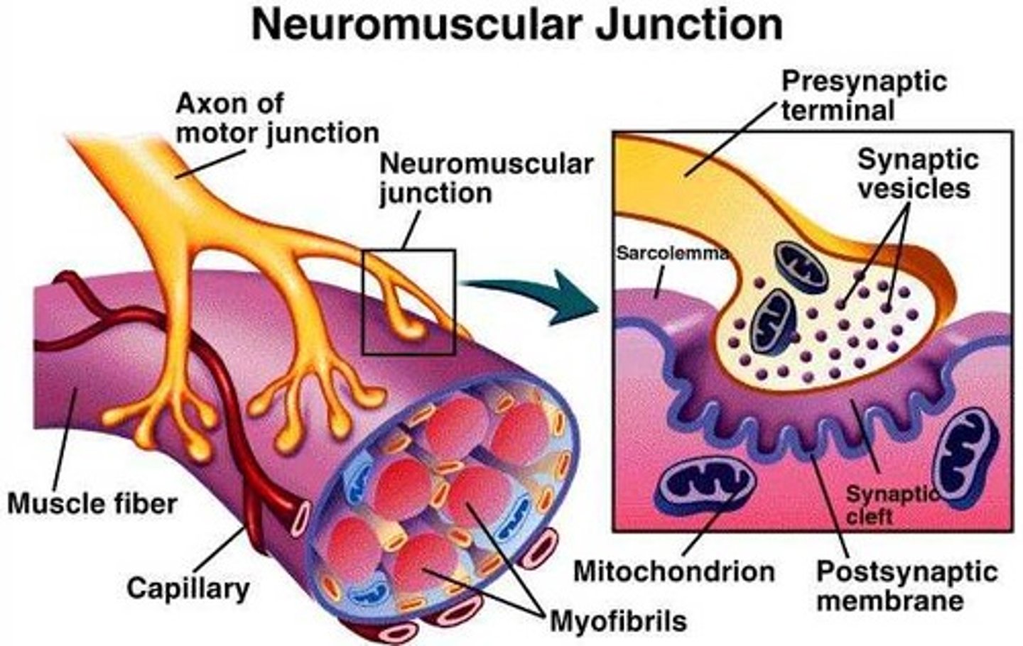
Neuromuscular junction/ motor end plate
Interface between the end of a motor neuron and a muscle fiber (synaptic area.) Acetylcholine and Cholinesterase are active in this region
Axon terminal
The endpoint of a neuron where neurotransmitters are stored
synaptic knob (terminal button)
Rounded areas (knobs) at the end of axon terminals that store neurotransmitters and assist in the transmission of neural information.
Synaptic cleft
The narrow gap that separates the presynaptic neuron from the postsynaptic cell.
Junctional folds
folds IN SARCOLEMMA at motor end plate that contain acetylcholine receptors; provide a large surface area for millions of ACh receptors located here
Endplate potential
the postsynaptic potential that occurs in the motor endplate in response to release of acetylcholine by the terminal button
Motor End Plate
specialized part of a muscle fiber membrane at a neuromuscular junction
Neuromuscular junction
region where a motor neuron comes into close contact with a skeletal muscle cell
Acetylcholinesterase
the enzyme that breaks down acetylcholine in the synaptic cleft
Excitation-contraction Coupling
The sequence of events from motor neuron's signaling to a skeletal muscle fiber to the contraction of the fiber's sarcomeres.
Creatine phosphate
An energy storage molecule used by muscle tissue. The phosphate from creatine phosphate can be removed and attached to an ADP to generate ATP quickly.
Creatine Kinase
enzymes that catalyzes the transfer of phosphate from CP to ADP
Motor unit
A motor neuron and all of the muscle fibers it innervates
Myogram
a chart of the timing and strength of a muscle's contraction
Muscle twitch
the response of a muscle to a single brief threshold stimulus
Latent period
time between application of a stimulus and the beginning of a response in a muscle fiber
Period of contraction
cross bridges are active, from the onset to the peak of tension development, and the myogram tracing rises to a peak
Period of relaxation
final phase, lasting 10-100ms, is initiated by reentry of Ca2+ into the SR; muscle tension decreases to zero and tracing returns to baseline
Treppe
the gradual increase in muscular contraction following rapidly repeated stimulation
Wave summation (temporal summation)
If stimulus frequency set at about 20 per second
Relaxation is not completed between twitches
Contractile forces add up to produce higher tensions
unfused/incomplete tetanus
Further increase in stimulus frequency causes muscle to progress to sustained, contraction generating more force with very brief periods of incomplete relaxation; frequency of stimulation allows only incomplete relaxation; generates submax. sustained contraction
fused/complete tetanus
when the muscle is stimulated so rapidly that no evidence of relaxation is seen and the contractions are completely smooth and sustained and generate maximum tension
Multiple motor unit summation
the more motor units activated the stronger the contraction
Sub-threshold stimuli
stimuli that produce no observable contractions
Threshold stimulus
stimulation level that must be exceeded to elicit a nerve impulse or a muscle contraction
Maximal stimulus
A stimulus which is strong enough to create action potentials in all the motor neurons innervating a whole muscle
Muscle tone
The state of partial contraction in a muscle, even when the muscle is not being used.
Isotonic contractions
muscle length changes and moves the load, the tension remains relatively constant through the rest of the contractile period; come in two flavors concentric and eccentric
Concentric contractions
those in which the muscle shortens and does work, such as picking up a book or kicking a ball;
Eccentric contractions
muscle generates force as it lengthens
Isometric contractions
muscular contraction that increases tension but does not produce movement.
Glycolysis
the breakdown of glucose by enzymes, releasing energy and pyruvic acid.
Lactic acid
Produced in muscle cells from the reduction of pyruvate (under anaerobic conditions) to regenerate NAD+ so that glycolysis can continue. A rise in lactic acid usually accompanies an increase in physical activity.
Anaerobic glycolysis
Energy-yielding conversion of glucose to lactic acid in various tissues, notably muscle, when sufficient oxygen is not available.
Aerobic respiration
Respiration in which oxygen is consumed and glucose is broken down entirely; water, carbon dioxide, and large amounts of ATP are the final products. This metabolic process in which pyruvate is broken down in the Kreb's cycle and the electron-carrier molecules produced are used to produce ATP through electron transport
Aerobic endurance
the length of time a muscle can continue to contract using aerobic pathways
Muscle fatigue
Inability of muscle to maintain its strength of contraction or tension after prolonged activity; may be related to insufficient oxygen, depletion of glycogen, and/or lactic acid buildup
Excess post-exercise oxygen consumption
additional oxygen consumed immediately after an exercise bout when the body is no longer exercising
Myoblasts
stem cells that fused to form each muscle fiber early in development
Myosatellite cell
stem cells that function in the repair of damaged muscle tissue
prime mover (agonist)
muscle that produces most of the force during a particular joint action
Antagonist
muscle that opposes or reverses a prime mover
Synergist
muscle that aids a prime mover in a movement and helps prevent rotation
Fixator
Muscle that immobilizes one or more bones, allowing other muscles to act from a stable base.