AFl, AF, HF, congenital heart diseases
 Call Kai
 Learn
Learn Practice Test
 Spaced Repetition
 Match
Match Flashcards
 Knowt Play
1/79

There's no tags or description
Looks like no tags are added yet.
Name | Mastery | Learn | Test | Matching | Spaced | Call with Kai | Chat |
|---|
No analytics yet
Send a link to your students to track their progress
80 Terms
26% of patients are completely asymptomatic
AF
Every 2nd or 3rd beat conducts to ventricles
AFl
Atria is beating 240-400 bpm
AFl
Rapid ventricular rate (150+ bpm)
AFl
In elderly, there is a 30% chance patients with AFl will develop ____
AF
Multiple p waves precede each QRS
AFl
P waves have saw tooth appearance
AFl
R-R intervals usually regular, may become irregular
AFl
Symptoms include: palpitations, shortness of breath, dizziness, syncope
AFl
Chaotic and unsynchronized quivering of atrial myocardium —> rapid, disordered atrial contractions
AF
Atrial rate of 400+ bpm
AF
No distinguishable p waves
AF
Irregularly irregular ventricular rhythm due to AV conduction
AF
Typically narrow QRS
AF
AF terminates spontaneously or with intervention within 7 days, recurrence with variable frequency
Paroxysmal AF
Continuous AF that is sustained > 7 days
Persistent AF
Long term AF >1 yr
Permanent AF
AF in the absence of rheumatic mitral stenosis, a mechanical or bioprosthetic heart valve, or mitral valve repair
Nonvalvular AF
Absence of identifiable disease
Isolated event, or intermittent occurrence
Younger patients, primarily male
Triggers: alcohol, drugs, caffeine, smoking, endurance sports, physical or mental stress, electrolyte abnormalities
Lone (idiopathic) AF
Symptoms:
Recurrent palpitations
Shortness of breath
Chest pain
Dizziness
Increased frequency of urination
Blackout spells
Congestive heart failure
AF
Treatment: stroke prevention, rhythm control, rate control
AF
Anticoagulation
Stroke prevention
Antiarrhythmic meds, cardioversion in addition to anticoagulation, AF ablation
Rhythm control treatment
Meds to control ventricular rate
AV node ablation and pacemaker implant
Rate control treatments
In people over 80, AF is a direct cause of 25% of ____
Strokes
% of blood ejected from the left ventricle with each ventricular contraction
Ejection fraction (EF)
Stroke volume divided by end diastolic volume
Ejection fraction (EF)
In normal hearts, __ ranges between 50-70%
Ejection fraction (EF)
Left side of heart is unable to deliver enough blood to vital body organs
LSHF
Systolic failure: LV loses ability to contract normally, heart cannot efficiently pump blood into circulation
HFrEF
EF =<40%
HFrEF
Diastolic failure: LV loses ability to relax normally (stiff muscle), heart cannot fill with blood during resting period between beats
HFpEF (P = preserved)
EF >= 50%
HFpEF (P = preserved)
In between HFrEF and HFpEF
HFmrEF (mildly reduced)
EF between 41-49%
HFmrEF (mildly reduced)
A previous EF was <= 40% and a follow up EF is >40%
HFimpEF (improved)
Symptomatic HF with baseline LVEF <= 40%, a 10-point inc from baseline LVEF, and 2nd measurement of LVEF >40%
HFimpEF (improved)
SV = EDV - ESV
Stroke volume = end-diastolic volume - end-systolic volume
EF = SV/EDV
Ejection fraction = stroke volume divided by end-diastolic volume
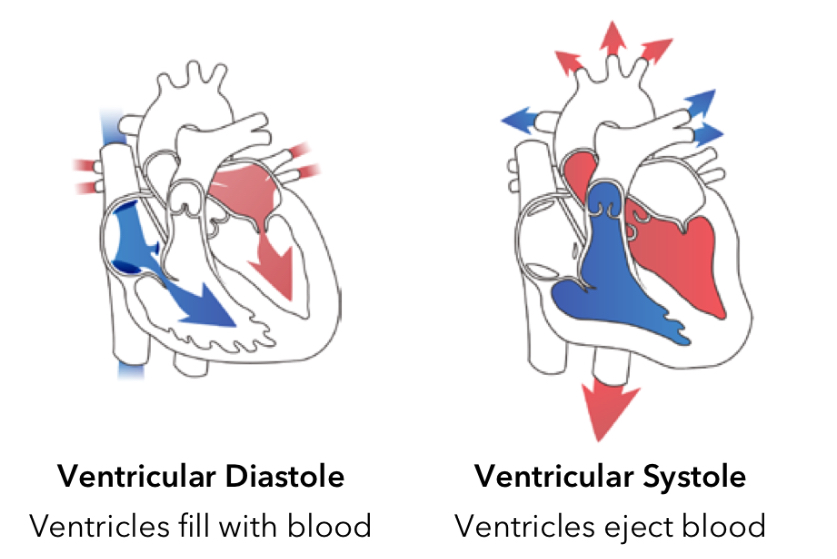
Normal heart (EF 50-70%)
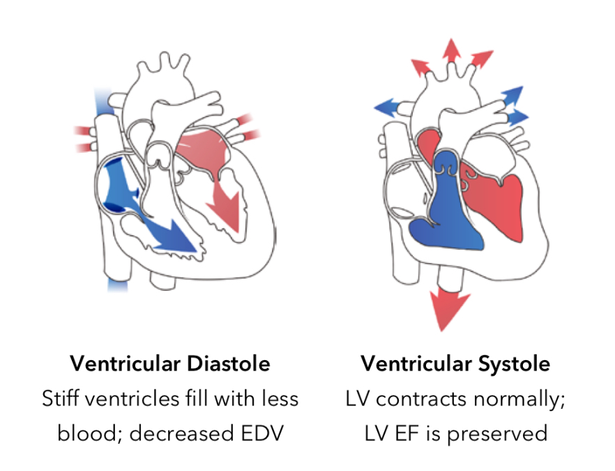
Diastolic dysfunction (HFpEF)
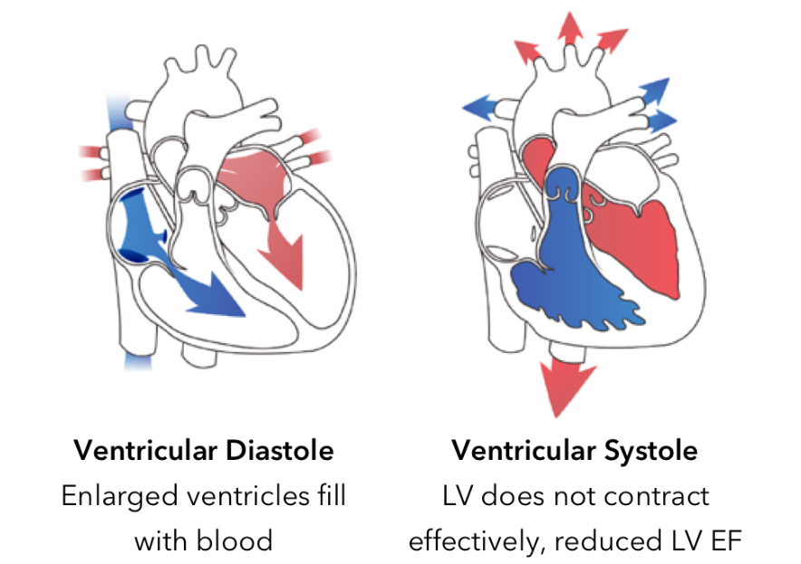
Systolic dysfunction (HFrEF)
RV too weak to pump enough blood to lungs —→ increases venous pressure which pushes fluid into surrounding body tissues —→ cause swelling in legs, abdomen, and body tissues
RSHF
Typically the result of LSHF but can be caused my pulmonary hypertension, pulmonary embolism, and COPD
RSHF
Main causes are:
Hypertension
Abnormal heart valve
CAD
MI
HF
Other causes are:
Congenital heart defects
Familial cardiomyopathy
Infection of heart or valves
Substance abuse
Chemotherapeutic drugs
Rheumatologist or autoimmune disorders
Endocrine or metabolic disorders
Heart rhythm related
Infiltration cardiac disease
Peripartum cardiomyopathy
Stress cardiomyopathy
RV pacing
Electrical dyssynchrony
HF
Shows:
Wide QRS (caused by electrical dyssynchrony)
Damage done to heart
ECG
Shows:
Size of heart
Congestion in lungs
X-ray
Shows:
Objective data about heart valves and function of walls
EF
Echocardiogram
Shows:
Ventricular stretching
Lab tests (beta natriuretic peptide)
No symptoms
Class I
Symptoms with moderate activity
Class II
Symptoms with minimal activity
Class III
Symptoms at rest
Class IV
At risk of heart failure, no structural damage, no symptoms
Stage A
Structural heart damage, no symptoms
Stage B
Structural heart damage, symptomatic
Stage C
End-stage symptoms of HF, normal treatment options do not work
Stage D
Activity, low salt diet, fluid intake, weight control
HF lifestyle changes
Ace-inhibitor, angiotensin receptor blocker, angiotensin receptor/neprilysin inhibitor, beta-blockers, aldosterone antagonist, vasodilators, SGLT-2 inhibitors, digitalis, diuretics
HF drug therapies
valve replacement, heart transplant
HF surgeries
For patients Class II, III, IV heart failure and conduction system defects
CRT device
For patients with class IV HF
Ventricular assist device
Characterized by a back leak of blood from systematic to pulmonary circulation
Left-to-right shunts
Atrial and ventricular septal defects, AV canal defects, patent ductus arteriosus
Types of left-to-right shunts
Some oxygen rich blood passes to right atrium —> extra volume causes damage to heart and lungs
Atrial septal defect (ASD)
75% of ___ occur in middle part of atrial septum
ASD
15-20% of newborns with Down Syndrome have complete ____
AV canal defect
Oxygenated LV and deoxygenated RV blood mix
Ventricular septal defect (VSD)
Can involve ASD, VSD, and tricuspid and mitral valve defects (or all 3)
AV canal defects
A patient with complete _____ has a hole in the center of the heart allowing blood flow between all chambers
AV canal defect
A blood vessel between the aorta and pulmonary artery that fails to close at birth —> increases blood flow to heart and lungs, causing pulmonary hypertension and an enlarged and weakened heart
Patent ductus arteriosus (PDA)
Deoxygenated blood bypasses lungs and flows directly into systemic circulation of the body
Right-to-left shunts
Transposition of the great arteries (TGA)
Tetralogy of Fallot (ToF)
Turn us arteriosus
Total anomalous pulmonary venous return (TAPVR)
Ebstein’s anomoly
Single ventricle defects
Types of right-to-left-shunts
Most common form of cyanotic congenital heart disease (represents ~10% of all cases of congenital heart disease)
ToF
a combination of VSD (hole in ventricular septal wall), PVS (narrowed pulmonary and outflow tract), overriding aorta (aorta is between LV and RV, over the VSD), and RV hypertrophy (thick RV muscle)
ToF
Narrowing of the cardiac valve
Valve stenosis
Valve does not close tightly enough to prevent blood from leaking backwards (aka insufficiency)
Valve regurgitation
May occur at any of the 4 valves
Valve stenosis and regurgitation
Present in 0.3-0.5% of gen pop and 4.5% in people with congenital heart diseases
Malformation of thoracic venous return
Persistent left SVC (PLSVC)