OCR Level 3 Cambridge Technicals in Health and Social Care - Unit 4 Anatomy and Physiology for Health and Social Care
1/103
There's no tags or description
Looks like no tags are added yet.
Name | Mastery | Learn | Test | Matching | Spaced |
|---|
No study sessions yet.
104 Terms
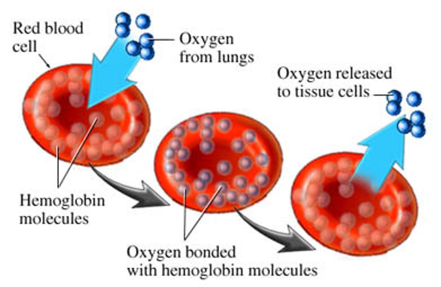
Erythrocytes
These are red blood cells and are made in the bone marrow and contain haemoglobin. They have a thin, biconcave shape so they can fit into small gaps within the capillaries and no nucleus which allows more oxygen to be transported within the cell. The haemoglobin exchanges materials at different parts of the body, for example it exchanges carbon dioxide for oxygen at the lungs.
Leucocytes
These cells are a part of the immune system and fight away foreign bodies to prevent infection and is the term for white blood cells as a collective.
Lymphocytes
These white blood cells are made in the lymphatic system. There are two types of this cell; B-cells and T-cells. The B-cells are produced in the bone marrow whilst T-cells are made in the thymus gland.
Plasma
This liquid forms around 55% of the total blood volume and allows the transportation of red blood cells, white blood cells, waste products, hormones and nutrients. It also helps with the temperature regulation of the body as it takes heat from the tissues, such as the muscles, and circulates this heat around the body.
Monocytes
They are the largest type and fight bacteria, viruses and fungi. They are originally formed in the bone marrow and are released to remove dead cells and bacteria.
Neutrophils
They are made in the bone marrow and are very mobile so they are able to move quickly. They are attracted to the chemicals released by the damaged cells and move around in large numbers to engulf the bacteria, causing the infection of the bacteria.
Platelets
These are produced in the bone marrow and help to clot the blood, when the skin is damaged or broken, to prevent blood loss. This stops the bleeding and allows the wound to heal.
Right Atrium
One of the four chambers and passes the deoxygenated blood through the tricuspid valve and into the right ventricle.
Tricuspid Valve
A valve that prevents the backflow of deoxygenated blood from the right ventricle into the right atrium.
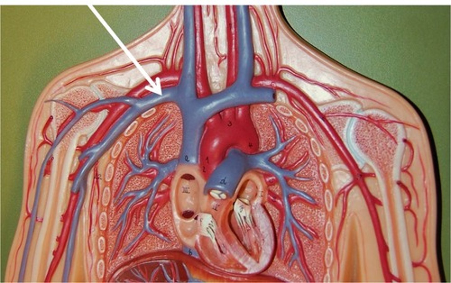
Vena Cava
This is the large vein that transports deoxygenated blood from the body and into the heart, specifically the right atrium.
Right Ventricle
Another one of four chambers and forces the deoxygenated blood through the pulmonary valve and to the lungs, via the pulmonary artery.
Pulmonary Artery
Transports deoxygenated blood from the right ventricle to the lungs to gain oxygen and become oxygenated blood to be circulated around the body.
Pulmonary Vein
Transports oxygenated blood from the lungs and back to the heart through the left atrium.
Semi-Lunar Valves (Pulmonary and Aortic)
Two sets of valves that prevents the backflow of deoxygenated blood between the right ventricle and the pulmonary artery and the backflow of oxygenated blood between the left ventricle and the aorta.
Left Atrium
One of the four chambers that receives oxygenated blood from the pulmonary vein and allows it to pass through into the left ventricle.
Bicuspid Valve
A valve that prevents the backflow of oxygenated blood from the left ventricle into the left atrium.
Left Ventricle
Another one of four chambers and receives oxygenated blood from the left atrium and forces out blood at a high rate, with the help of the thick cardiac muscle, so it can circulate the body and back through the Aorta.
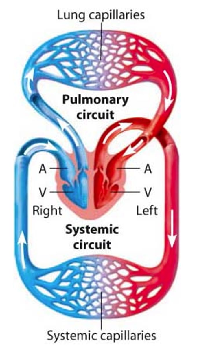
Double Pump
This is what the heart acts as, which means that there are two separate circulatory systems. These systems are called the systematic and the pulmonary. The systematic system contains the left side of the heart and the arteries, which carry oxygenated blood to organs and tissues. Whereas the pulmonary system contains the right side of the heart and the veins, which carry deoxygenated blood back to the heart from organs and tissues.
Coronary Arteries
These are smaller arteries that branch from the aorta to supply the heart with oxygenated blood, so it is able to continue pumping.
Aorta
This is the largest artery within the body and has a thick wall so it can withstand the high blood pressure, due to its need to be strong so the blood can travel around the body and back to the heart.
Cardiac Cycle
The average heart rate for an adult is around 60-80 BPM (Beats Per Minute). This means the cardiac cycle is repeated around 70 times in a minute. When the heart contracts, this is the systole and when it relaxes, this is the diastole.
Atrial Systole
The contraction of both the right and left atria, which is one of the stages of the cardiac cycle.
Ventricular Systole
The contraction of both the right and left ventricles, which is one of the stages of the cardiac cycle.
Complete Cardiac Diastole
The relaxation of the atria and the ventricles, which is one of the stages of the cardiac cycle.
Sinoatrial Node
This is located in the upper wall of the right atrium and is responsible for setting the rhythm of the heart (like a pacemaker). It also ensures that both the atria contract simultaneously.
Atrioventricular Node
This is located on the bottom wall of the right atrium and is responsible for delaying the the electrical impulses of the SA node. This allows enough time for the blood in the atria to leave into the ventricles.
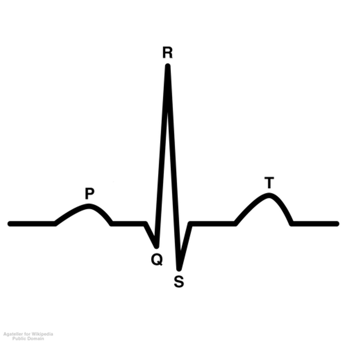
ECG
This is a test that can be used to see the electrical activities of the heart. It contains five waves; P, Q, R, S and T. The P wave shows the atrial contraction, the QRS waves show the ventricular contraction (systole) and the T wave shows the ventricles relaxing (diastole). The ECG trace can show which part of the heart is being problematic, if the heart is out of rhythm and disordered.
Purkyne Fibres
These are specialised cardiac muscle fibres that rapidly transmit electrical impulses from the AV node to the ventricles. These fibres are located in the inner ventricular walls.
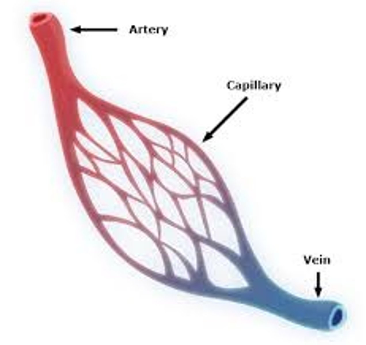
Arteries
They carry blood away from the heart. The walls have several layers of elastic fibres and thick, smooth muscle and has a small lumen. This is because they have to carry blood at a high pressure (except from the pulmonary artery, as the capillaries in the lungs are delicate) so that it can go around the whole body and back to the heart. Apart from the pulmonary artery that carries deoxygenated blood to the lungs to become oxygenated, the blood it carries around the body is oxygenated so it can exchange oxygen for any waste products, such as carbon dioxide, from the tissues.
Veins
They have a large lumen and thin elastic fibres and smooth muscle walls, and carry blood back towards the heart. They also contain valves as they ususally are under a low blood pressure, so they prevent the backflow of blood throughout the veins, in order for the blood to return to the heart. Excluding the pulmonary vein which carries oxygenated blood from the lungs to the heart, veins carry deoxygenated blood from the body, which includes waste products, such as carbon dioxide, from the bodily tissues so that it can be exhaled during respiration.
Capillaries
They are very small blood vessels with walls that are one cell thick. This means that they are able to diffuse materials such as water, nutrients, waste, oxygen and carbon dioxide efficiently, through its walls. This is key in the respiratory system as this allows for the transference of oxygen and carbon dioxide, allowing for more bodily processes, such as the production of energy in the muscles. They are the connection between the arteries and the veins.
The Lymphatic System
This consists of lymph, lymph vessels, lymph nodes, lymph organs e.g. spleen, bone marrow. The lymph is the filtered fluid drained by the lymphatic system and is formed by plasma containing white blood cells. It helps to remove waste and infectious organisms for the tissues of the body. This system is the drainage and filtration system for the body and removes excess fluid from the bodily tissues. It also absorbs fatty acids and transports fats into the bloodstream so that it can be absorbed by the small intestines. Additionally, it produces white blood cells as part of the immune system, which then allows the production of antibodies.

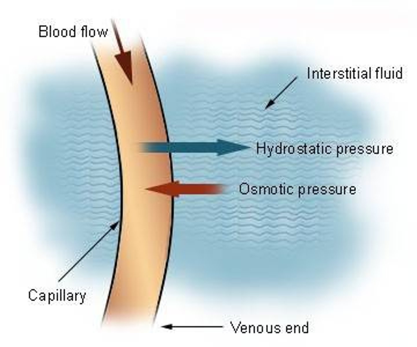
Tissue Fluid
This (also known as interstitial fluid) is the fluid between cells in the body and carries oxygen and other nutrients to tissue cells. It is formed from the filtration of blood from the capillaries, as a result of hydrostatic pressure.
Blood Proteins
They (also known as plasma proteins) consist of albumins, globulins and fibrinogen. Albumins are in the most abundance and determine the viscosity of the plasma and to osmotic pressure. This pressure retains the fluid within the blood vessels. If the blood protein levels decrease, the osmotic pressure will also decrease, which would cause the fluid to leak out of the blood vessels and accumulate in the surrounding tissues. If this does happen, it would cause oedema. Globulins are the secondmost abundant and include the immunoglobulins/antibodies. They are essential for protection of the body, as part of the immune system and are made by the white blood cells (lymphocytes). Fibrinogen allows the blood to clot (coagulation) at appropriate times, so as to not cause damage to the body e.g. stroke.
Hydrostatic Pressure
This is the pressure from heart contractions that forces the water and dissolved substance of the blood plasma out past the capillary walls into the surrounding tissues, which forms the tissue fluid.
Hypertension
Symptoms - blood pressure of 140/90mmHg (normal is between 90/60mmHg and 120/80mmHg). Otherwise no noticeable symptoms.
Biological Explanation - this is the pressure your blood is pumped around the body, which is represented by the number on top; the systolic pressure and the lower number being the diastolic pressure, this being the resistance to the blood flowing in the blood vessels. The high blood pressure can damage the blood vessels.
Cause - age, family history, high salt intake, lack of exercise, being overweight/obese, smoking, drinking alcohol excessively over a long period of time or some conditions, such as kidney disease, diabetes and hormonal problems e.g. hyperthyroidism.
Hypertension Monitoring, Treatment etc.
Monitoring - regular readings will need to be made on the blood pressure machine (sphygmomanometer) at home or in the doctor's office.
Treatment - change in diet (low fat, low salt) and lifestyle choices (drinking alcohol and smoking less if they don't choose to quit), exercise, regular sleep, reduce stress and medications such as ACE inhibitors, which lower blood pressure, and beta-blockers, which lower heart rate.
Impact - it could lead to more health conditions such as Coronary Heart Disease (CHD), strokes and kidney disease.
Coronary Heart Disease
Symptoms - angina, which can then include breathlessness, dizziness, chest pain, and a heart attack, which also includes light headedness, feeling weak, sweating, chest pain and shortness of breath.
Biological Explanation - CHD is when the coronary arteries of the heart become blocked with fatty deposits (atherosclerosis). When they become fullt blocked, it would cause a heart attack and cause permanent damage to the heart tissues and could be fatal if left untreated.
Cause - smoking, lack of exercise, obesity, high cholesterol levels, hypertension, diabetes, age, gentics and gender.
Coronary Heart Disease Monitoring, Treatment etc.
Monitoring - blood tests can be done to check the levels of fats, cholesterol, sugar and proteins present in the blood, an ECG can show if there is any damage to the heart and any signs of CHD.
Treatments - change in diet (low fat), exercise, reduce or stop smoking, good amount of sleep, medications such as nitrates (to relax the coronary arteries, which allows more blood to pass through), ACE inhibitors (which lowers blood pressure) and surgical procedures such as an angioplasty where they insert a balloon and a stent on a long wire into the groin or wrist to the affected coronary artery and a stent is infalted and placed (used with an angiogram using contrast dye in the arteries) and a coronary artery bypass is where they use a bypass machine and attach it to the aorta so the blood doesn't flow into the coronary arteries and a small artery, usually from the groin, is used as a graft on the heart so they are able to restore goo blood flow into the heart.
There is no cure for CHD.
Larynx
Connects the back of the nose to the trachea.
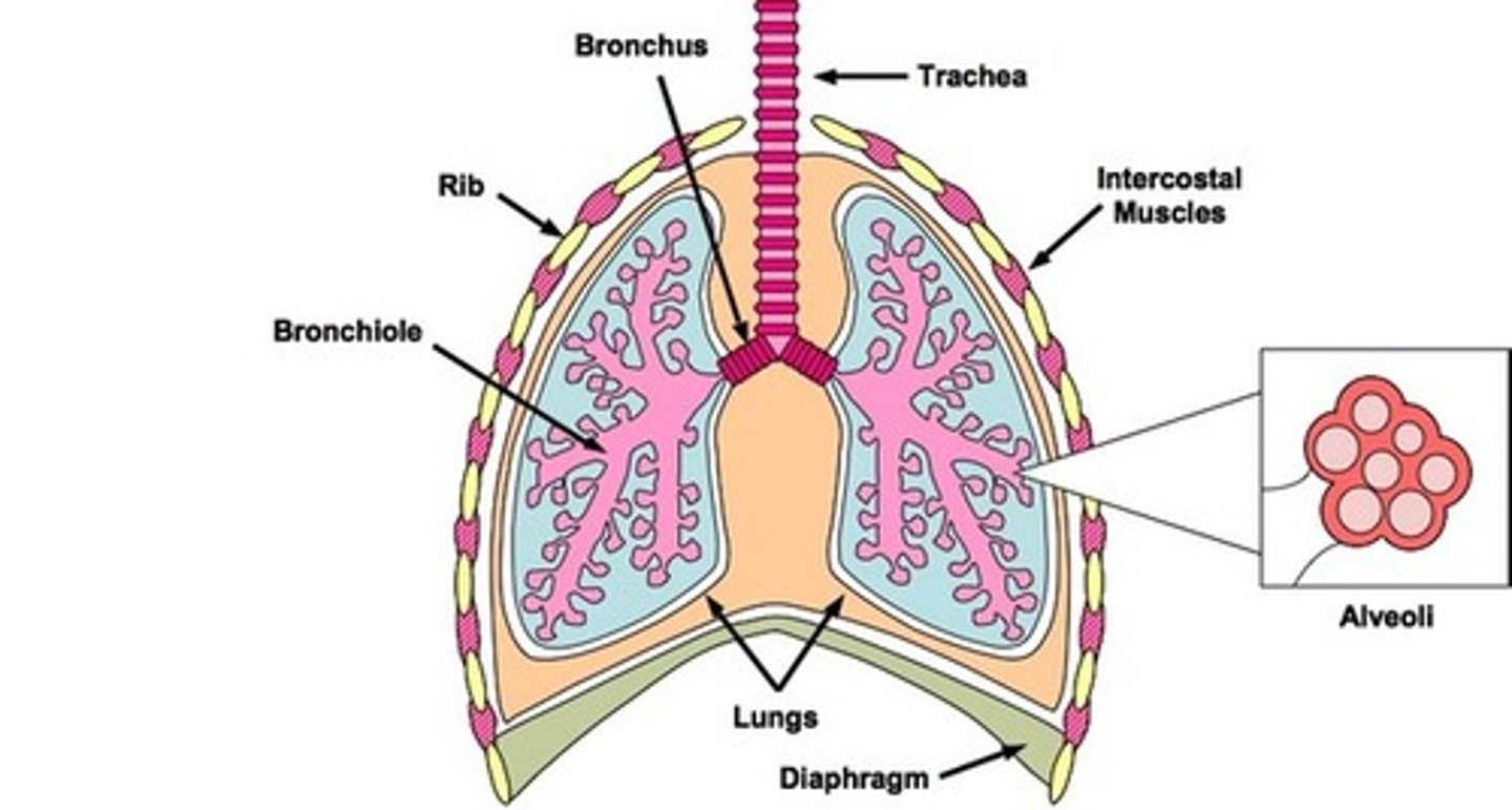
Trachea
Also known as the windpipe, it is supported by C-shaped cartilage to stay open and allow air into the lungs.
Bronchi
These are two branches that stem from the trachea and also have C-shape cartilage that allows air to enter both the left and right lungs.
Bronchioles
These are small branches that come from the bronchi and are in a high quantity spread all around the lungs.
Alveoli
They are small sacs at the end of every bronchiole and are surrounded by capillaries, so that oxygen can enter the bloodstream and carbon dioxide can enter into the lungs to be exhaled out of the body.
Central Nervous System (CNS)
This contains the brain and the spinal cord and allows the communication of the brain, through the spinal cord, to different parts of the body.
Spinal Cord
This is protected by specialised bones called vertebrae. It runs through the centre of these bones and transmits electrical impulses through nerves from the brain to different parts of the body.
Autonomic Nervous System (ANS)
They control and regulate automatic actions as they are done unconsciously to keep us alive, such as our heart rate and gut movements.
Somatic Nervous System (SNS)
They contain sensory and motor neurones. These nerve pathways work together in order to do an action, such as picking up an object. It can also be protective as it allows a fast reaction in an emergency situation, such as when something is very hot.
Sensory neurones transmit information from the senses TO the brain.
Motor neurones transmit information to the muscles FROM the brain.
Peripheral Nervous System (PNS)
This contains all of the nerves outside of the CNS and relays information between the CNS and the body. This includes the ANS and the SNS.
Cerebral Cortex
This is the outermost layer of the brain and is responsible for thinking and processing sensory information from the body. It contains four lobes, each processing different pieces of information. It is made of tightly packed neurons.
Cerebellum
This is positioned at the lower back of the brain. This coordinates and regulates muscle activity for gross and fine motor skills, such as walking and writing. It also helps in maintaining balance.
Frontal Lobes
This carries out a higher level mental processes, such as thinking, decision making and planning. This is located at the very front of the brain.
Corpus Callosum
This acts as a bridge between the two hemispheres of the brain, in order to communicate between them. It is located in the centre of the brain and is made of nerve tissue.
Hypothalamus
This is responsible for the regulation of body temperature, appetite and thirst. It is located at the lower, off-centre part of the brain.
Medulla Oblongata
It regulates life-sustaining functions such as breathing, swallowing and heart rate. It is located on the brain stem, on the front.
Meninges
There are three membranous layers surrounding the brain and the spinal cord, and acts as a barrier from the rest of the body, to prevent infection.
Neuron Structure
KEY
Neuron
Axon
Dendrites
Myelin Sheath
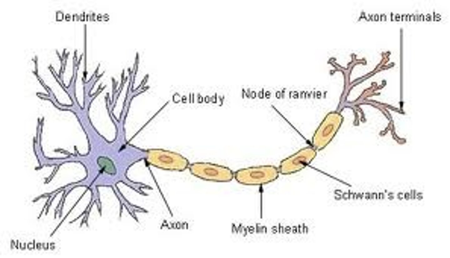
Neuron
These are specialised nerve cells that allow the transmission of electrical impulses from one part of the body to another.
Axon
They are long and thread-like part of the neuron and allow the electrical impulses to be carried away from the cell body. There is only one of this per neuron.
Dendron
As plural is dendrites, They are short branches that stem out of the cell body and receives electrical impulses from other neurons, carrying them towards the cell body and can be as many as a 1000 of these per neuron.
Myelin Sheath
It is a fatty white substance that surround the axon, in order to protect it from damage, allowing electrical impulses to be transmitted quickly and efficiently.
Cortex
This is the outer layer of the kidney.
Medulla
This is the inner region of the kidney that contains many nephrons.
Renal Artery
This supplies the kidney with oxygenated blood.
Renal Vein
This carries the blood that has been filtered by the kidney.
Calyx
These are chambers within the kidney where urine passes through.
Ureters
These are tubes that carry the urine from the kidney and into the bladder.
Bladder
This stores the urine produced.
Urethra
This allows the urine to pass through to exit the body.
Osmoregulation
This is one of two main functions of the kidney, where it maintains the water balance of the body. This is done by controlling the concentration of water in the plasma of the blood. This allows a constant balance of water within the body. It also controls the salt levels and if water is no put back into the blood, it will be excreted in urine.
Urea Removal
Another main function of the kidneys where it excretes a waste product called urea.
Glomerulus
This is a cluster of capillaries surrounded by Bowman's capsule in the nephron and allows the ultrafiltration to happen.
Bowman's Capsule
This surrounds the glomerulus in the nephron and serves a structural function for the cluster of capillaries.
Collecting Duct
This is where the filtered waste product accumulates and urine is collected.
Distal Convoluted Tubule
This is where salt and water are reabsorbed between the collecting duct and the loop of henle.
Loop of Henle
This is a U-shaped structure surrounded by capillaries that recollects water and salt from the urine.
Pupil
This is the opening in the middle of the eye, seen as a black hole. This regulates the amount of light enters the eye and the retina receives.
Iris
This is the area with colour, surrounding the pupil whilst also controlling its size. This controls the amount of light entering the eye through the pupil.
Tear Gland
It continuously lubricates the eye and is located on one side of the eye. It also protects the eye by flushing any small foreign particles from the eye, such as dirt.
(Vitreous and Aqueous) Humours
A clear gel that fills the centre of the eye and provides structure. It also receives the light and allows it to pass to the retina and protects the eye from damage and shock.
Conjunctiva
This is a mucous membrane that covers the outer back of the eye and the inside of the eyelids. It prevents the entry of microbes entering the eye, which could cause infection and also helps to produce tear fluid.
Cornea
It is a transparent layer that covers the pupil and iris. It protects the eye from large particles (i.e. dirt) and microbes and also helps in focusing the light that enters the eye.
Retina
This is a sensory membrane filled with rods and cones, which are light sensitive cells, on the inner back of the eye. It transforms the light into electrical impulses for the brain.
Mascula
It is one of the central layers of the eye, which lies behind the retina. They are responsible for central vision and detailed colour images. They contain many photoreceptor cells that detects light and sends electrical impulses to the brain.
Optic Nerve
This is a collection of nerve cells that connect the eye to the brain. They transmit the electrical signals of the eye to the brain so it can be turned into an image and contains not rods or cones (aka the "blindspot").
Ciliary Muscle
A small muscle on either side of the lens and allows it to change shape to focus an image.
Suspensory Ligaments
These are small ligaments that attach the ciliary muscle to the eye, allowing it to change the lens' shape.
Lens
This is located behind the pupil and focuses the rays of light that enters the eye through the pupil, by refraction. It changes the focal distance of the eye so it is able to focus on objects at various distances.
Pinna
This is the outermost part of the earand acts as a funnels for sound, assisting it into the ea and is part of the outer ear.
Auditory Canal
It is also part of the outer ear and directs sound towards the middle ear.
Ear Drum
This is a thin layer of tissue, located in the middle ear, and receives sound as vibrations, causing itself to vibrate.
Malleus, Incus and Stapes
These are small bones within the middle ear and carry he vibrations it receives from the ear drum and into the inner ear.
Cochlea
This contains a jelly-like fluid in a coiled tube, located within the inner ear and allows the vibrations to pass through the liquid and is converted to electrical impulses.
Organ of Corti
They are located within the inner ear and are lined with tiny hair-like structures that convert these sound vibrations into electrical impulses.
Eustachian Tube
This connects the middle of the ear to the throat, ensuring that the pressure inside of the middle ear is the same as the pressure outside of the ear.
Round Window
It is a drum-like membrane that allows vibrations from the round window to pass through to the cochlea, found in the inner ear.
Auditory Nerve
A bunch of nerves that transmit the electrical impulses from the cochlea to the brain, and is located in the inner ear.
Semi-Circular Canals
These are small tubes filled with a fluid that aids in the balance of the body and work as a motion centre, without being involved in the hearing process. They are located in the inner ear and are lined with cilia and their movements are communicated with the brain to maintain balance.
Ampullae
They are in the semi-circular canals of the inner ear that surrounds sensory receptors and are responsible for sensory related movements such as spatial awareness and pressure change.
Glaucoma
Symptoms - no symptoms to begin with and develops over many years, affects the peripheral vision first, blurred vision, rainbow-coloured circles around lights and blindness.
Cause - a buildup of pressure in the eye when fluid from the aqueous humour is unable to drain itself, increasing the pressure on the optic nerve, age, ethnicity (African, Caribbean and Asian are higher risk), family history and other conditions such as diabetes.
Monitoring - regular eye tests.
Treatment - daily eye drops, laser treatments to open blocked drainage tubes or reduce fluid production and surgery to improve drainage.
Care Needs -