neuro exam 1
 Call Kai
 Learn
Learn Practice Test
 Spaced Repetition
 Match
Match Flashcards
 Knowt Play
1/82

There's no tags or description
Looks like no tags are added yet.
Name | Mastery | Learn | Test | Matching | Spaced | Call with Kai | Chat |
|---|
No analytics yet
Send a link to your students to track their progress
83 Terms
Dorsal, ventral, caudal, rostral on the brain

superior, inferior, medial, lateral
Exterior Views of the Brain
Superior - you are superior and looking down on the brain
Inferior - you are inferior and looking up at the brain
Lateral- side view of the brain
Medial- toward the midline
sagittal (midsagittal), coronal, horizontal, and transverse planes


Neurons
Brain cells that send signals to create all of our experiences
Three main components of neurons
The three main parts of a neuron are the dendrites, the cell body (soma), and the axon.

Direction of information flow for neurons
Dendrites → Cell body → Axon → Axon terminals
(Dustin Calls Anna Auntie Today)
Myelin Sheath
a protective, fatty-protein layer that wraps around the long fibers of nerve cells.
protects axons
Nodes of Ranvier
segments in the myelin sheath
message can jump from node to node very quickly
What does myelin do?
Myelin sheath allows action potentials to jump from one node of Ranvier to the next. It's faster and more efficient and necessary for long motor neurons.
Damaged myelin impairs nerve conduction (as in multiple sclerosis)
Damaged neuron/myelin= distorted messages
What happens at the synapse?
At the synapse, one neuron passes a signal to another neuron.
Efferent vs Afferent Neurons
Outgoing MOTOR response- EFFERENT NEURONS
Incoming SENSORY response- AFFERENT NEURONS
Grey matter vs. white matter
Grey matter- neuron cell bodies, generate signals
White matter- neuron cell axons, carry signals
-In the brain, grey matter outside and white matter inside
-In the spinal cord, white matter outside and grey matter inside
The CNS has about the same amount of…
glial cells and neurons
Lateralization of somatosensory and motor function
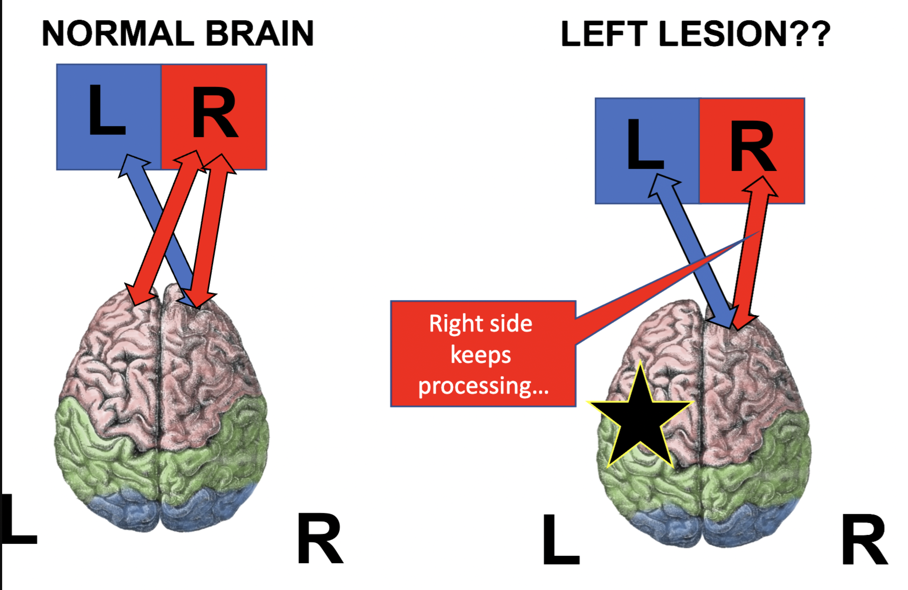
Motor and sensory functions are controlled contralaterally.
Right side of brain → controls left side of body
Left side of brain → controls right side of body
Somatotopy with overlap (i.e., motor homunculus)
About 90% of motor and sensory fibers cross/decussate at the brainstem in the medulla
Laterality of brain organization (left hemisphere versus right functions)
Left hemisphere → language
Right hemisphere → emotions, visuospatial skills, musical skills, paralinguistic functions, and attention
Sensorimotor control is contralateral.
Everything below the neck is controlled by the opposite side of the brain.
Each Brodmann area has a unique…
cytoarchitecture and function
Important Brodmann’s Areas
BA01 - Primary somatosensory cortex (S1)
BA02 - Secondary somatosensory cortex (S2)
BA03 - Tertiary somatosensory cortex (S3)
BA04 - Primary motor cortex (M1)
BA17 - Primary visual cortex (V1)
BA18 - Secondary visual cortex (V2)
BA41 - Primary auditory cortex (A1)
BA42 - Secondary auditory cortex (A2)
BA 22 - Wenicke's area
BA 44 - Broca's
major gyri and sulci and their FUNCTIONS
??? Come back later
If a given lobe, gyri, Brodmann area is damaged, what will the symptoms be?
Frontal lobe damage → problems with executive functions, personality, planning, impulse control, or motor function
Precentral gyrus / BA 4 damage → motor problems
Left inferior frontal gyrus / BA 44 damage → problems with speech production, associated with Broca's aphasia
Parietal lobe damage → sensory/perceptual problems and possible visuospatial problems
Postcentral gyrus / BA 1, 2, 3 → sensory problems
Right parietal damage → possible left neglect
Temporal lobe damage → auditory, language, memory, or olfactory problems
BA 41/42 damage → auditory processing problems
Left BA 22/Wernicke's area damage → problems with language comprehension/auditory word recognition
Right temporal damage → can cause problems such as amusia
Occipital lobe damage → visual problems
Difference between CNS and PNS and their subsystems
CNS = Central Nervous System
Brain
Spinal cord
PNS = Peripheral Nervous System
All neurons/fibers outside the CNS
Includes nerves that exit the brainstem and spinal cord
Commisural fibers
connects the 2 hemispheres of the brain
between hemispheres= INTERHEMISPHERIC
most interhemispheric connections are between homotopic areas
association fibers
connect cortical areas in the same hemisphere of the brain *** (ex right frontal lobe connected to right temporal lobe)
Confined within hemisphere= INTRAHEMISPHERIC
short - bend around a sulcus, connect adjacent gyri
long - connect more distant cortical areas
main commisural fibers
Corpus callosum → association cortices ↔ opposite hemisphere
Anterior commissure → olfactory + part of limbic system
Hippocampal/posterior commissure → part of limbic system.
main Association fibers:
Superior longitudinal fasciculus (SLF) → all four lobes within each hemisphere
Arcuate fasciculus → Broca's ↔ Wernicke's
Inferior longitudinal fasciculus (ILF) → temporal ↔ occipital
Uncinate fasciculus → anterior temporal ↔ VPFC
Cingulum → medial frontal/parietal ↔ temporal

Superior Longitudinal Fasciculus (SLF)*
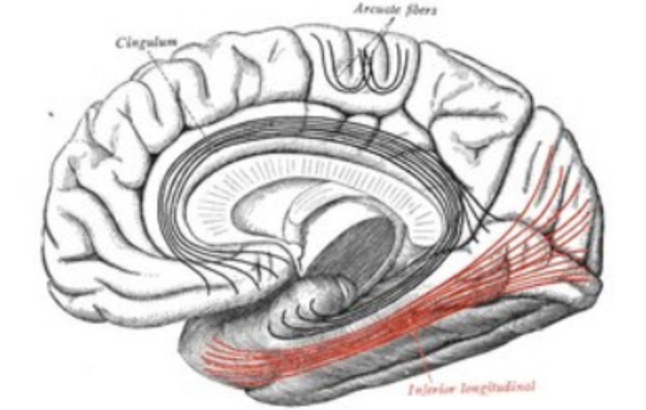
Inferior Longitudinal Fasciculus (ILF)

Arculate fasciculus (connects broca’s and wernicke’s)

Uncinate fasciculus

Cingulum
split brain syndrome
What: No communication between the left and right hemispheres
Cause: Complete sectioning of the corpus callosum
Symptoms: Usually normal cognitive function and behavior; problems mainly appear during specific tests or actions. Some people may have difficulty naming or identifying something that is presented to only one side of the body/visual field.
alien hand conflict
What: A hand seems to act on its own, as if it has a mind of its own.
Cause: Damage/disruption to brain areas involved in voluntary movement and communication between brain regions.
Symptoms: The person feels they cannot control the hand. The hand may perform actions without the person's intention, and the person may describe the hand's actions as if someone else is controlling it.
how are alien hand and split brain different?
Split-brain: The two brain hemispheres cannot communicate normally because the corpus callosum has been cut. Symptoms show up mainly during certain tasks.
Alien hand: A hand seems to act on its own, and the person feels like they cannot control it.
Main difference: Split-brain is about loss of communication between the hemispheres, while alien hand is about uncontrolled, seemingly independent hand movement
amygdala
Emotions, especially fear and emotional responses
Basal Ganglia:
Movement control and coordination of voluntary movement
Central sulcus:
Separates the frontal and parietal lobes; separates motor and sensory areas
Cerebellum:
Balance, coordination, and fine motor control
Cingulate gyrus:
part of the limbic system.
Emotion, motivation, and behavior
Hypothalamus:
Homeostasis; regulates hunger, thirst, body temperature, sleep, and hormones
Insula:
Taste, body sensations, and awareness of internal body states
Limbic system:
Emotion, memory, and motivation
Lateral sulcus:
Separates the temporal lobe from the frontal and parietal lobes
Lateral ventricles:
CSF-filled spaces in the brain
Longitudinal fissure:
Separates the left and right cerebral hemispheres
Thalamus:
Relays sensory information to the cerebral cortex
left hemisphere functions
• Processing speech sounds
• Basic langauge comprehension
• Motor speech
• Facial recognition
• Motor control for the right side of the body
right hemisphere functions
• Visuoapatial processing
• Emotion
• Supralinguistics (sarcasm, humor, tone)
• Music
left neglect- what’s involved, why more common than right neglect?
Left neglect: Not paying attention to or being unaware of things on the left side of space.
Brain area: Usually caused by damage to the right parietal lobe.
Why more common than right neglect? The right hemisphere pays attention to both sides of space, while the left hemisphere mainly attends to the right side. So right-hemisphere damage can cause left neglect, but left-hemisphere damage is less likely to cause right neglect.
glial cells
Support cells that provide
Structure
Protection
Waste management
There are approximately the same number of neurons as glial cells in the CNS
what does myelin do?
Myelin acts as a protective, insulating layer around nerve fibers that speeds up electrical signals in the nervous system
electrical vs chemical gradients
Electrical signals (action potential) - within the neuron
Chemical signals (neurotransmitters) - between neurons in the synaptic space

Which ions are in greater/lesser concentration inside vs. outside cell for a neuron at rest?
More sodium (Na+) and calcium (Ca2+) outside the cell
More potassium (K+) inside the cell
What happens to the ions inside/outside the neuron from rest, to stimulation, during firing, and then during the absolute and relative refractory periods?
At rest, Na⁺ and Ca²⁺ are higher outside and K⁺ is higher inside.
During depolarization, Na⁺ moves into the neuron.
During repolarization, K⁺ moves out.
During hyperpolarization, K⁺ continues to leave briefly, making the inside more negative.
The Na⁺/K⁺ pump then helps restore the resting ion gradients.
What are the channels and pumps doing?
Na⁺ channels open → Na⁺ enters
K⁺ channels open → K⁺ leaves
Na⁺/K⁺ pump → restores Na⁺ and K⁺ gradients
diagram of the action potential

resting membrane potential
The inside of the cell is negatively charged compared to the outside when the neuron is at rest.
hyperpolarization
The difference in electrical charge between the inside and outside of the cell becomes greater as the inside of the cell becomes more negative, thus increasing the polarization.
polarization
Inside is more negative than outside.
depolarization
the difference in electrical charge between the inside and outside of the cell becomes smaller (and may disappear altogether or reverse) as the inside of the cell becomes more positive, thus decreasing the polarization
aka Inside becomes less negative/more positive.
repolarization
The process by which the resting (baseline) electrical polarization of a neuron is re-established.
All or nothing principle of action potential
An action potential either happens completely or does not happen. The neuron must reach threshold to fire.
What are the absolute and relative refractory periods?
Absolute refractory period: Neuron cannot fire again.
Relative refractory period: Neuron can fire again, but needs a stronger stimulus.
ARP is about 3–4 ms; RRP follows and lasts about 5–10 ms.
What role does the sodium-potassium pump play?
Uses ATP to move Na⁺ out and K⁺ in, helping restore the resting ion gradients
Signal Propagation
The action potential travels down the axon by activating one section after another. In myelinated axons, it jumps from node to node. Larger axons and myelinated axons are faster.
What happens at the synapse?
Action potential arrives → Ca²⁺ enters → neurotransmitter is released → neurotransmitter binds to the next neuron → next neuron is excited or inhibited.
Craniectomy vs craniotomy
Craniotomy: A piece of the skull is temporarily removed and then put back.
Craniectomy: A piece of the skull is removed and not immediately put back.
Parkinson’s Disease
Degeneration of the substantia nigra → dopamine insufficiency.
Symptoms: resting tremor, too much or too little movement, masked face, progressive disorder.
Multiple Sclerosis:
Autoimmune disease that damages myelin, slowing or blocking signals.
Symptoms: walking difficulties, cognitive changes, vision issues, and possible swallowing/speech problems.
Myasthenia Gravis:
Autoimmune disruption of acetylcholine (ACh) receptors → not enough receptors open.
Causes progressive muscle weakness with activity that improves with rest.
Frontal Lobe- (movement, planning, decision-making, speech)
Precentral gyrus — Gyri — voluntary movement
Superior frontal gyrus — Gyri — executive functions
Middle frontal gyrus — Gyri — planning/executive functions
Inferior frontal gyrus — Gyri — speech (Broca’s area)
Central sulcus — Sulci — separates frontal and parietal lobes
Precentral sulcus — Sulci — separates motor areas
Parietal lobe — (sensation, spatial awareness, sensory processing)
Postcentral gyrus — Gyri — sensation
Superior parietal gyrus — Gyri — visuospatial processing
Inferior parietal gyrus — Gyri — higher-level processing
Supramarginal gyrus — Gyri — memory and language
Angular gyrus — Gyri — reading and writing
Intraparietal sulcus — Sulci — separates superior/inferior parietal areas
Corpus callosum
Connects the left and right cerebral hemispheres.
Thalamus
Relays sensory information to the cerebral cortex.
Hypothalamus
Controls homeostasis, hormones, and basic drives.
Midbrain
Controls movement and processes visual and auditory reflexes.
Pons
Regulates breathing and sleep, and relays information within the brain.
Medulla
Controls vital autonomic functions like breathing and heart rate.
Cerebellum
Controls balance, coordination, and smooth voluntary movement.
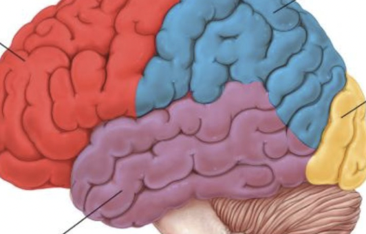
parietal, frontal, occipital, temporal

Frontal lobe
Brain region responsible for voluntary movement, planning, decision-making, and speech.
parietal lobe
Brain region responsible for sensation, spatial awareness, and sensory processing.
Temporal lobe
Brain region responsible for hearing, language processing, and memory.
Occipital lobe
Brain region responsible for visual processing and vision.