Transcranial Doppler (TCD)
 Call Kai
 Learn
Learn Practice Test
 Spaced Repetition
 Match
Match Flashcards
 Knowt Play
1/51

There's no tags or description
Looks like no tags are added yet.
Name | Mastery | Learn | Test | Matching | Spaced | Call with Kai | Chat |
|---|
No analytics yet
Send a link to your students to track their progress
52 Terms
Anterior Circulation of the Circle of Willis
Carotid siphon - parasellar, genu, and supraclinoid segments
ICA branches - ophthalmic & posterior communicating
MCA
Anterior cerebral - pre & post-communicating
Anterior communitating
Posterior Circulation of the Circle of Willis
Vertebral
Basilar
Posterior Cerebral - pre & post-communicating
Vertebrobasilar Circulation of the Circle of Willis
Vertebral - posterior inferior cerebellar
Basilar - anterior inferior cerebellar & superior cerebellar
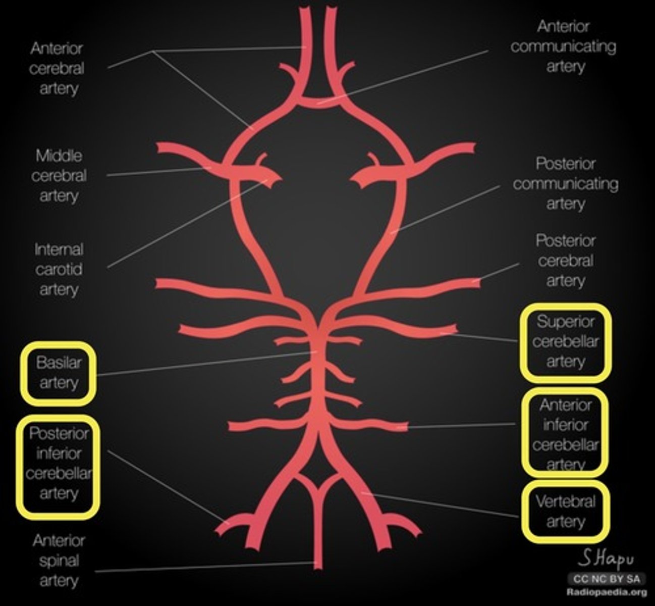
Common Anatomical Variants in Cranial Vessels
Hypoplasia of posterior communicating A
Hypoplasia of pre-communicating posterior cerebral A
Hypoplasia &/or Duplication of anterior communicating A
Hypoplasia of pre-communicating anterior cerebral A
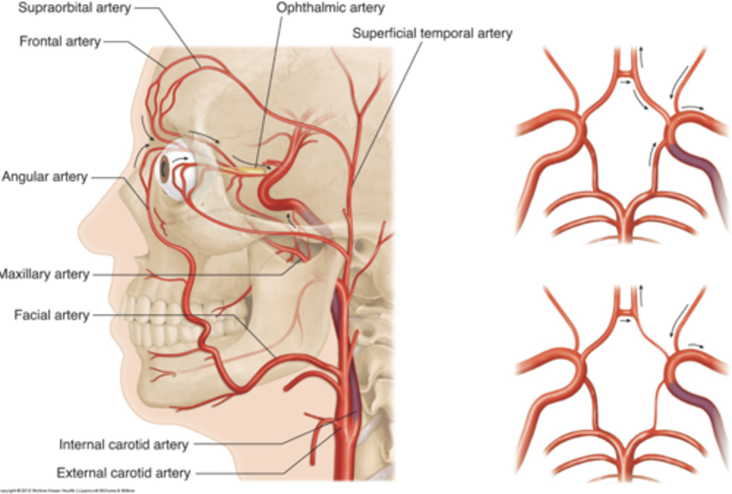
Collateral Flow Patterns of Cranial Vessels
ECA to ICA via reversed ophthalmic A
Crossover collateral via anterior communicating A
Posterior-to-anterior collateral via posterior communicating A
Disadvantages to TCD
Extracranial status
CO2 status
Intracranial pressure
Limited windows
Blind spots
Pilot error (6 month learning curve)
TCD Indications
Vasospasm - trauma or aneurysm
Vertebral basilar insufficiency
Intraoperative monitoring
Intracranial pressure
Brain death
PFO - right to left shunts
Sickle Cell
Sub arachnoid hemorrhage - head trauma, aneurysms, AVM
TCD Risk Factors
Diabetes
Hypertension
High cholesterol
Tobacco abuse
Cardiac disease or hx of heart attack
Claudication
Stroke or TIA
Previous vascular procedures - stents or bypass
TCD Relevant Patient History
Face, leg, or arm numbness, tingling, weakness
Problems speaking/understanding speech
Vision problems - blindness or double vision
Gait disturbance or leg weakness
Vertigo or fainting
Severe headaches
Equipment for Non-Imaging TDC
Dedicated non-imaging System
1-2 MHz transducer
Equipment for TDC Duplex Imaging
Ultrasound system
1-5 MHz sector transducer
Equipment for TCD Monitoring
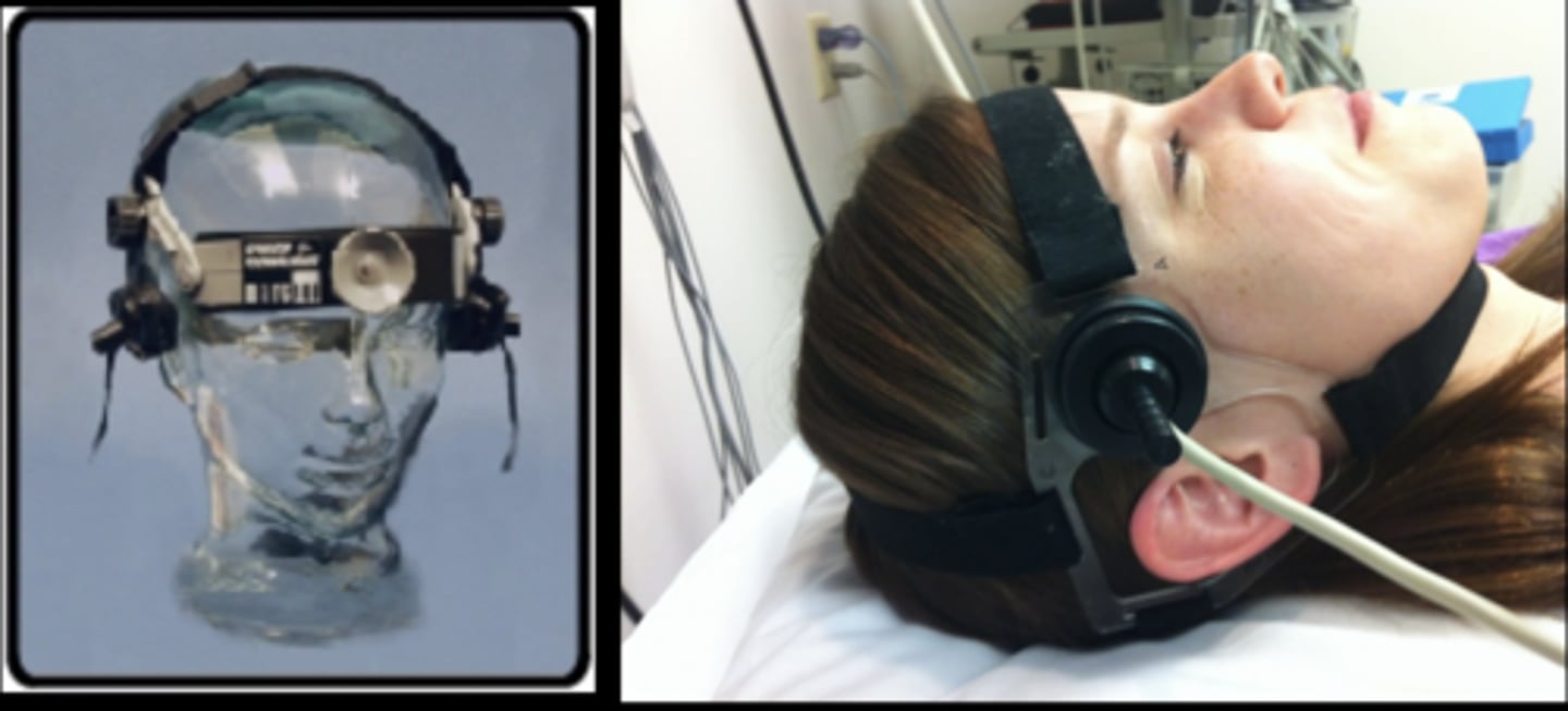
How to Identify Vessels on TDC
Scanning window/approach
Sample volume depth
Bloodflow direction - antegrade/retrograde
Spatial anatomical relationship to other vessels
Flow velocity
TCD Velocity Measurements
Time average peak velocity (TAPV)
Mean of the max velocity
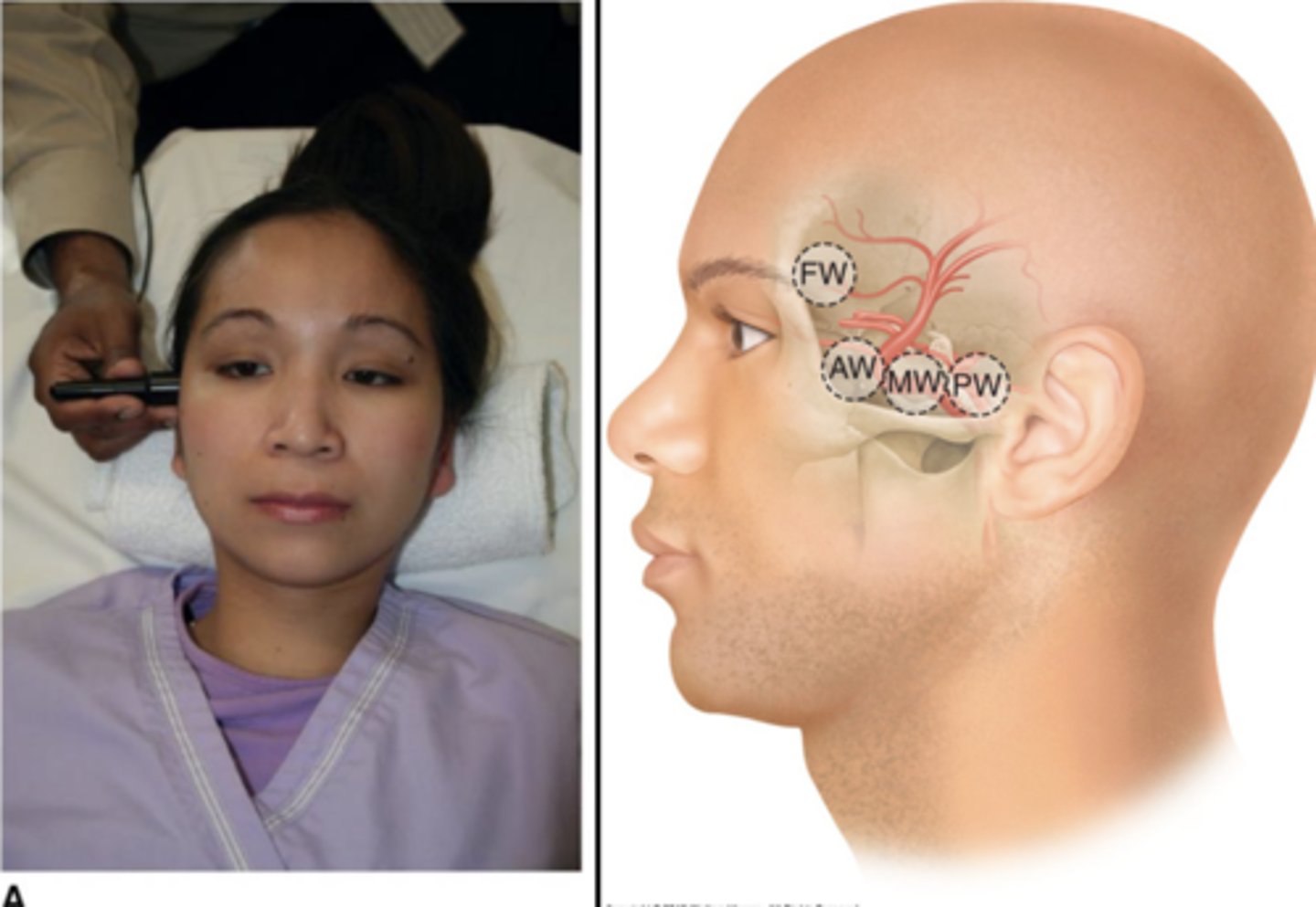
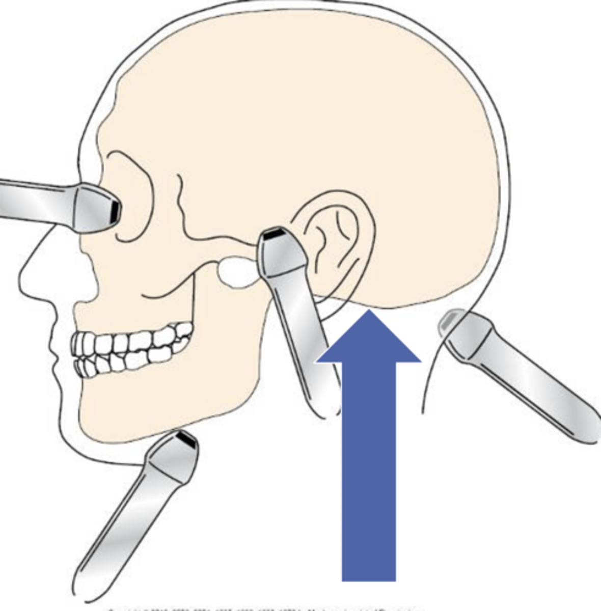
Transtemporal TCD
Over temporal bone
Frontal, middle, anterior, and posterior windows
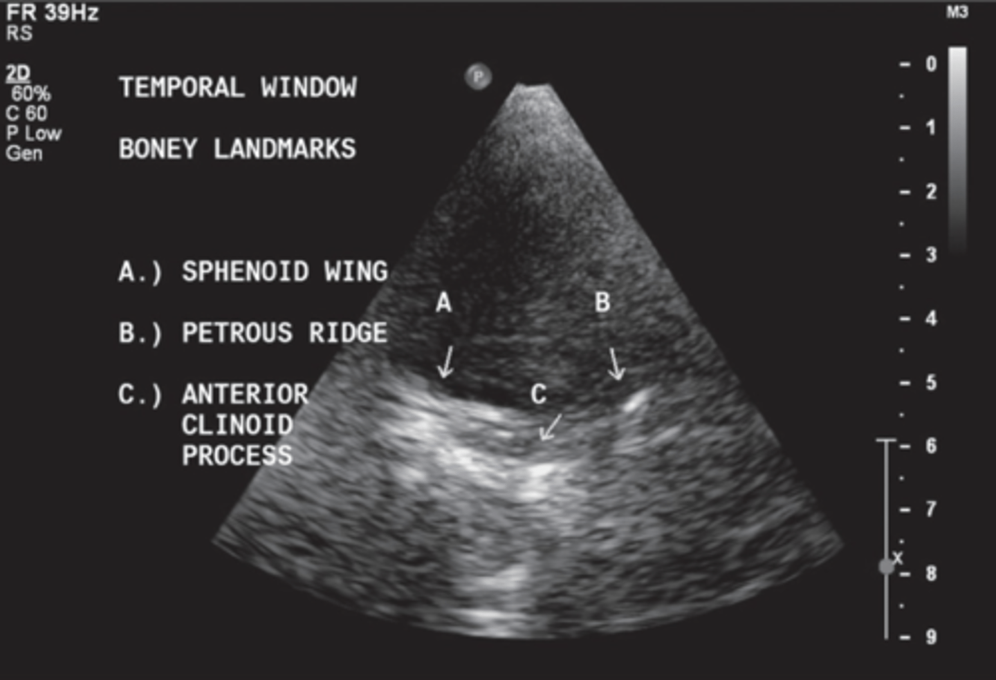
Transtemporal TCD Bony Structures
Sphenoid
Petrous ridge
Anterior clinoid process
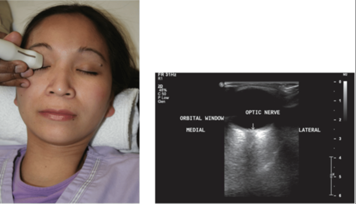
Transorbital TCD
Through orbital place, optic canal, and superior orbital fissure
Acoustic Output for Transorbital TCD
MI < 0.23
Transforaminal/Sub-Occipital TCD
Through foramen magnum
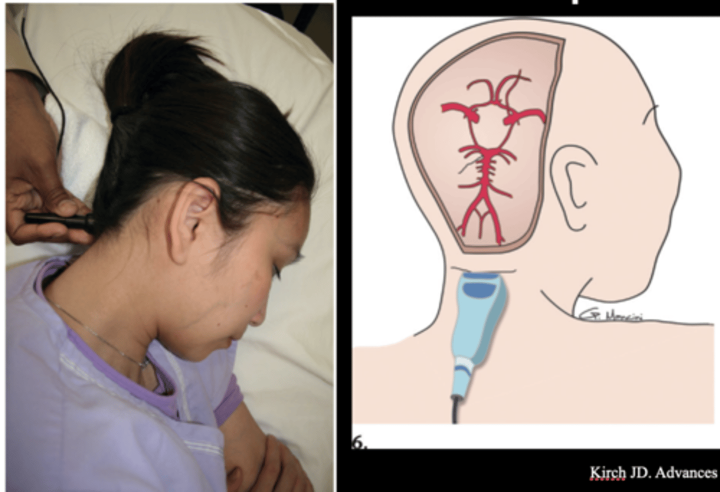
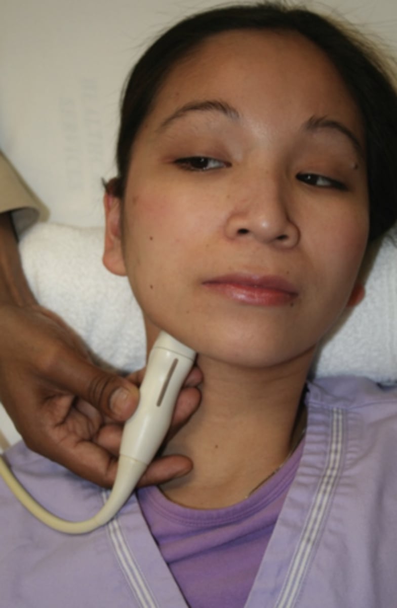
Submandibular TCD
Under jaw angled cephalad
*images ICA
Atlas Loop TCD
Behind ear between mastoid & sternocleidomastoid muscle
1.25 cm below mastoid process
Terminal Internal Carotid A (tICA)
Temporal window
60 - 70 mm depth
Bidirectional flow (red or blue)
39 +/- 9 cm/sec
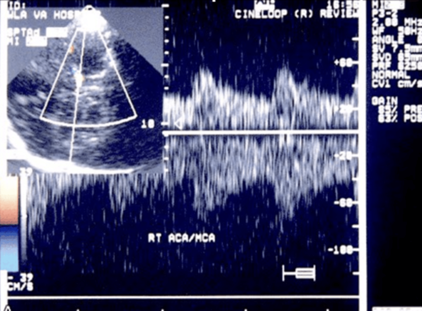
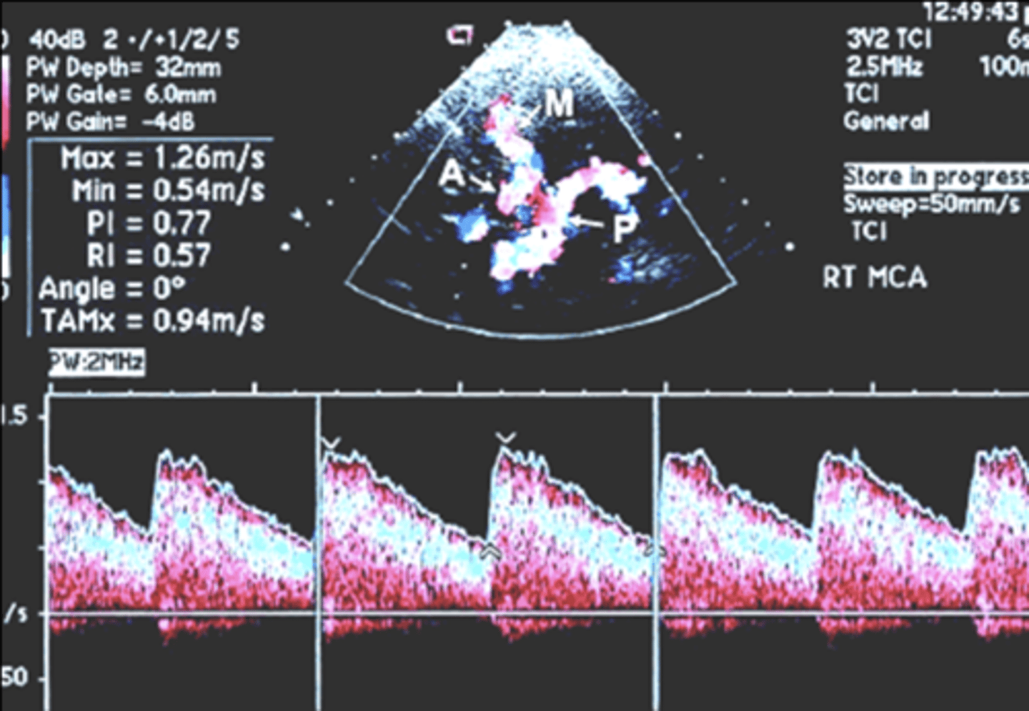
Middle Cerebral A (MCA)
Temporal window
30 - 60 mm depth
Flow towards tx (red)
55 +/- 12 cm/sec
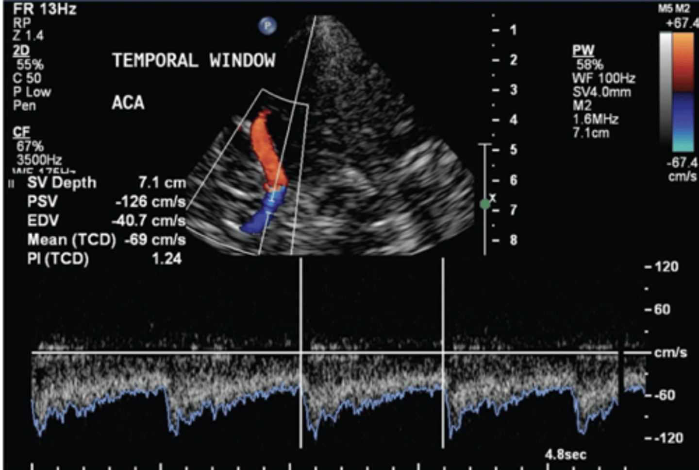
Anterior Cerebral A (ACA)
Temporal window
60-75 mm depth
Flow away from tx (blue)
50 +/- 11 cm/sec
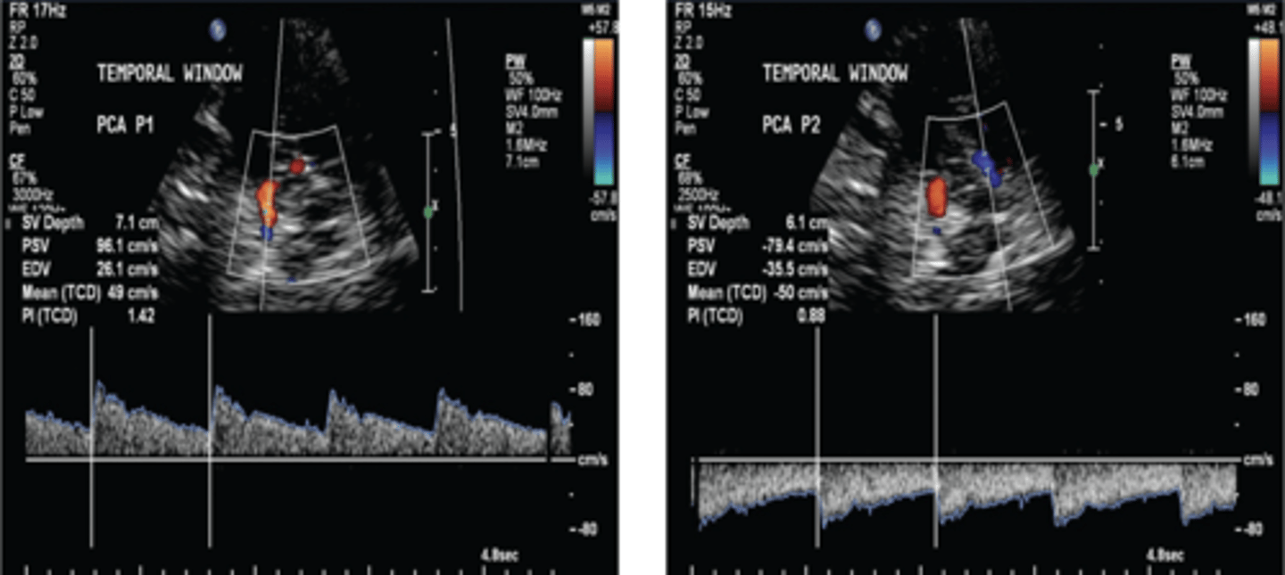
Posterior Cerebral A (PCA)
Temporal window
60 - 80 mm depth
Pre-communicating: flow towards tx (red)
Post-communicating: flow away from tx (blue)
39 +/- 10 cm/sec
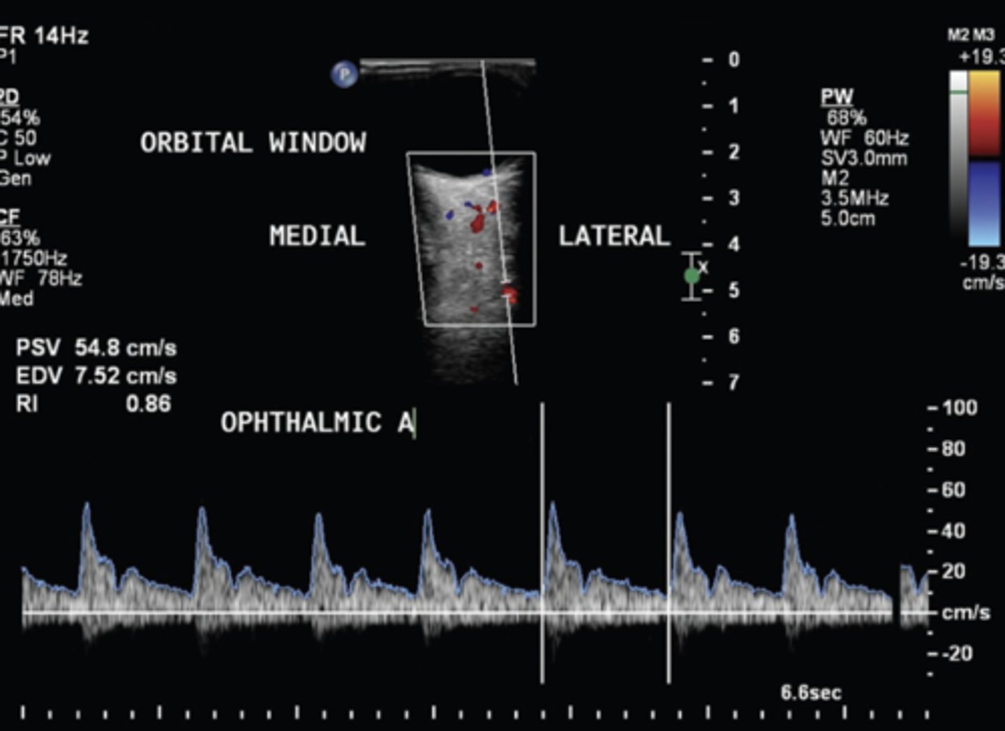
Ophthalmic A
Orbital window
35 - 55 mm depth
Flow towards tx (red)
21 +/- 5 cm/sec
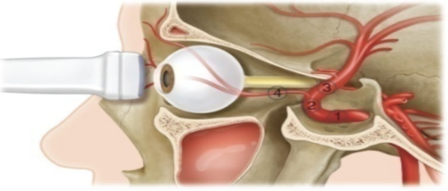
Carotid Siphon
Orbital window
60 - 80 mm depth
41 +/- 11 cm/sec
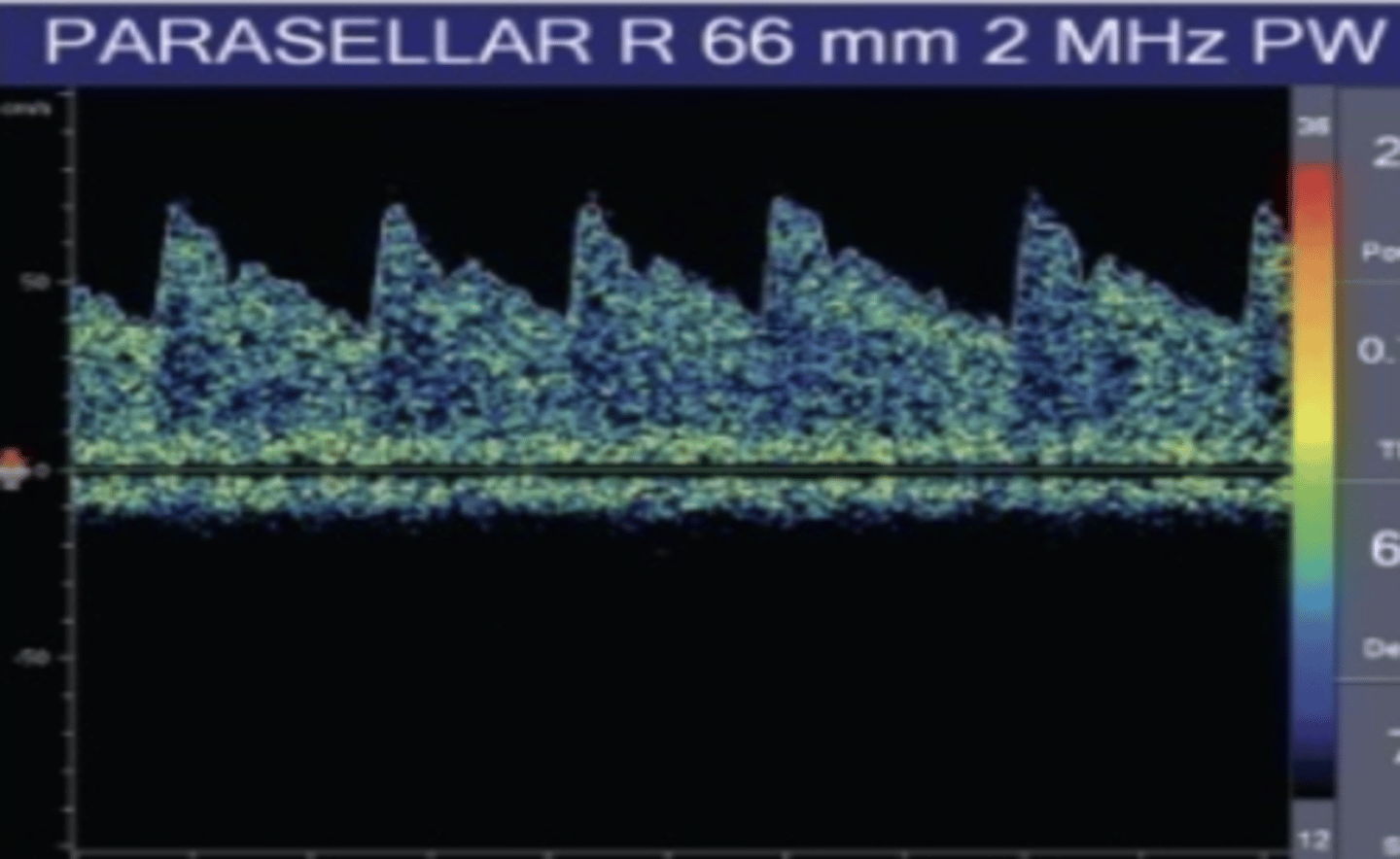
Parasellar Segment of Carotid Siphon Flow
Towards tx (red)
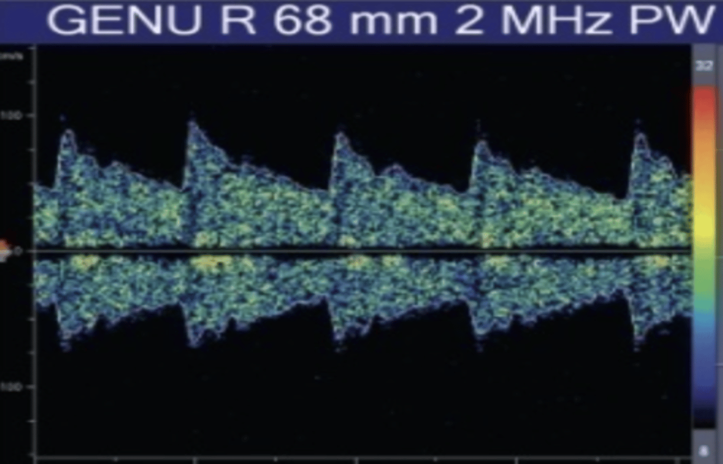
Genu Segment of Carotid Siphon Flow
Bidirectional (red or blue)
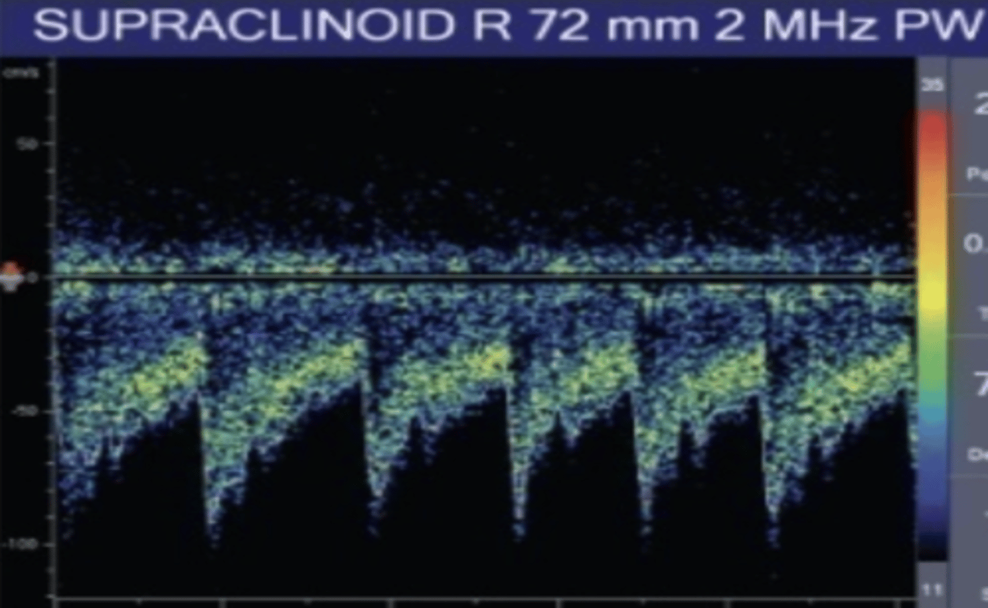
Supraclinoid Segment of Carotid Siphon Flow
Away from tx (blue)
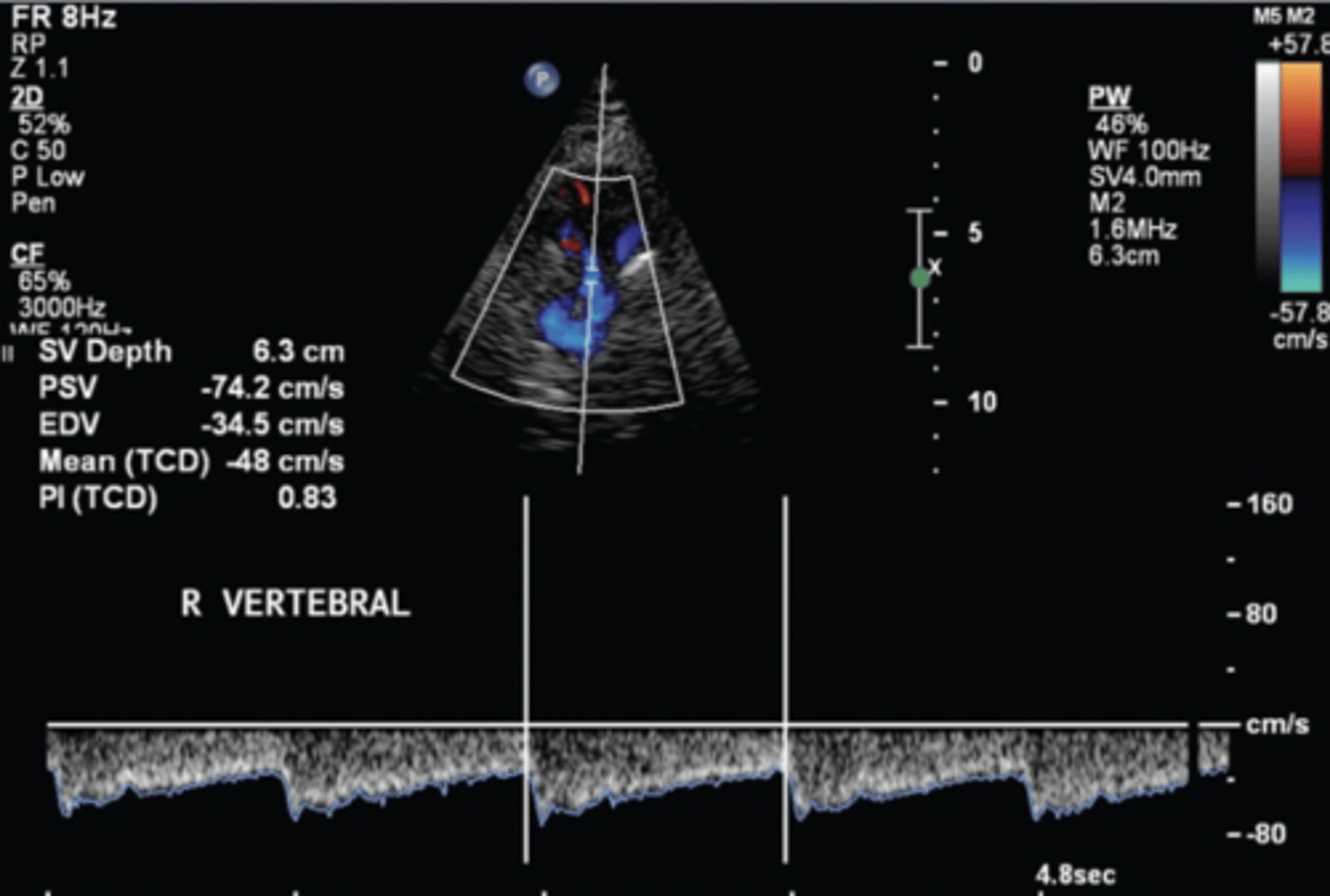
Vertebral A
Transforaminal/sub-occipital window
60-90 mm depth
Flow away from tx (blue)
38 +/- 10 cm/sec
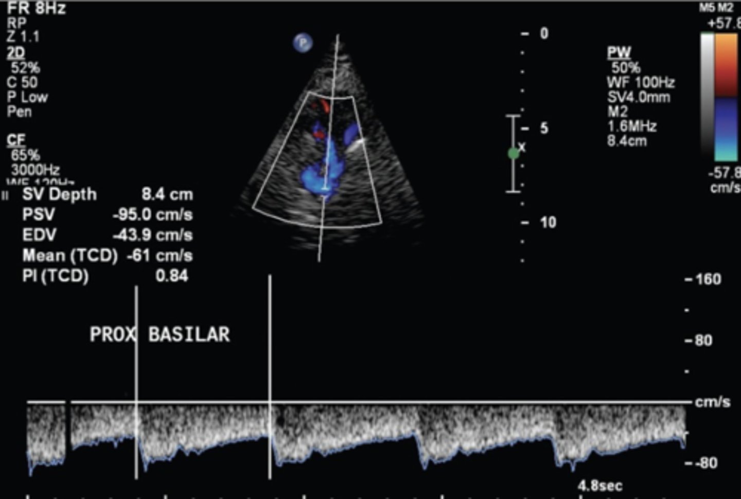
Basilar artery
Transforaminal/sub-occipital window
70-120 mm depth
Flow away from tx (blue)
41 +/- 10 cm/sec
Intracranial/Extracranial Stenosis
Most commonly caused by cardiac disease & atherosclerosis
Matches stenotic flow profile - pre-stenotic high resistance with post-stenotic turbulence & drop in velocity
Intracranial/Extracranial Occlusion
Most commonly caused by embolism
Flow returns to normal within 6 hours - good prognosis
Flow remains decreased after 12 hours - poor recovery
Vasospasm
Transient narrowing of cerebral arteries - mainly at skull base
Commonly due to subarachnoid hemorrhage (SAH)
Can lead to stroke - delayed cerebral ischemia (DCI)
Progression of Vasospasm
Begins 3-4 days after SAH
Peaks 6-8 days after
Resolves in 2-4 weeks
Sviri Ratio
Used for posterior circulation vasospasm
Average of bilateral vertebral A velocities ÷ highest basilar A mean velocity
Severe MCA/ICA Vasospasm Velocity
> 200 cm/s
Lindegaard Ratio
Used for anterior circulation
Differentiates increased flow volume (hyperemia) from vasospasm
MCA mean velocity ÷ submandibular ICA mean velocity
Severe MCA/ICA Vasospasm Ratio
> 6
Detecting PFO Via TCD
Agitated saline with micorbbubles area seen as "hits" or microembolic signals (MES) during/after injection if a PFO is present
Perform valsalva if needed
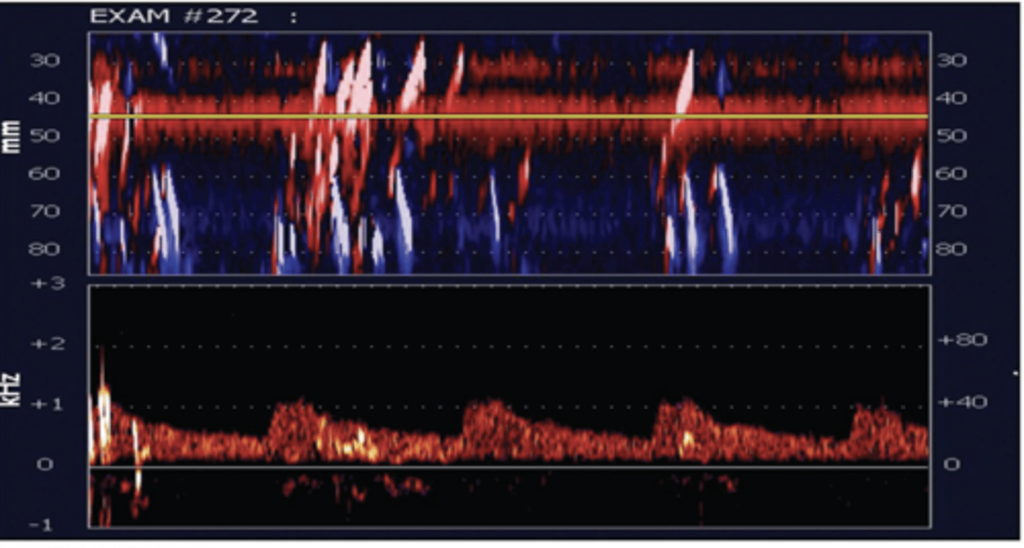
Low-Grade Shunt
1-10 hits
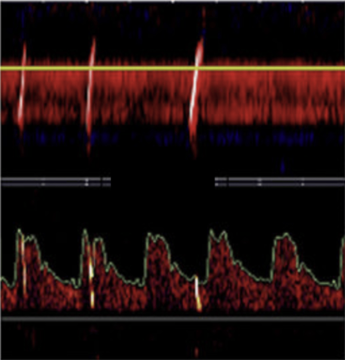
Medium-Grade Shunt
> 10 hits without curtain effect
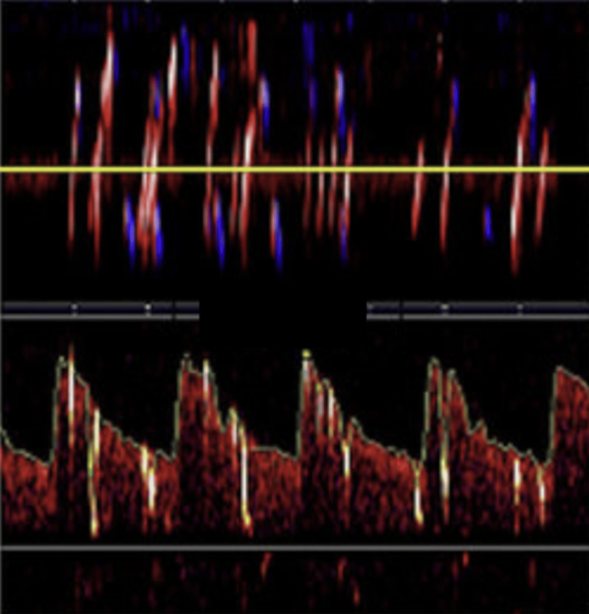
High-Grade Shunt
Curtain effect
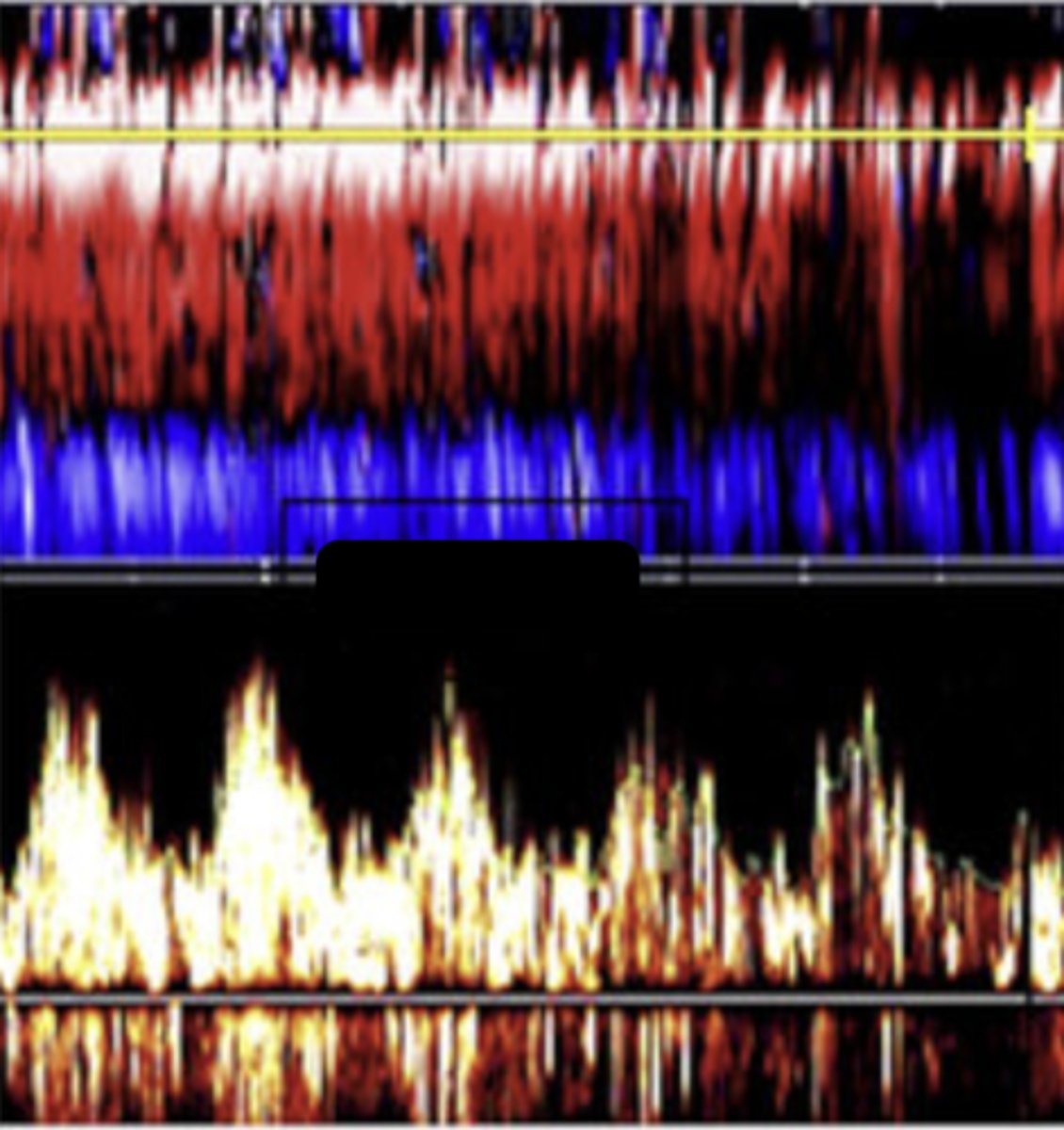
Vasomotor Reserve
Brain's ability to dilate/constrict vessels in response to CO2 changes (holding breath) to maintain stable blood flow
Reduced with stenosis, small artery disease, concussion
Functional Testing via TCD
Assessment of cerebral ischemia with high grate stenosis/occlusion
Assess vasomotor reserve
Cerebral Circulatory Arrest (CCA)
Brain death - ECG criteria must also be met
Early - low diastolic flow
Final stage - persistent oscillating high resistance waveforms with no diastolic flow
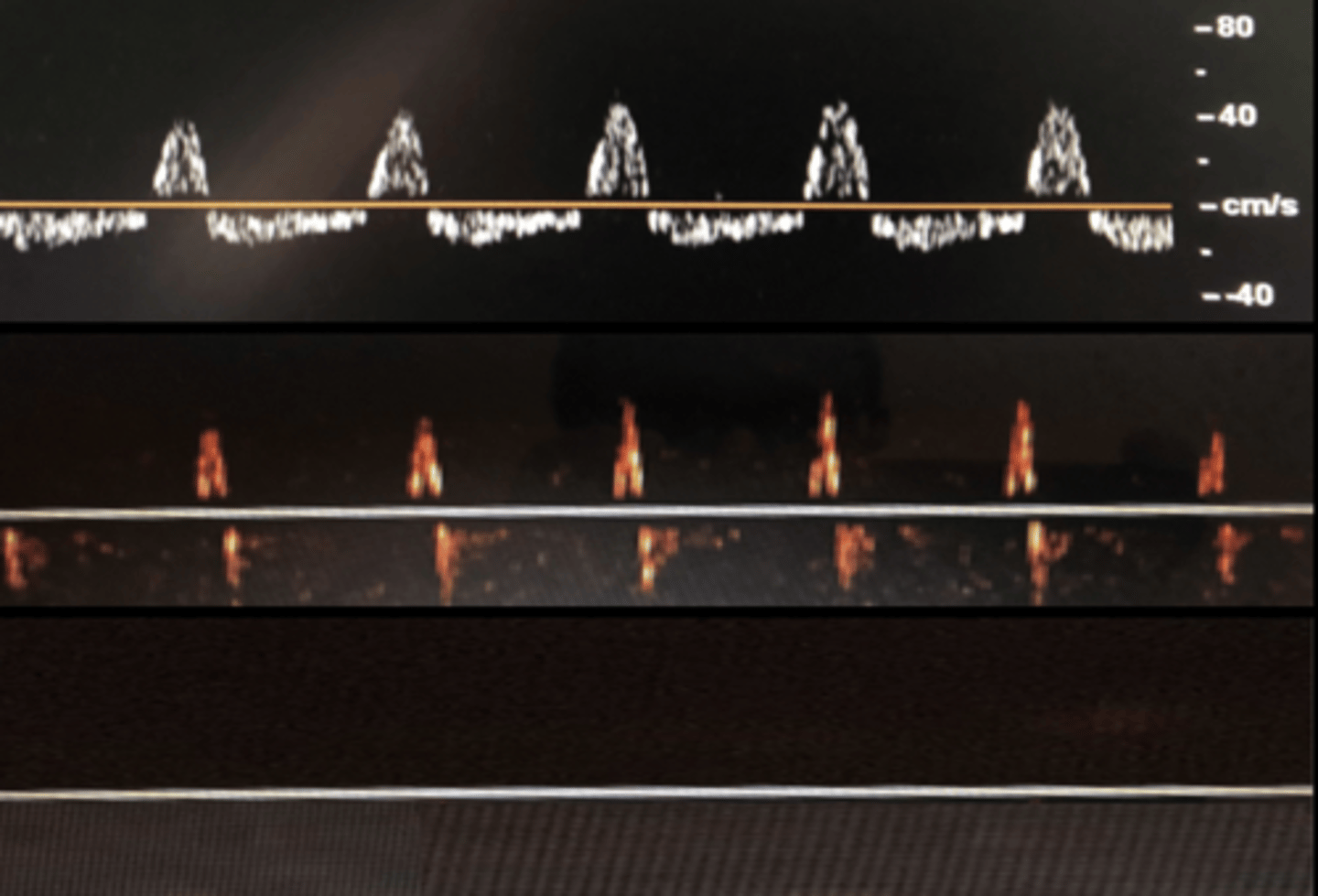
Advantages of TCD for CCA Diagnosis
Portable
Non-invasive
Repeatable
Does not require contrast
Not affected by drugs that suppress the CNS
Disadvantages of TCD for CCA Diagnosis
Need experienced operators
Inadequate acoustic windows in up to 20% of the time
False negatives in anoxic patients
False positives
Patient's with sickle cell anemia are prone to...
MCA & ACA strokes
MCA Mean Velocity in Sickle Cell Anemia Patients on TCD
> 200 cm/sec
MCA Mean Velocity in Sickle Cell Anemia Patients with Stoke on TCD
190-250 cm/sec