Blood Supply to CNS and Cerebrospinal Fluid (Week 1, Mod 8)
1/38
Earn XP
Description and Tags
Name | Mastery | Learn | Test | Matching | Spaced |
|---|
No study sessions yet.
39 Terms
Though the brain is only 2% of our body weight, how much of our cardiac output does it receive?
Brain receives about 20% of our total cardiac output
What area of the brain has the most blood supply? What arterial structure is it supplied by?
Hypothalamus has the richest blood supply in the brain
Supplied by the cerebral arterial circle; is SURROUNDED by this, located on the ventral aspect of the forebrain
What are the 5 main arteries that form the cerebral arterial circle?
1) Rostral communicating arteries
2) Right and left rostral cerebral arteries
3) Internal carotid artery
4) Right and left CAUDAL communicating arteries
5) Basilar artery
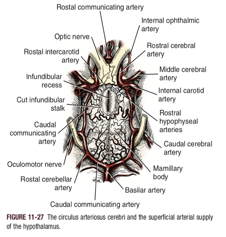
What 5 pairs of arteries supply the brain? Where do they arise from?
1) Rostral cerebral artery - arises from cerebral arterial circle, from the internal carotid artery
2) Middle cerebral artery - Same as ^^
3) Caudal cerebral artery - arises from the cerebral arterial circle, comes from the caudal communication artery ← internal carotid artery
4) Rostral cerebellar artery - arises from cerebral arterial circle; comes from caudal communicating artery
5) Caudal cerebellar artery - arises from the basilar artery
Arterial circle
The basilar artery… runs at the BASE of the brain
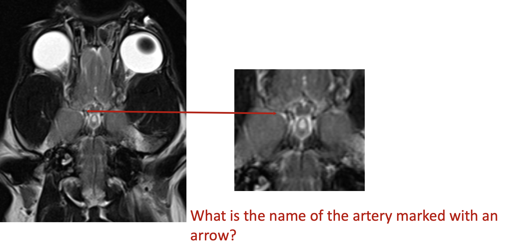
Middle cerebral artery… runs through the MIDDLE of the brain
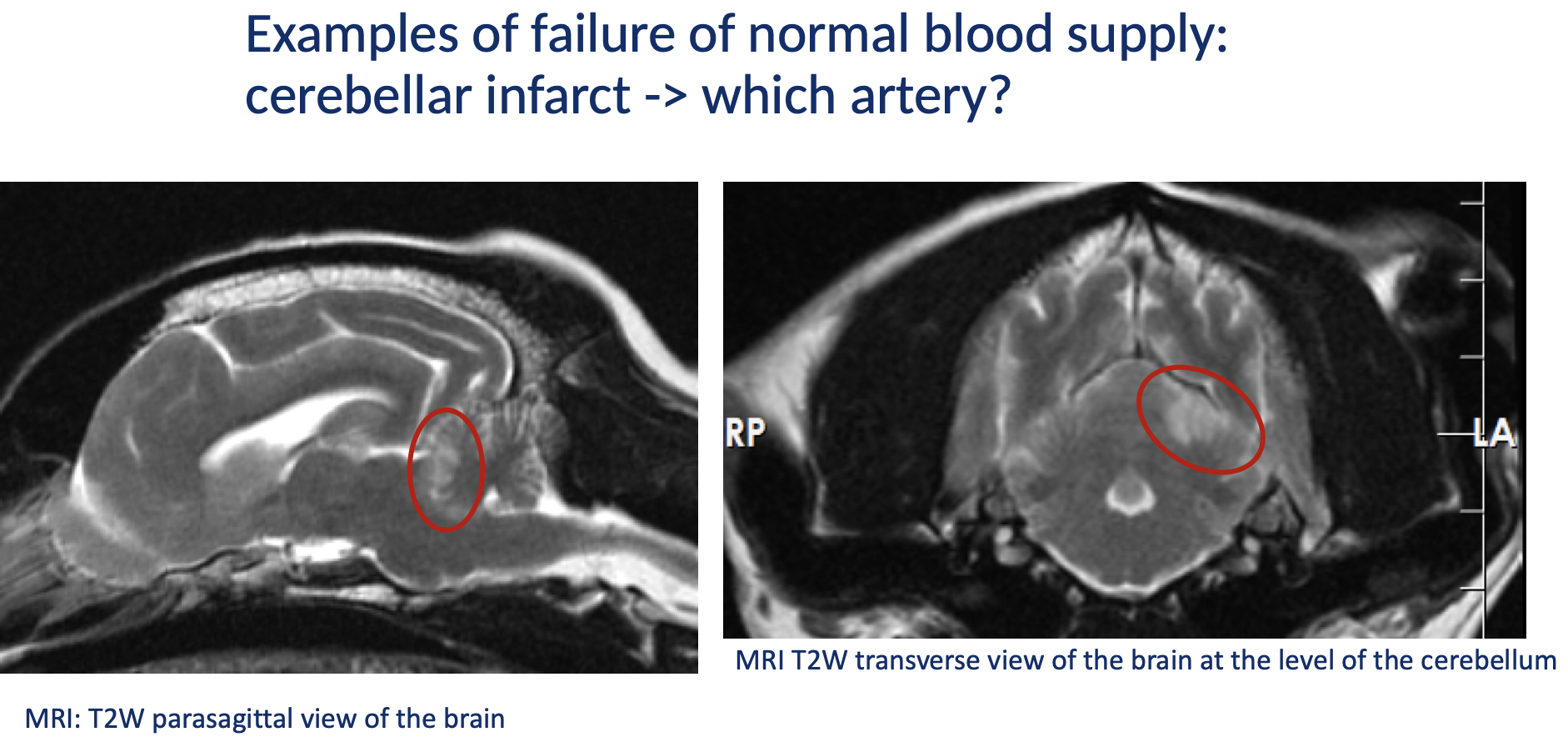
Cerebellar infarction; possibly affects the rostral cerebellar artery
What are the 4 incoming arterial branches that supply the cerebral arterial circle?
Channel 1 - internal carotid artery
Channel 2 - Basilar artery
Channel 3 - Maxillary artery
Anastomoses with the internal carotid artery via anastomosin rammus
Channel 4 - Vertebral artery
What arterial channels in the horse, dog, and human are the main blood suppliers for the cerebral arterial circle?
Internal carotid artery
Basilar artery
Vertebral artery
What arterial channels in the sheep and cat are the main blood suppliers for the cerebral arterial circle?
Maxillary artery
Vertebral artery
***Is different because 2/3s of the internal carotid artery is obliterated in the first few weeks / months of birth
What arterial channels in the cow are the main blood suppliers for the cerebral arterial circle?
Is a mixture of vertebral and maxillary blood via the ANASTOMOSIN RAMUS OF THE MAXILLARY AND VERTEBRAL ARTERY
What is the “rete mirabile” in the ruminant? What is its function?
Is a mesh network of anastomoses that fills the cavernous sinus…
Possibly involved in THERMOREGULATION
Apparently, the heat exchange occurs between rete blood and cavernous blood (rete blood is cooled before it enters the brain)
What three main segmental arteries supply blood to the different segments of the spine?
1) Lumbar arteries → supply blood to the lumbar spine
2) Intercostal arteries → supply blood to the thoracic spine
3) Subclavian arteries → supply blood to the cervical spine
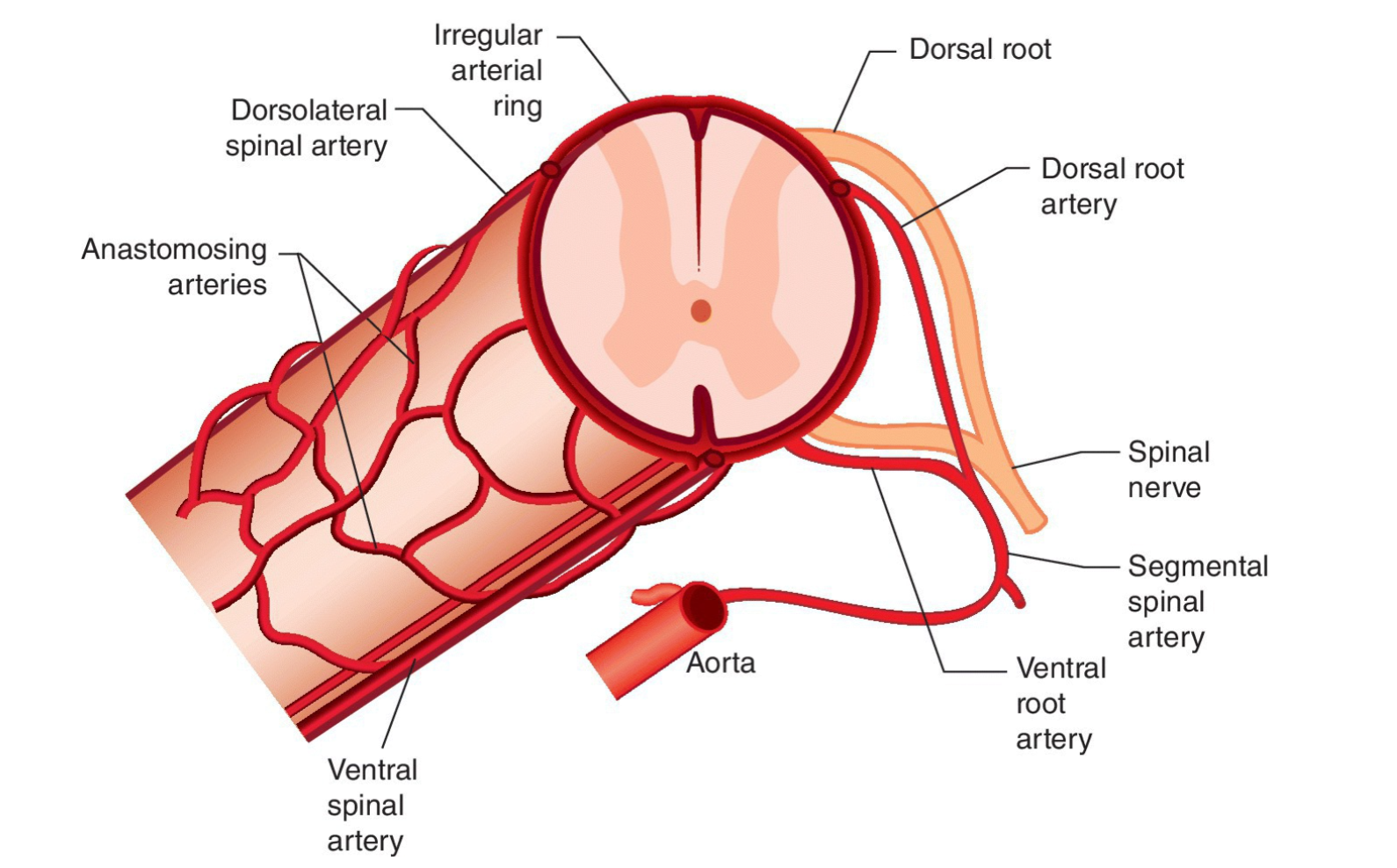
Describe the pathway of blood flow to the spine via SUPERFICIAL spinal arteries…
Aorta → segmental spinal artery → Dorsal + ventral root arteries → irregular arterial ring (composed of 2 dorsolateral spinal arteries and a SINGLE ventral spinal artery) → anastomosing arteries (netting of arteries that spreads around entire structure
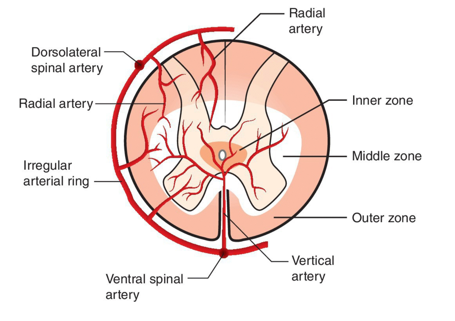
What are the 2 deep spinal cord arteries? Which of these supplies primarily gray matter, and which supplies primarily white matter?
1) The Vertical Artery -
Supplies most of the GRAY matter and reaches peripherally into the white matter
2) The Radial Arteries -
Supplies the WHITE matter and outer regions of the gray matter
What are the 3 “vascular zones” in the spine, and which of the 2 deep arteries are they each supplied by?
1) Inner zone - supplied by VERTICAL arteries only
Makes sense… inner zone should only have gray matter
2) Middle zone - supplied by BOTH vertical and radial arteries
3) Outer zone - supplied by RADIAL arteries only
Again, makes sense
What is a fibrocartilage embolism?
Is a condition secondary to an embolism of the spinal cord artery within the intervertebral disc material
Causes an INFARCTION of the region of the spinal cord supplied by that artery
Results in acute or peracute onset of paraparesis / tetraparesis; is non-painful but regressive
How are the veins in the CNS physically different from veins elsewhere in the body?
The veins of the CNS LACK:
Tunica media
Tunica adventitia
DO NOT HAVE VALVES
Where does venous drainage take place in the brain? Be specific.
Drainage happens within the SINUSES between the two layers of dura mater… the external periosteal layer and the internal meningeal layer
What are the 3 sinuses that drain the dorsal area of the brain?
Dorsal System of Sinuses:
Dorsal sagittal sinus
Straight sinus
Transverse sinus
What are the 3 sinuses that drain the ventral area of the brain?
Ventral System of Sinuses:
Cavernous sinus
Dorsal petrosal sinus
Ventral petrosal sinus
What sinus connects both the DORSAL system and the VENTRAL system of the sinuses?
The SIGMOID sinus
What is the falx cerebri in the brain?
A fold of the dura mater that descends into the longitudinal fissure to separate the brain’s left and right hemispheres
Describe the dorsal sagittal sinus… where can it be found in the brain? How does it act as a venous drainage system?
Is a single sinus which lines in the FALX CEREBRI and drains the dorsal region of the forebrain by the dorsal cerebral veins
Describe the STRAIGHT sinus… where can it be found in the brain? How does it act as a venous drainage system?
Is a single sinus which lines the CAUDAL part of the falx cerebri (rostral to the tentorium cerebelli) and drains the dorsal regions of the forebrain via the great cerebral vein
Describe the transverse sinus… where can it be found in the brain? How does it act as a venous drainage system?
Is a PAIRED SINUS
The left and right transverse sinuses are joined in the midline to form a confluence of sinuses
Here, the dorsal sagittal sinus is received before it continues laterally, then dividing into the temporal and sigmoid sinuses
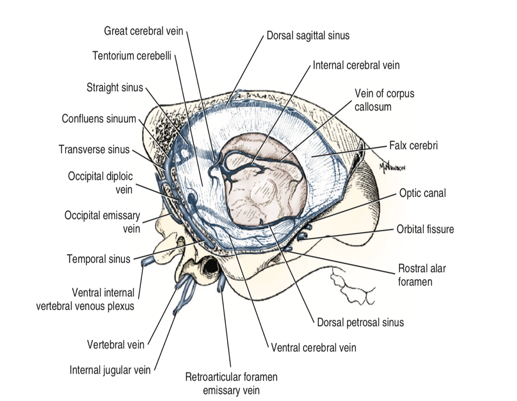
Describe the cavernous sinus… where can it be found in the brain? How does it act as a venous drainage system?
Is a PAIRED SINUS that surrounds the hypophysis. It drains from the PETROSAL sinuses, which drain the dorsal region of the brain
Describe the dorsal and ventral petrosal sinuses… where can they be found in the brain? How do they act as a venous drainage system?
The dorsal petrosal sinus drains the ventral region of the brain, while the VENTRAL petrosal sinus acts as a connection between the cavernous sinus to the sigmoid sinus
Describe the SIGMOID sinus… where can it be found in the brain? How does it act as a venous drainage system?
Has an S shape; receives the dorsal system (via transverse sinus) and the ventral system (via petrosal sinuses) and connects DIRECTLY with the spinal system (via vertebral plexus). Drains into the maxillary vein, then into systemic circulation
Describe the venous drainage of the SPINAL CORD, from smallest vein to systemic circulation...
Note: essentially follows arterial blood supply
Vertebral spinal vein → Vertebral venous sinus → Spinal veins → Vertebral veins, Azygos veins, Caudal vena cava → Systemic circulation
What 3 veins drain the cervical region, the thoracic region, and lumbar region of the spine?
Cervical region → VERTEBRAL vein
Thoracic region → AZYGOS vein
Lumbar region → CAUDAL VENA CAVA
What is cerebrospinal fluid? What is it mad up of, and how is it produced?
Is a clear, colourless fluid that surrounds entire CNS (located in subarachnoid space)
Originates from capillaries throughout the CNS
CSF produced by selective ultrafiltrate of blood plasma (+ active transport mechanisms)
Major production site are choroid plexi of lateral, third and fourth ventricles
What are the 5 main functions of the CSF?
1) Physical support (buoyancy)
2) Protection against trauma
3) Modulates pressure changes within the skull
4) Nutrition (transport of metabolites, nutrients, and neurotransmitters)
5) Chemical buffer (maintains ionic balance)
What are the 4 aspects of the choroid plexus (ventricular system) that we NEED to know?
1) Lateral ventricle
2) Third ventricle
3) Fourth ventricle
4) Mesencephalic aqueduct
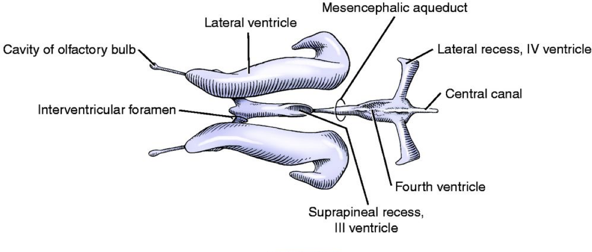
How is CSF moved through the brain? Describe its pathway, starting from its production point in the choroid plexus…
CSF flow is caused by blood pulsations and motile cilia of the surface of ependymal cells
Passes from the lateral ventricles through the interventricular foramina (*) to the 3rd ventricle
Flows through the mesencephalic aqueduct and into the 4th ventricle
Then flows into the central canal of the spinal cord, OR through the lateral apertures (**) to the subarachnoid space
With each arterial pulsation, the CSF pressure rises and surges towards the lateral apertures
In the spinal cord, CSF flows in the central canal and into the subarachnoid space
The central canal is continuous with the fourth ventricle, surrounded by gray matter and lined with ependymal cells
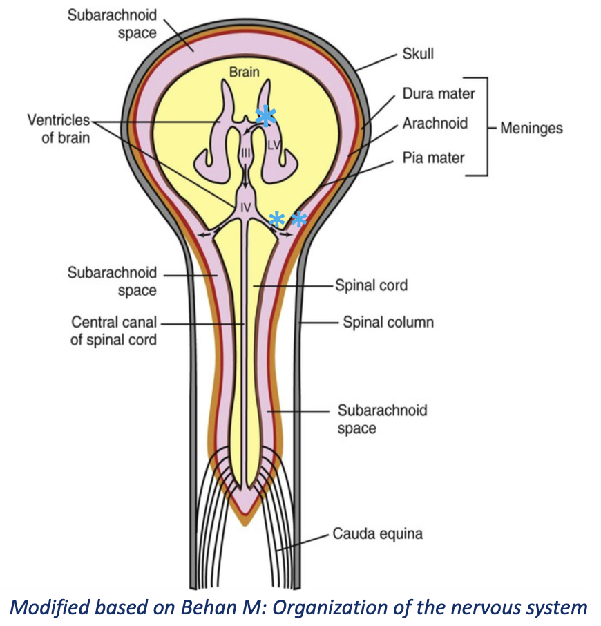
How is CSF absorbed? What structures assist in this?
CSF is absorbed by arachnoid vili in venous sinuses primarily. Also is absorbed by:
Venules in the sub-arachnoid space
Lymphatic vessels of cranial and spinal nerve roots
When the pressure of CSF in the arachnoid space becomes GREATER than it is in the venous sinuses, the arachnoid vili (see image) will open and allow CSF to drain into the sinus
Collapses after this, not allowing back flow

Why would sampling CSF be beneficial diagnostically?
CSF is in close relationship with brain and spinal cord parenchyma
In some disease processes such as inflammation, trauma or occasionally tumours, the CSF composition may be altered
➔ CSF sampling may help in diagnosis of inflammatory or neoplastic conditions
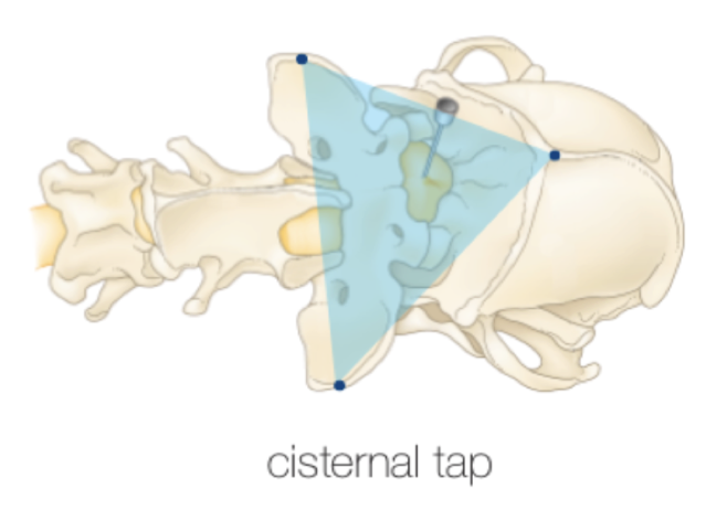
What are the general steps of collecting CSF from the cisterna magna in a cisternal tap?
Flexion of the neck
Enter in the midline between the skill and C1
Stop advancing when entering the CSF space