Lecture 4 - Breast Imaging
 Call Kai
 Learn
Learn Practice Test
 Spaced Repetition
 Match
Match Flashcards
 Knowt Play
1/41

There's no tags or description
Looks like no tags are added yet.
Name | Mastery | Learn | Test | Matching | Spaced | Call with Kai | Chat |
|---|
No analytics yet
Send a link to your students to track their progress
42 Terms
What is the sensitivity and specificity of mammogram
Sensitivity: 75-85%
Specificity: 90-97%
What is mammography effective for
Masses
Architectural distortion
Asymmetries
Suspicious calcifications
What’re the views in mammography
CC: cranial caudal
MLO: medial lateral oblique
Why is multimodality imaging important in mammography
Pathology may appear different in different modalities
US can determine if mass is solid/fluid filled
Dose justification - young patients may be referred to MRI/US
Breast density
What’re some ways breast pathology can present
Asymmetric lesion - on one boob but not the other
Architectural distortion
Calcifications
Smooth wall benign lesions
Invasive irregular wall lesions
Malignant lesion with lymph node, skin or nipple changes
What’re the 4 quadrants for lesion localisation
5: upper outer
6: upper inner
7: lower outer
8: lower inner
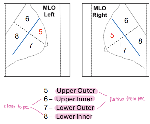
What’re the regions for lesion localisation

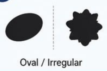
Which shape is benign/malignant
Oval: benign
Irregular: malignant

Which margin is benign/malignant
Circumscribed: benign
Spiculated: malignant

Which density is benign/malignant
Low: benign
High: malignant
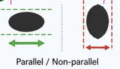
Which orientation is benign/malignant
Parallel: benign
Non-parallel: malignant
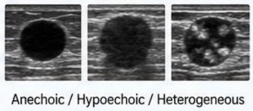
Which echogenicity is benign/malignant
Anechoic: benign
Hypoechoic: malignant
Heterogenous: malignant

Which posterior acoustic feature is benign/malignant
Enhancement: benign
Shadowing: malignant
What’re some common benign masses
Cyst
Fibroadenoma
Intramammary lymph node
Lipoma
Hamartoma
What’re the characteristics of a benign fibroadenoma
Fibrous non-cancerous solid growth
Painless
Smooth wall
Common
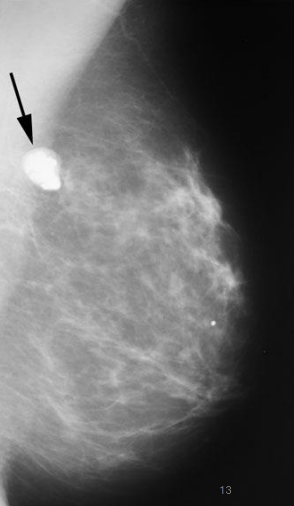

Name this pathology
Benign bibroadenonma
What’re the characteristics of a benign breast cyst
Fluid filled sac
Painful and affected by menstrual cycle
Can be aspirated
Better seen on US - determine nature of cyst
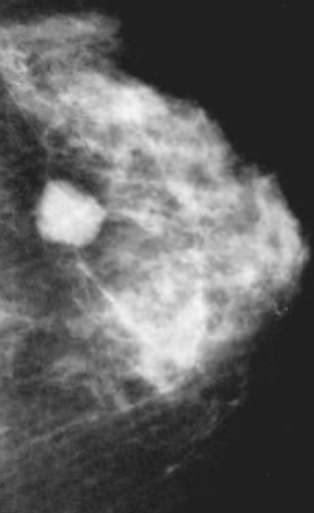

Name this pathology
Breast pathology
What’re the characteristics of micro calcifications
Benign
Tec cups
Pearls
Vascular markings
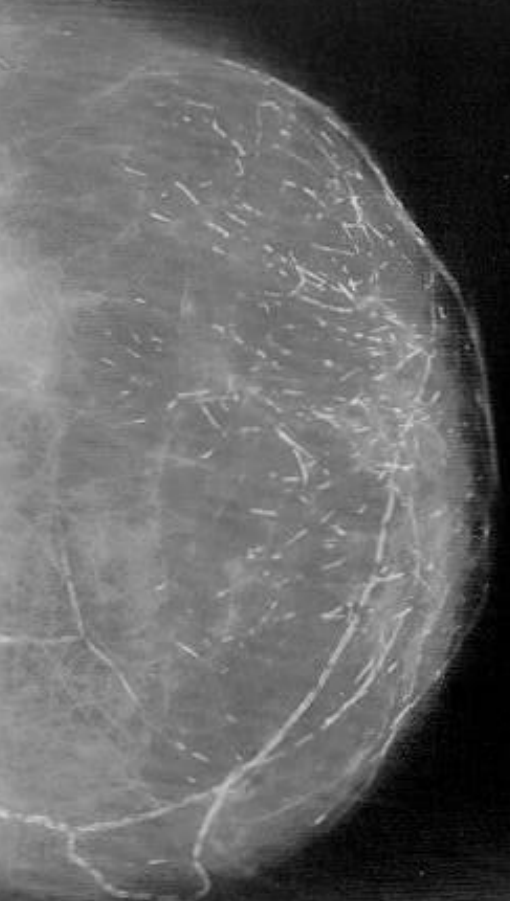
What’re the characteristics of malignant microcalcifications
Clustered
Irregular shape and size
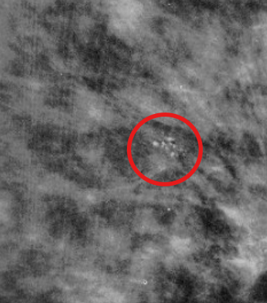

Benign or malignant
Likely malignant
Poorly defined spiculated mass
Dense
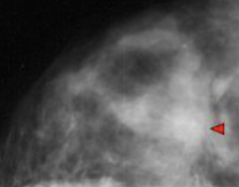

Benign or malignant
Likely malignant
Defined spiculated mass
Dense
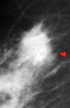

What kind of mass is this
Stellate mass (invasive lobular carcinoma with radiating margins)
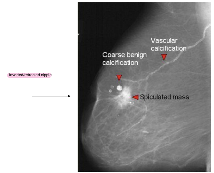
What kind of mass is this
Invasive ductal carcinoma (NOS)
What extra view can you preform in mammography
Spot view
What is breast density
Relative amounts of fibroglandular tissue and fatty tissue
more fibroglandular tissue = more dense
How does far appear in mammograms
Dark
How does fibroglandular tissue appear on mammograms
White
What are the BI-RADS density categories
A: Almost entirely fatty
B: Scattered areas of fibroglandular density
C: Heterogeneously dense
E: Extremely dense
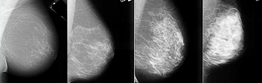
How does breast density affect mammography
Dense tissue can obscure malignancy
Dense tissue and cancer appear radiopaque
Breast density is a risk factor for breast cancer
What is the masking effect
Similar cancer within a dense breast
What’re imaging options for dense breast
Breast tomosynthesis
US
Contrast enhanced mammography
MRI
Describe tomosynthesis
Multiple low-dose projections across angular range
Images reconstructed into thin slices
Reduces tissue superimposition compared with conventional 2D mammography
MLO and CC

What’re the advantages of tomosynthesis
Reduces tissue overlap
Improves cancer detection and lesion characterisation
Particularly useful in dense breasts
Reduces recalls and false positives
Provides better lesion localisation
What’re the disadvantages of tomosynthesis
Longer interpretation time
Larger image storage requirements
Higher cost/additional training needs
Higher radiation dose when combined with2D mammography
Potential motion artefact
What is a needle biopsy
Needle is used to draw sample fluid and small amounts of tissue from a lump
What is a core biopsy
Needle is used to scoop out tissue sample - gives more information
What is a lumpectomy
Surgeon removes lesion only
What is a mastectomy
Whole breast is removed
What is the dose of mammography comparable to
Flight from Australia to London
3 months of natural exposure
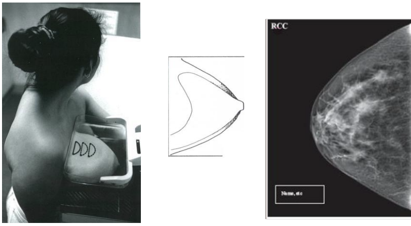
Positioning for CC view
Boob is centred in the middle with the nipple in profile
Pec muscle seen indicating that the boob has been positioned as far forward
Medial and lateral portions of breast seen
Well compressed and well exposed
Positioning for MLO view
Boob pulled forward and the parenchyma is spread out
Pec muscle is seen as wedge down to the level of the nipple
Nipple in profile
Abdominal wall is includes