tissues + hair structures
1/61
There's no tags or description
Looks like no tags are added yet.
Name | Mastery | Learn | Test | Matching | Spaced | Call with Kai | Chat |
|---|
No analytics yet
Send a link to your students to track their progress
62 Terms
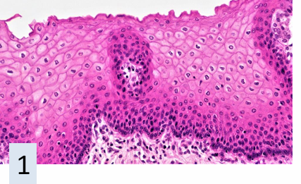
stratified squamous epithelial tissue; Multiple layers of cells with a surface of dead, keratin-filled cells that protect against abrasion and water loss.
what is this tissue
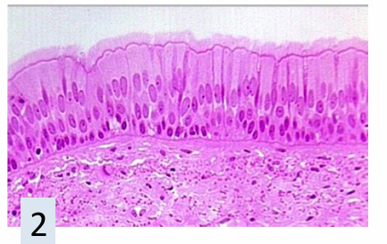
pseudostratified columnar ; Type of tissue made of a single layer of irregularly shaped cells
what is this tissue
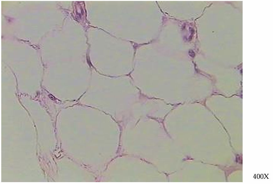
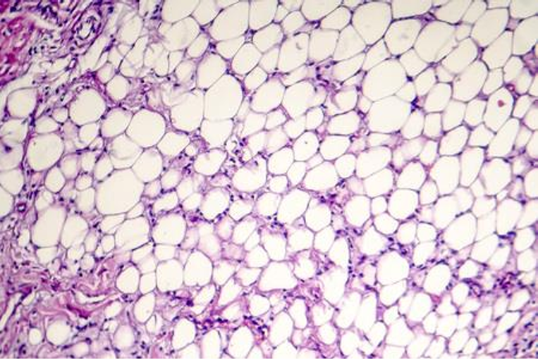
loose connective adipose at-storing tissue that cushions organs, insulates the body, and stores energy.
what is this tissue
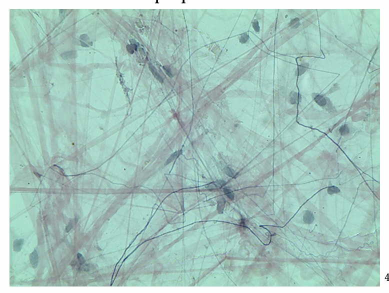
areolar connective tissue Loose tissue that wraps organs, cushions tissues, and holds tissue fluid.
what is this tissue
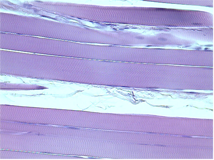
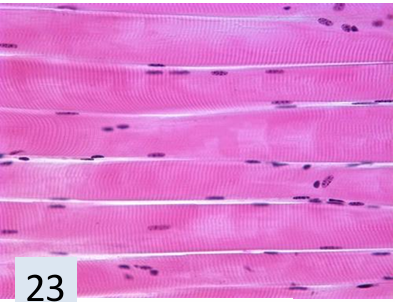
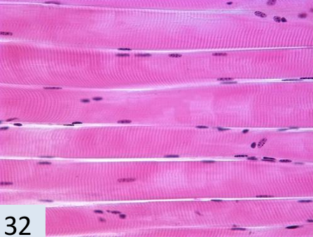
skeletal muscle Voluntary, striated muscle attached to bones that produces body movement.
what is this tissue
collagen fibers; the primary structural protein in the extracellular matrix of connective tissues. Acting as the body's scaffolding, these tough, rope-like fibers are packed with tensile strength to give skin, bones, tendons, and ligaments their shape, flexibility, and structural support
what part is this
mesenchyme a type of loosely organized embryonic connective tissue made of undifferentiated, fluidly moving cells.
what is this tissue
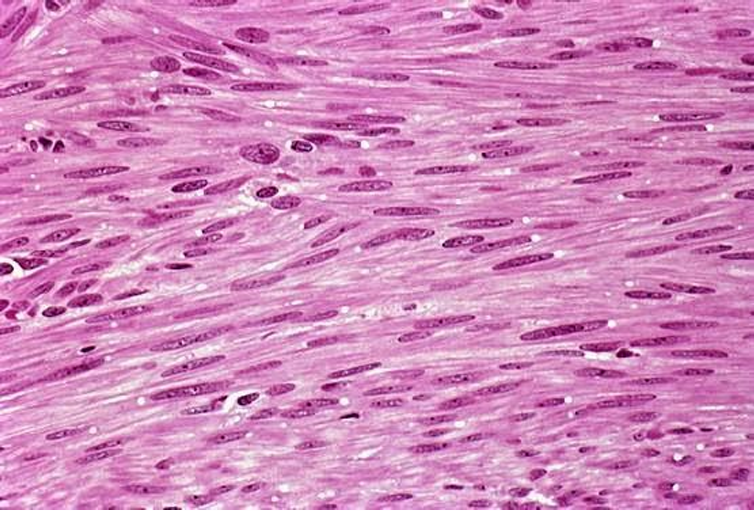
smooth muscle ; Involuntary, nonstriated muscle found in the walls of hollow organs that moves substances through the body.
what is this tissue
smooth muscle; Involuntary, nonstriated muscle found in the walls of hollow organs that moves substances through the body.
what is this tissue
lamella
a thin layer, membrane, or plate of tissue, especially in bone.

central canal;
what part of the bone tissue is this; the longitudinal CSF-filled space within the spinal cord, extending from the caudal angle of the fourth ventricle through the medulla oblongata and the entire spinal cord to the conus medullaris, where it widens into the terminal ventricle
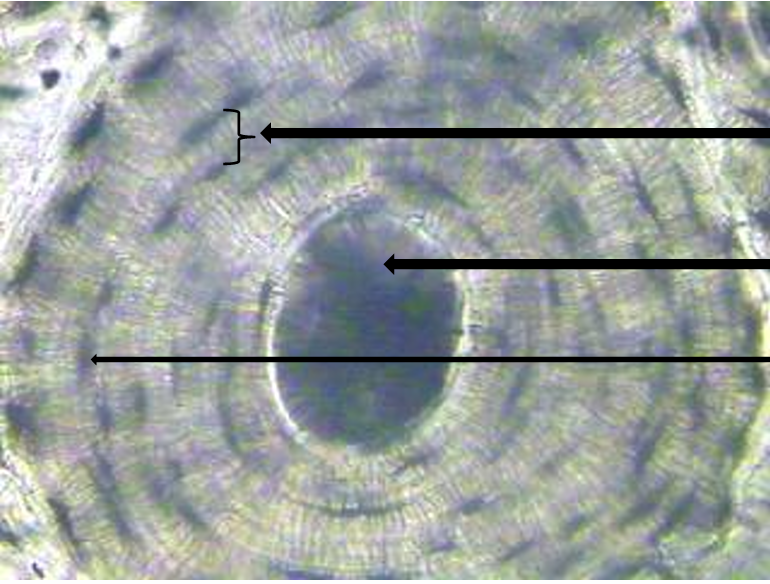
lacunae;
what part of the bone tissue is this; small cavity, pit, or discontinuity in an anatomical structure

loose connective tissue, reticular; Loose tissue that wraps organs, cushions tissues, and holds tissue fluid.
what tissue is this
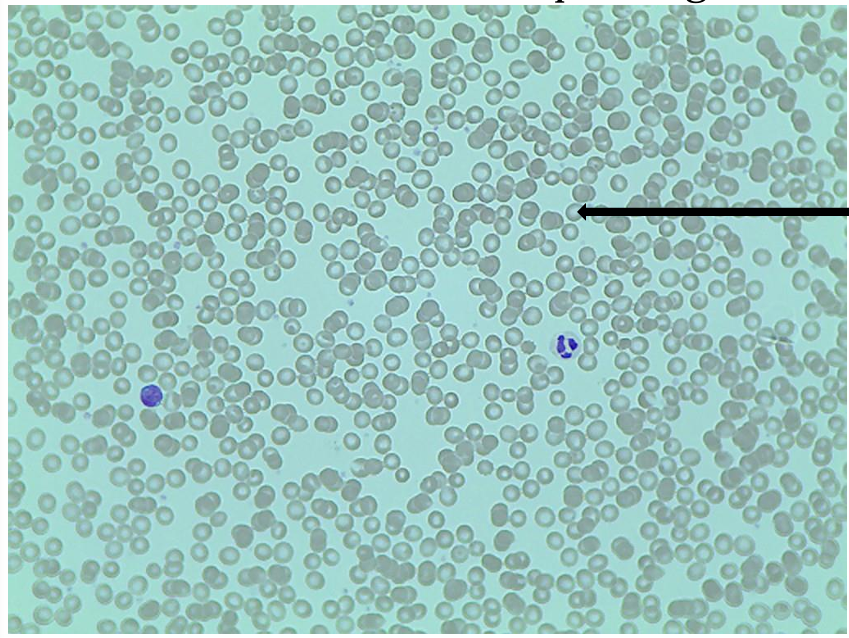
red blood cell; responsible for carrying oxygen from the lungs to the rest of the body
what cell is this?
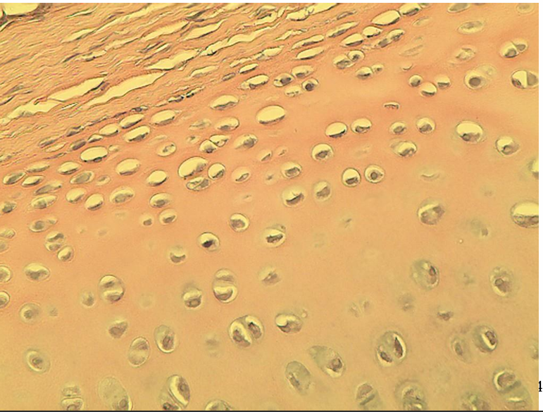
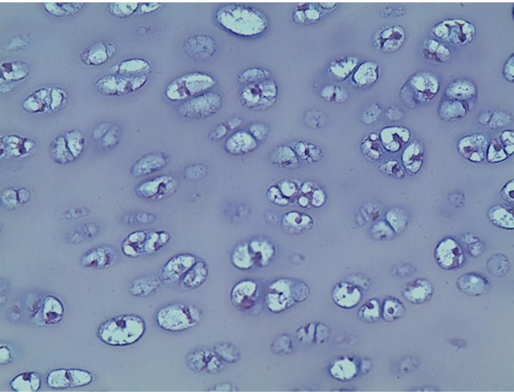
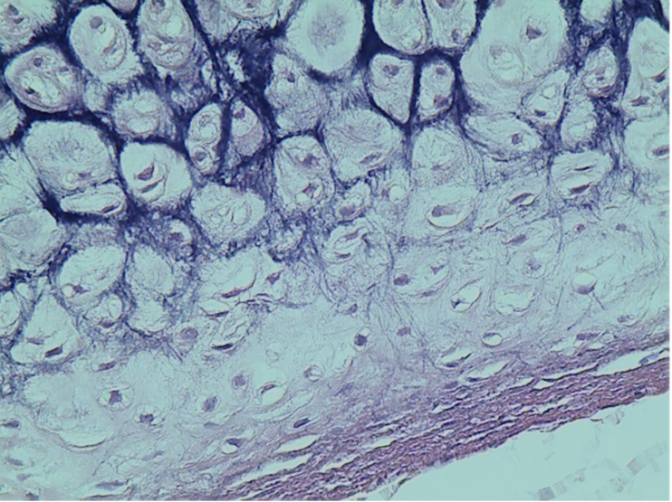
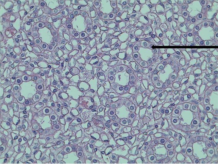
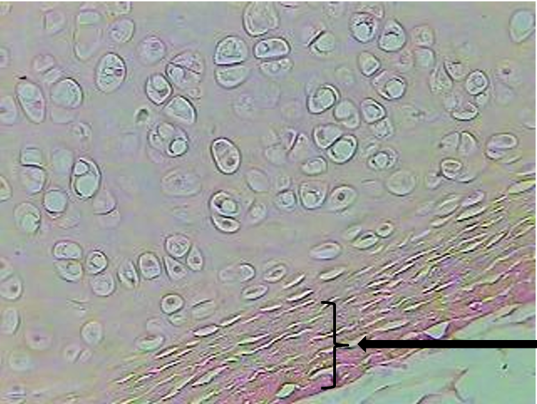
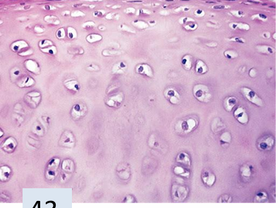
hyaline cartilage ( no fiber cartilage on exam) Smooth, glassy cartilage that supports structures and reduces friction in joints
what tissue is this?
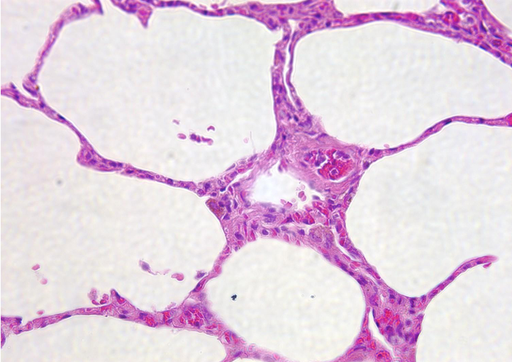
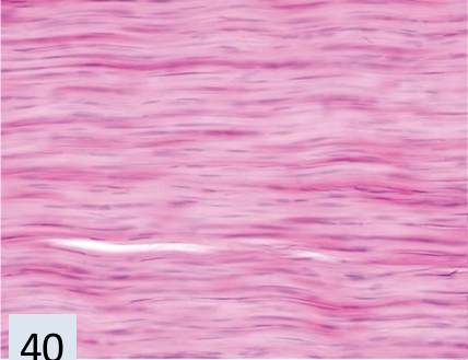
simple squamous epithelium; A single layer of flat cells specialized for diffusion and filtration
what tissue is this?
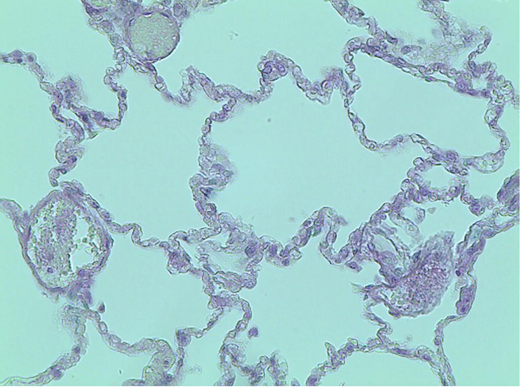
simple squamous epithelium; A single layer of flat cells specialized for diffusion and filtration
what tissue is this?
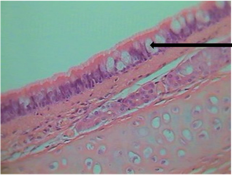
goblet cell; specialized, cup-shaped epithelial cells found lining the respiratory and intestinal tracts
what cell is this?
hyaline cartilage; Smooth, glassy cartilage that supports structures and reduces friction in joints.
what tissue is this?
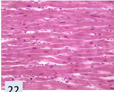
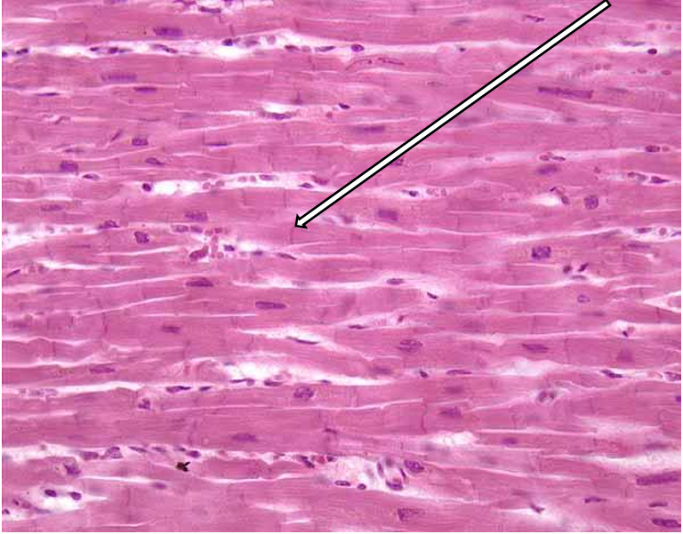
cardiac muscle; Involuntary, striated muscle found only in the heart that pumps blood.
what tissue is this?
intercalated discs; specialized, zigzag-shaped junctions that physically connect adjacent cardiac muscle cells (cardiomyocytes) in the heart
what part of the muscle tissue is this?
cardiac muscle; Involuntary, striated muscle found only in the heart that pumps blood.
what tissue is this?
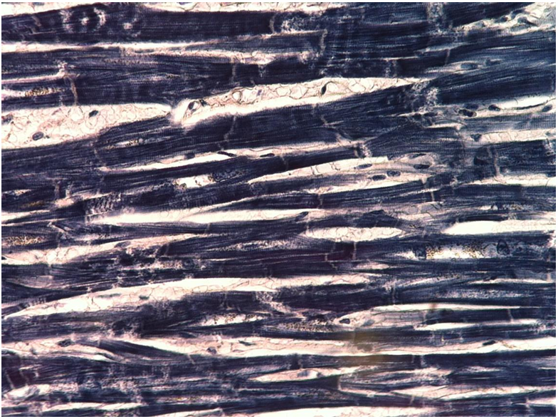
skeletal muscle; Voluntary, striated muscle attached to bones that produces body movement.
what tissue is this?
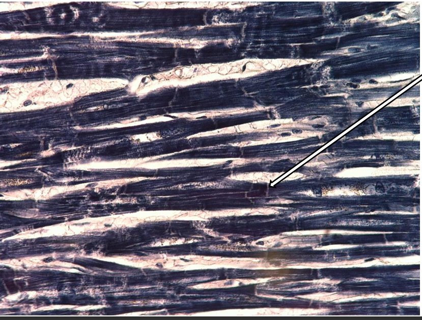
intercalated discs; specialized, zigzag-shaped junctions that physically connect adjacent cardiac muscle cells (cardiomyocytes) in the heart
what tissue is this?
loose connective tissue adipose; Fat-storing tissue that cushions organs, insulates the body, and stores energy.
what tissue is this?
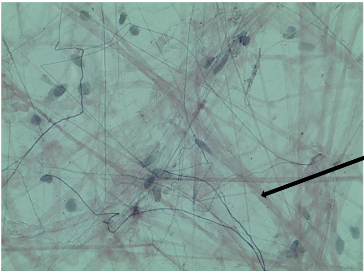
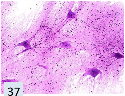
functional pathways or mechanisms by which the nervous system receives, integrates, and transmits information
What are the arrows pointing to? neuro processes
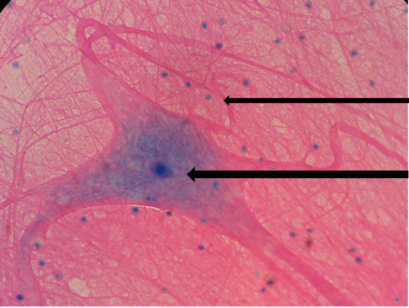
cell body of a neuronalso known as the soma, is the spherical part of the neuron that contains the nucleus and other major organelle
What are the arrows pointing to?

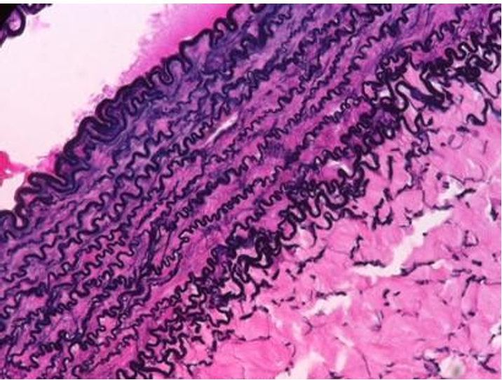
cartilage elastic; Flexible cartilage rich in elastic fibers that maintains shape.
what tissue is this?
stratified cuboidal epithelium; A single layer of cube-shaped cells specialized for secretion and absorption.
what tissue is this?
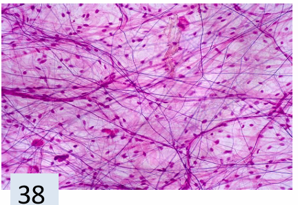
fibroblast; the primary cells of connective tissue in animals, responsible for synthesizing collagen, elastin, and the extracellular matrix
identify this part of connective tissue
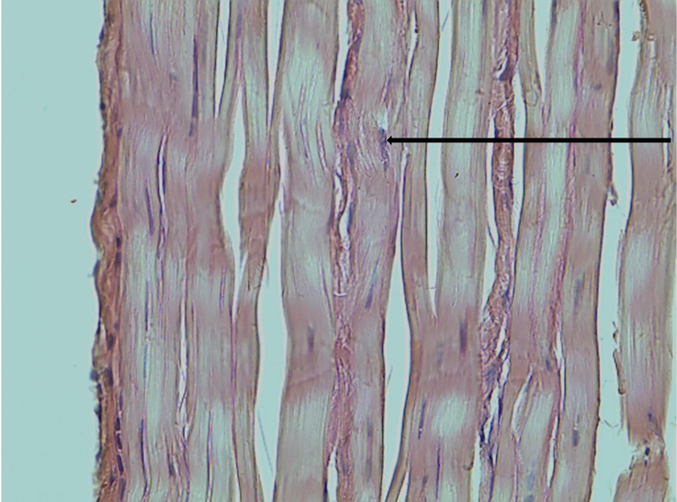
dense regular ct Parallel collagen fibers provide strong attachment in one direction; found in tendons and ligaments.
what tissue is this?
skeletal; Voluntary, striated muscle attached to bones that produces body movement.
what tissue is this?
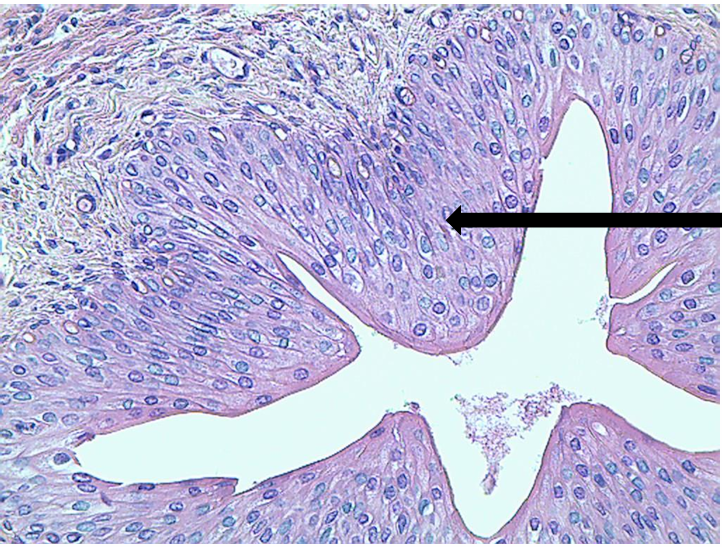
transitional epithelium; Stretchable epithelium that allows urinary organs to expand and recoil.
what tissue is this?
simple columnar; A single layer of tall cells specialized for absorption and secretion; may contain goblet cells and microvilli.
what tissue is this?
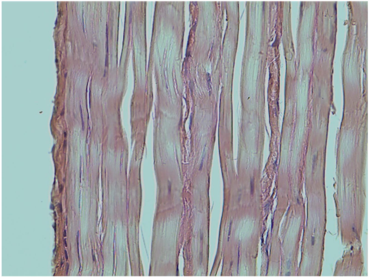
dense regular connective tissue; Parallel collagen fibers provide strong attachment in one direction; found in tendons and ligaments.
what tissue is this?
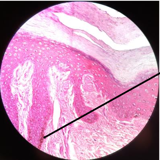
perichondrium; a dense, fibrous membrane that covers the exterior surface of most cartilage in the body (except for articular cartilage in joints and fibrocartilage).
what part is this?
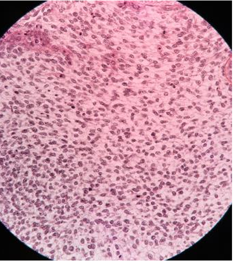
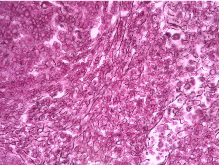
nervous tissue; the primary, specialized tissue of the nervous system. It generates, receives, and transmits electrical impulses across the body, allowing for communication, sensory perception, and motor coordination
what tissue is this?
connective tissue areolar; Loose tissue that wraps organs, cushions tissues, and holds tissue fluid.
what tissue is this?
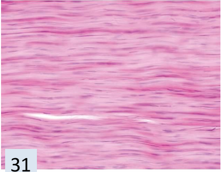
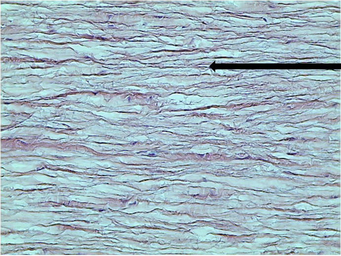
dense elastic; Dense tissue rich in elastic fibers that allows tissues to stretch and recoil.
what tissue is this?
dense regular; Parallel collagen fibers provide strong attachment in one direction; found in tendons and ligaments.
what tissue is this?
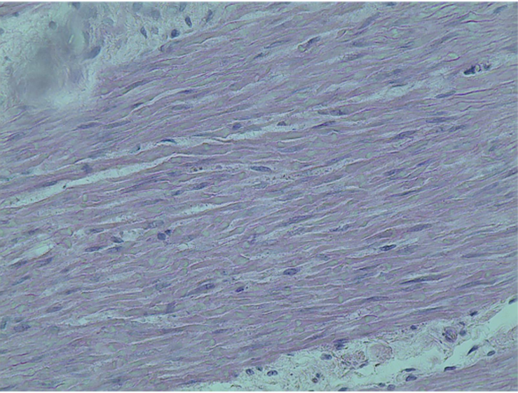
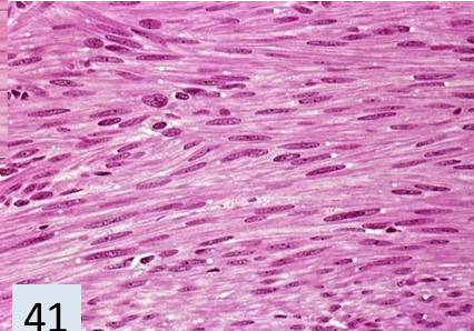
smooth; Involuntary, nonstriated muscle found in the walls of hollow organs that moves substances through the body.
what tissue is this?
dense regular; Parallel collagen fibers provide strong attachment in one direction; found in tendons and ligaments.
what tissue is this?
hyaline cartilage
what tissue is this?
stratified squamous; Multiple layers of living cells that protect moist surfaces from abrasion. flat
what tissue is this?
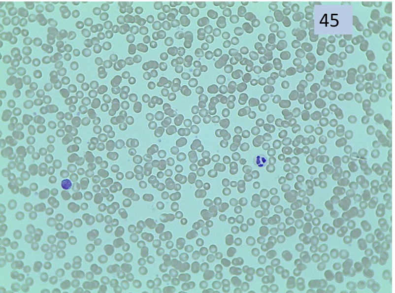
blood; Connective tissue that transports oxygen, nutrients, hormones, wastes, and immune cells.
what tissue is this?
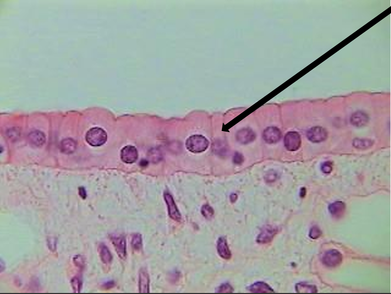
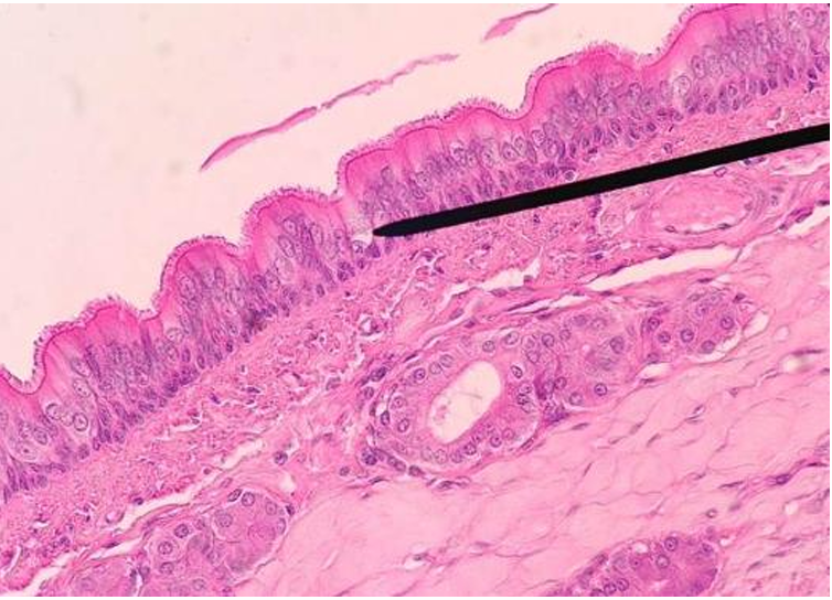
pseudostratified columnar; a type of tissue made of a single layer of irregularly shaped cells. Because the cells vary in height and their nuclei sit at different levels, it creates the optical illusion of being multilayered (stratified).
what tissue is this?
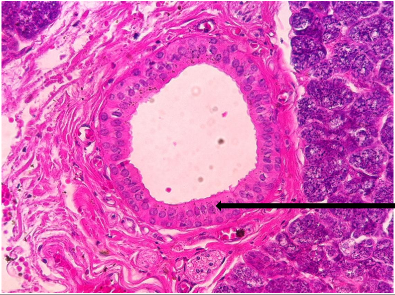
stratified cuboidal; Two or more layers of cube-shaped cells that protect and secrete in gland ducts.
what tissue is this?
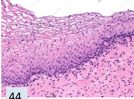
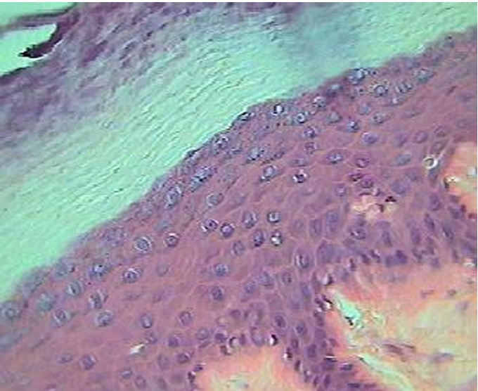
stratified squamous keratinized; Multiple layers of cells with a surface of dead, keratin-filled cells that protect against abrasion and water loss.
what tissue is this?
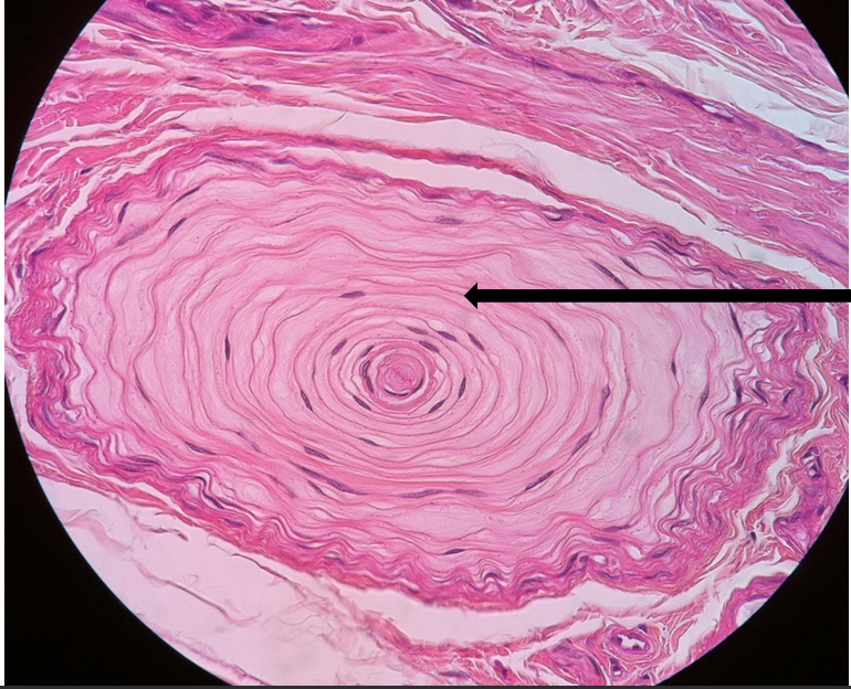
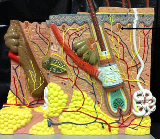
lamellar corpuscle; is an oval-shaped, rapidly adapting mechanoreceptor. Found deep in the skin, joints, and internal organs, its unique, onion-like layers of connective tissue allow it to detect high-frequency vibrations
what tissue is this?
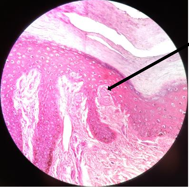
tactile corpuscle; an encapsulated, rapidly adapting mechanoreceptor in the skin. Located primarily in hairless areas like the fingertips and lips, it is highly sensitive to light touch, texture changes, and low-frequency vibrations
what part of the hair system is this?
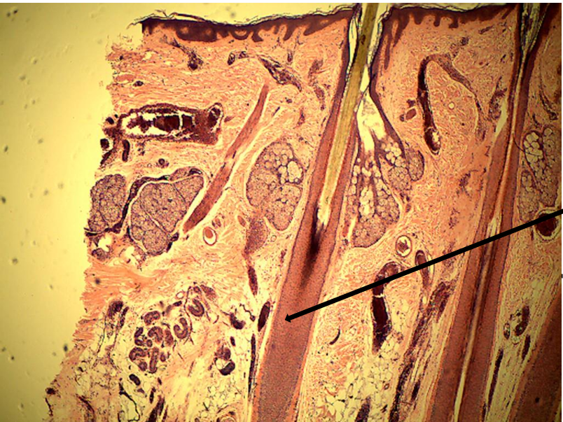
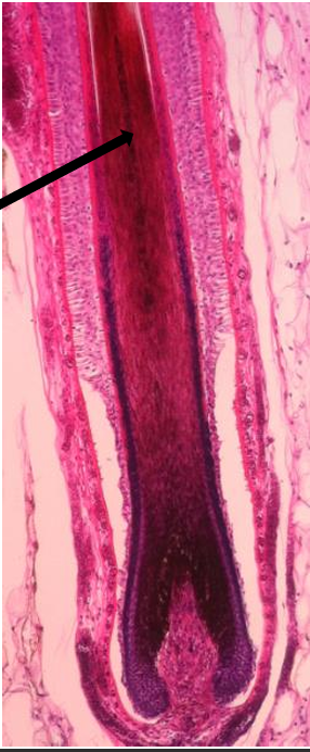
hair follicle; a tunnel-like, tube-shaped structure in your skin that anchors each individual hair in place and produces the hair strand. It extends from the top layer of your skin (epidermis) down into the deeper layers, serving as the biological engine that grows, nourishes, and shapes your hair.
what part of the hair system is this?
medulla; the innermost, soft core of the hair shaft, acting like a marrow or pith. It is typically found in thicker, coarser hair and is often completely absent in fine or light-colored hair
what part of the hair system is this?
papillary layer; the thin, superficial layer of the dermis, sitting directly beneath the epidermis
what tissue is this?
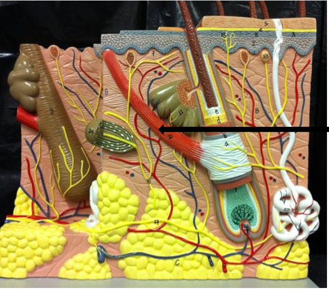
arrector pili; a tiny, microscopic band of smooth muscle that connects a hair follicle to the underlying skin tissue (the dermis).
what part of the hair system is this?
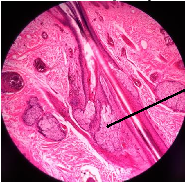
sebaceous gland; microscopic oil glands in the skin that secrete an oily, waxy substance called sebum
what part of the hair system is this?
tactile corpuscle; an encapsulated, rapidly adapting mechanoreceptor in the skin. Located primarily in hairless areas like the fingertips and lips, it is highly sensitive to light touch, texture changes, and low-frequency vibrations
what part of the hair system is this?
specialized cells in the body responsible for producing melanin, the pigment that determines the color of your skin, hair, and eyes
melanocytes
dermis areolar ct
what tissue is this?
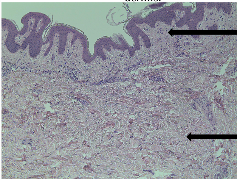
Identify the tissues that make up the layers of the dermis dense irregular
A connective tissue with densely packed collagen fibers arranged in many directions, providing strength and resistance to stretching from multiple directions. It is found in the dermis of the skin, organ capsules, and joint capsules.

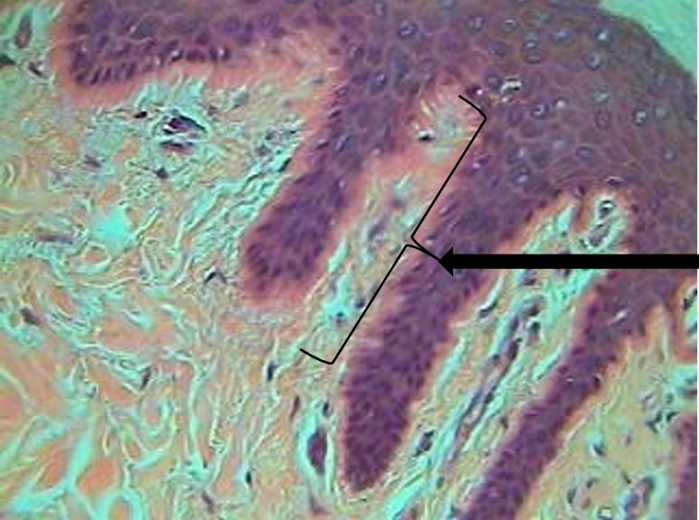
stratum corneum; The outermost layer of the epidermis made of dead, flattened, keratin-filled cells that protect the body from injury, water loss, and pathogens.
Identify the following layers of the epidermis

stratum granulosum; The layer where keratinocytes begin to die and accumulate keratin and waterproofing granules, forming a protective barrier.
Identify the following layers of the epidermis
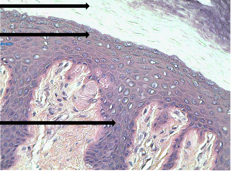
The layer of living keratinocytes connected by strong desmosomes, giving cells a "spiny" appearance and providing strength and flexibility to the skin.
Identify the following layers of the epidermis spinosum
