Overview of the Muscular System and Its Functions
1/164
There's no tags or description
Looks like no tags are added yet.
Name | Mastery | Learn | Test | Matching | Spaced | Call with Kai |
|---|
No analytics yet
Send a link to your students to track their progress
165 Terms
Muscle Tissue
One of four primary tissue types for contraction.
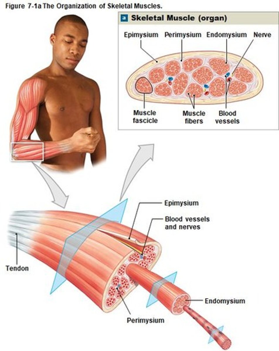
Skeletal Muscles
Composed of skeletal muscle tissue; attached to bones.
Muscular System
Includes about 700 skeletal muscles in the body.
Muscle Functions
Move skeleton, maintain posture, generate heat.
Tendons
Fibrous tissue attaching muscle to bone.
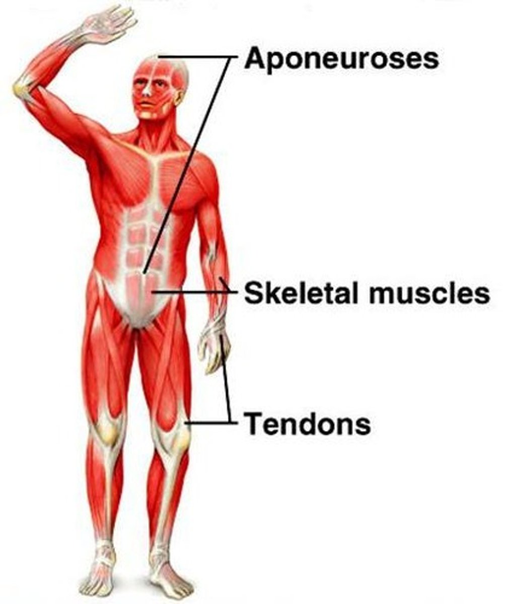
Aponeurosis
Broad sheet connecting muscles to each other.
Epimysium
Connective tissue covering the entire muscle.
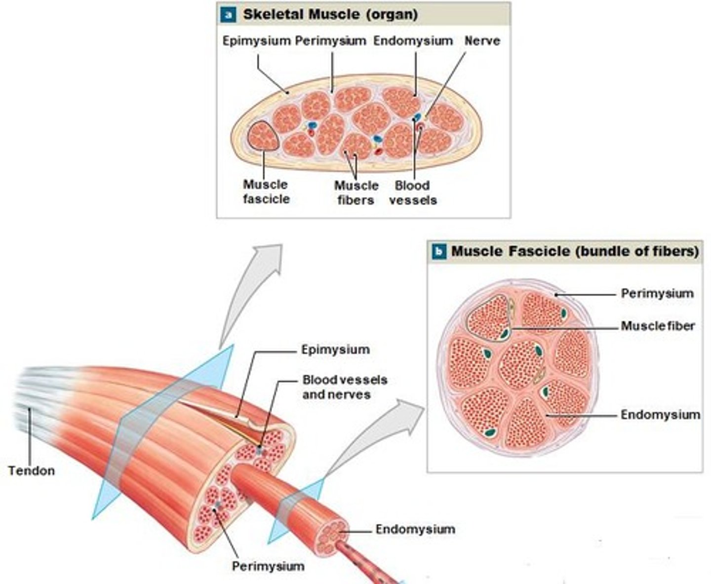
Perimysium
Divides muscle into fascicles; contains blood vessels.
Endomysium
Covers each muscle fiber; ties fibers together.
Muscle Fiber
Individual skeletal muscle cell; multinucleate structure.
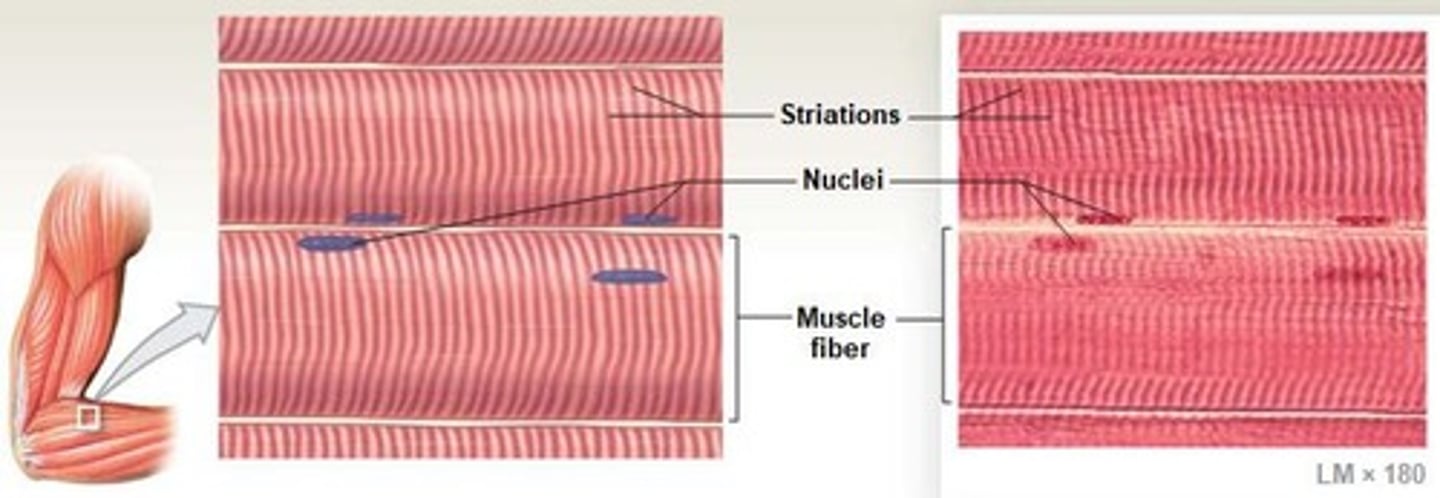
Skeletal Muscle Fibers
Long, striated fibers with repeating patterns.
Sarcolemma
Plasma membrane surrounding muscle cell's cytoplasm.
Transverse Tubules
Network allowing impulses to reach muscle fiber interior.
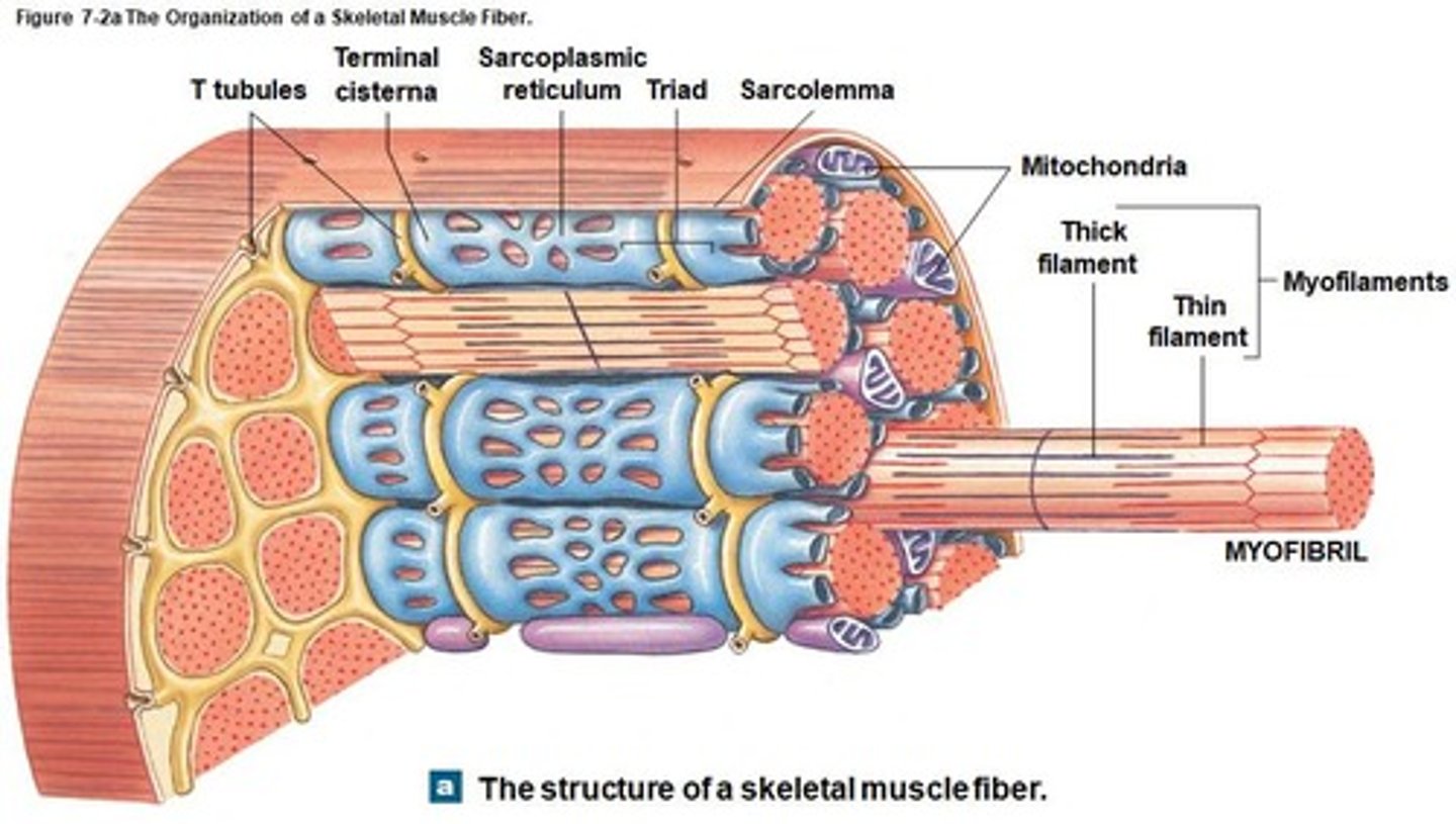
Myofibrils
Cylinder-shaped structures causing muscle fiber contraction.
Sarcoplasmic Reticulum
Smooth ER surrounding myofibrils; stores calcium ions.
Terminal Cisternae
Expanded SR portions adjacent to T tubules.
Triad
Two terminal cisternae and one T tubule combination.
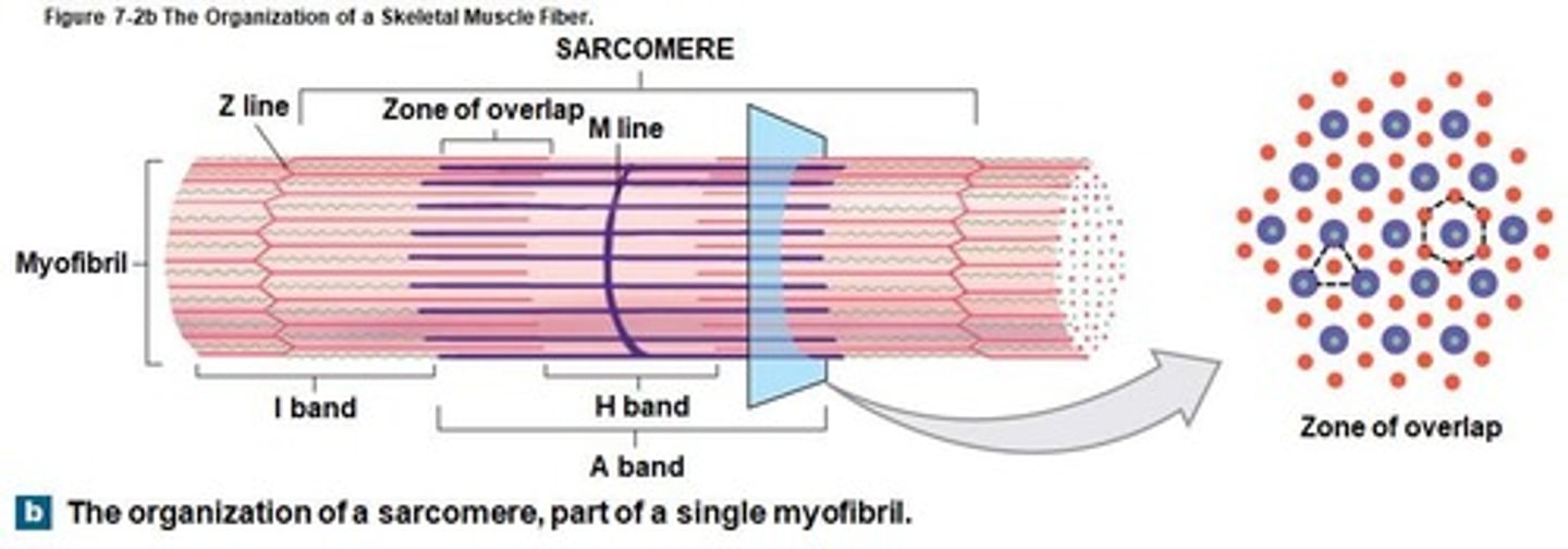
Sarcomeres
Smallest functional units of skeletal muscle fibers.
Myofilaments
Thick and thin filaments within myofibrils.
Striated Appearance
Pattern from arrangement of thick and thin filaments.
Voluntary Control
Skeletal muscles require CNS stimulation for contraction.
Blood Vessels
Extensive network supplying energy to skeletal muscles.
Muscle Contraction Heat
Generated by muscle contractions to maintain body temperature.
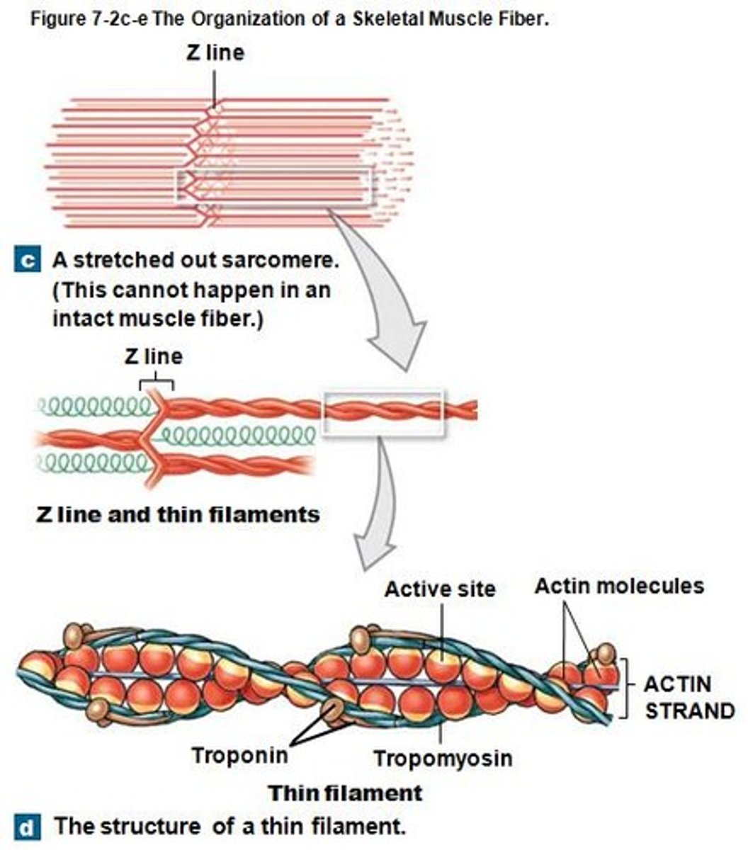
Z lines
Boundaries of each sarcomere in muscle fibers.
M line
Center of sarcomere connecting thick filaments.
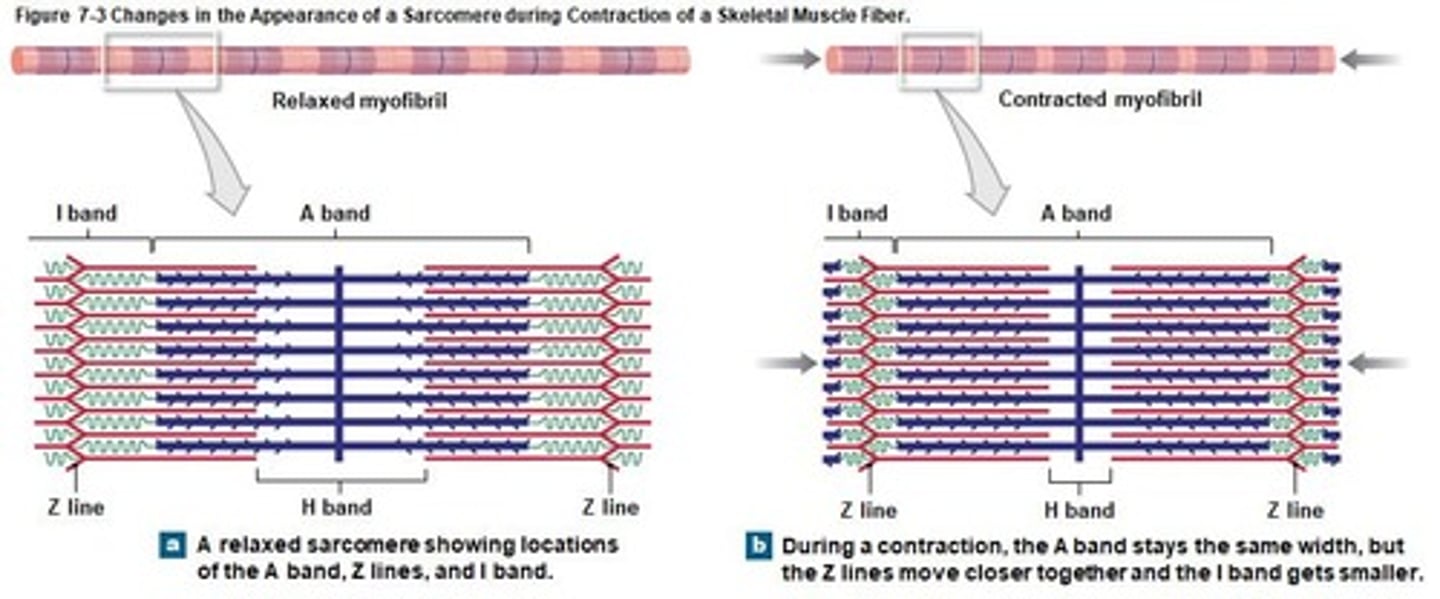
A band
Darker region with thick filaments and overlap.
I band
Lighter region containing only thin filaments.
H band
Region with only thick filaments when relaxed.
Thin filaments
Actin strands with active sites for myosin.
Thick filaments
Composed of myosin molecules with heads and tails.
Sliding Filament Theory
Explains sarcomere contraction by filament sliding.
Cross-bridges
Connections formed by myosin heads binding to actin.
Contraction Cycle
Process of muscle contraction involving cross-bridge cycling.
Neuromuscular Junction
Connection between motor neuron and muscle fiber.
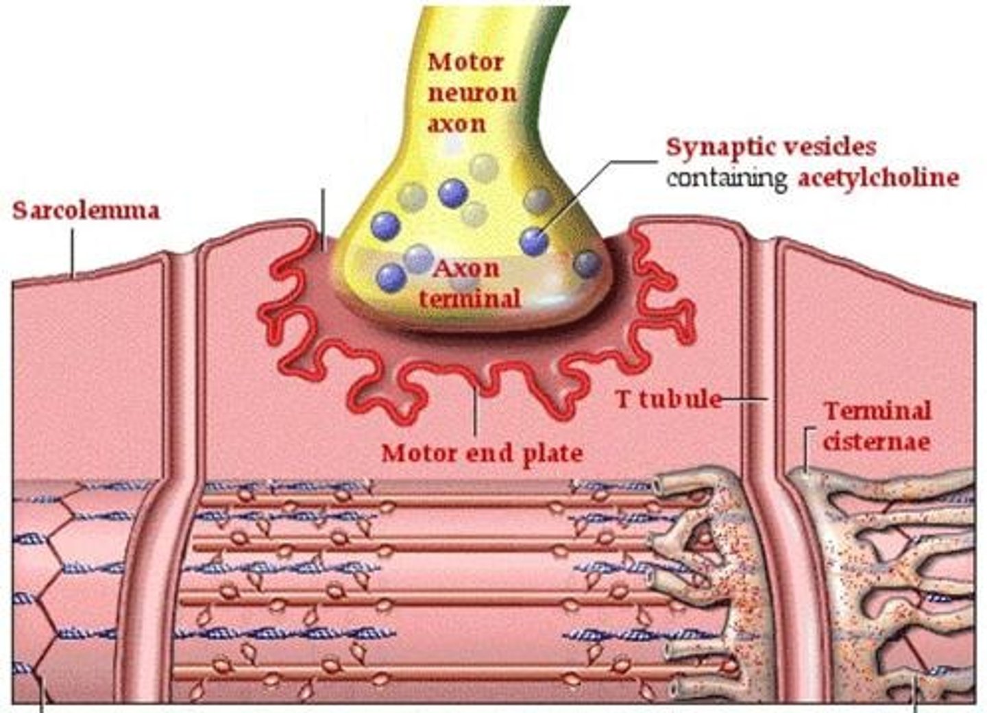
Acetylcholine (ACh)
Neurotransmitter released at the neuromuscular junction.
Synaptic cleft
Gap between axon terminal and muscle sarcolemma.
Tension
Force produced by muscle fiber contraction.
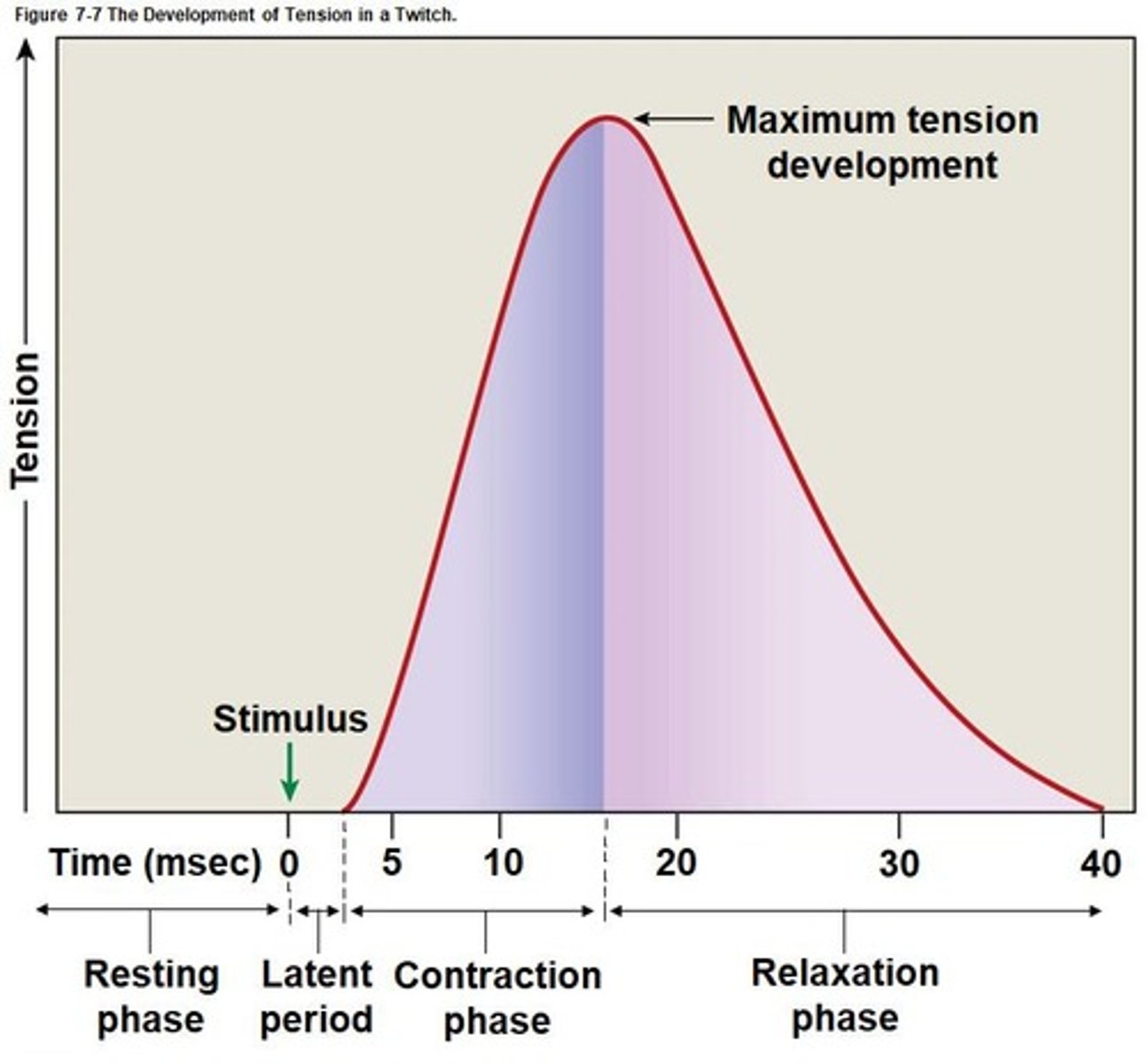
Muscle Twitch
Single stimulus-contraction-relaxation cycle in muscle.
Myogram
Graph showing tension development during a twitch.
Resting length
Initial length of muscle fiber before contraction.
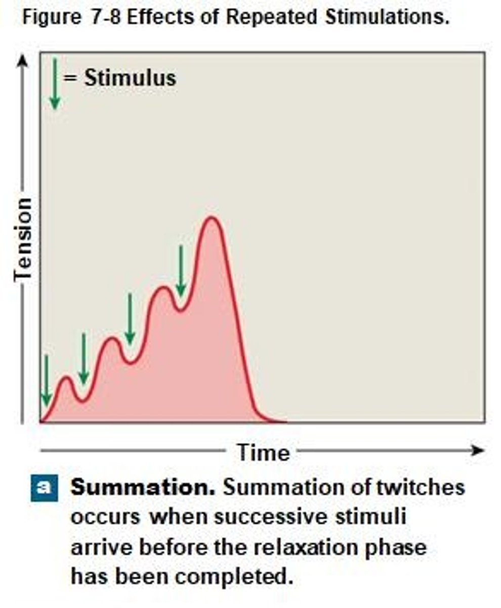
Frequency of stimulation
Rate affecting calcium ion concentration in fibers.
Sarcomere shortening
Causes muscle fiber contraction and tension production.
Active sites
Locations on actin for myosin head binding.
Tropomyosin
Covers active sites on actin at rest.
Troponin
Holds tropomyosin in position on actin.
Compression
Push applied to an object by muscle.
Latent period
Initial phase of muscle twitch lasting about 2 msec.
Contraction phase
Period of maximum tension within about 15 msec.
Relaxation phase
Tension decreases as calcium levels drop, lasts 25 msec.
Summation
Addition of twitches for stronger muscle contraction.
Incomplete tetanus
Rapid contractions produce near-peak tension.
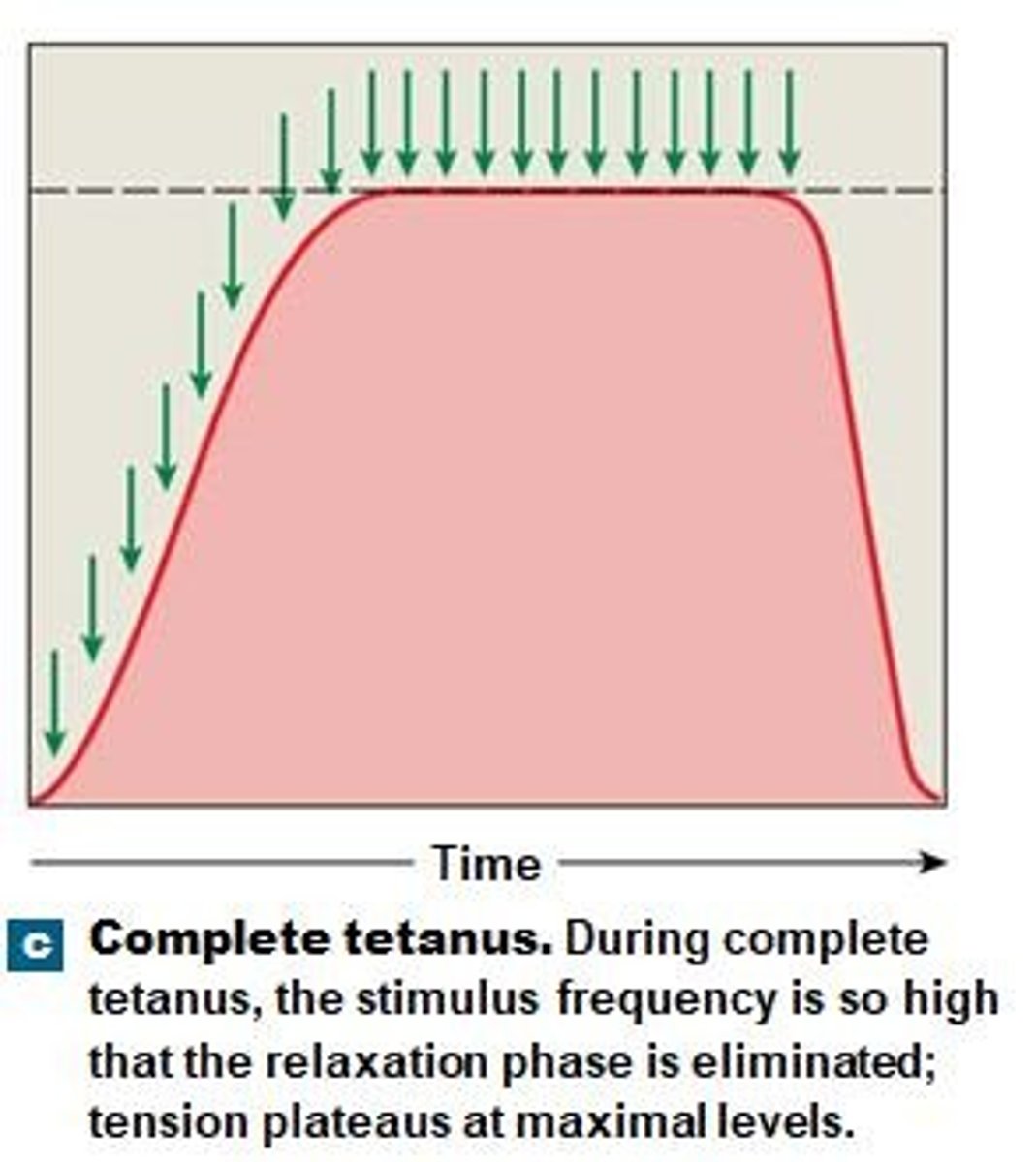
Complete tetanus
Continuous stimulation eliminates relaxation, maximum tension achieved.
Motor unit
Single motor neuron and all controlled muscle fibers.
Recruitment
Activation of more motor units for increased tension.
Muscle tone
Resting tension stabilizing bones and joints.
Atrophy
Muscle wasting due to lack of stimulation.
Isotonic contraction
Muscle length changes while tension remains constant.
Isometric contraction
Muscle length remains unchanged under tension.
Energy storage (CP)
Excess ATP converted to creatine phosphate for energy.
Creatine phosphokinase
Enzyme regulating ATP recharging from creatine phosphate.
Calcium ion concentration
High levels lead to complete tetanus.
Cross-bridge interaction
Binding of myosin and actin during contraction.
Passive muscle elongation
Muscle returns to length via passive forces.
Active mechanism
No active process exists for muscle elongation.
Skeletal muscle fibers
Controlled by motor neurons for movement.
Motor neuron control
Neurons can control hundreds to thousands of fibers.
Precise movements
Involve small motor units with few fibers.
Gross movements
Involve large motor units with many fibers.
Aerobic Metabolism
Requires oxygen, produces ATP in mitochondria.
ATP Production
95% of resting muscle ATP needs met.
End Products of Aerobic Metabolism
Produces ATP, water, and carbon dioxide.
Pyruvate in Citric Acid Cycle
About 15 ATP produced per pyruvate.
Glycolysis
Breaks glucose down to pyruvate in cytoplasm.
Anaerobic Process
Glycolysis can occur without oxygen.
ATP Yield from Glycolysis
Only yields 2 ATP per glucose.
Muscle Fatigue
Muscle fails to perform due to energy depletion.
Causes of Muscle Fatigue
Depletion of reserves or pH decline from H+.
Recovery Period
Restores muscle to pre-exertion conditions.
Oxygen Debt
Increased breathing to restore oxygen levels.
Lactate Conversion
Lactate is converted back to glucose.
Muscle Performance Measurement
Measured in force and endurance.
Fast-Twitch Fibers
Reach peak tension in 0.01 sec or less.
Characteristics of Fast Fibers
Large diameter, few mitochondria, fatigue rapidly.
Slow-Twitch Fibers
Contract slowly, fatigue resistant, high oxygen supply.
Capillary Network
Oxygen supply is greater due to this.
Myoglobin Function
Stores oxygen for muscle use.
Muscle Fiber Type Variation
Percentage varies among skeletal muscles.
White Muscles
Dominated by fast fibers, appear pale.
Red Muscles
Dominated by slow fibers, appear reddish.
Muscle Conditioning
Training increases power and endurance.
Anaerobic Endurance
Supported by glycolysis and existing ATP.
Aerobic Endurance
Supported by mitochondrial activity during activity.
Cardiac Muscle Tissue
Muscle found only in the heart.
Cardiac Muscle Cells
Small cells with one central nucleus.
Intercalated Discs
Connect cardiac muscle cells for communication.
Gap Junctions
Allow rapid communication between cardiac cells.
Automaticity
Ability to contract without neural stimulation.
Pacemaker Cells
Specialized cells that regulate heart contraction timing.
Calcium Ion Role
Increases permeability for contraction in cardiac cells.
Aerobic Metabolism
Cardiac muscle cells primarily rely on oxygen.