TCP Week 6 (Trauma)
 Call Kai
 Learn
Learn Practice Test
 Spaced Repetition
 Match
Match Flashcards
 Knowt Play
1/169

There's no tags or description
Looks like no tags are added yet.
Name | Mastery | Learn | Test | Matching | Spaced | Call with Kai | Chat |
|---|
No analytics yet
Send a link to your students to track their progress
170 Terms
primary survey for maxillofacial trauma (ABCDEs)
Airway: Assess movement of air/ obstruction
Disability: Examine pupillary response (are they equal? are they reactive?)
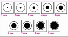
the most important consideration for maxillofacial trauma is
AIRWAY MANAGEMENT
- have anesthesia on hand for trauma
- if facial trauma, evaluate patient for head injury
Maxillofacial, neck, or laryngeal trauma:
- What can cause airway obstruction?
- Initially assess?
- Intubate when?
Maxillofacial, neck, or laryngeal trauma can cause airway obstruction: B/L mandible fractures can cause airway obstruction
Initially assess "the talking patient": Appropriate response means a patent airway, intact ventilation, and a perfused brain
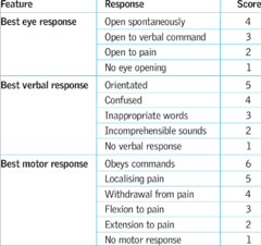
Patients with AMS or facial trauma require a definitive airway until fully assessed: GCS less than 8, intubate
picture of GCS score again
definitive airway steps & 3 types?
1. Tube placed in the trachea
2. Cuff inflated below the vocal cords
3. Connected to a form of oxygen-enriched ventilation
4. Secured with tape / tube holder
3 types:
- Nasotracheal
- Orotracheal (MC)
- Surgical airway
secondary survey (history of MOA)
More detailed exam after primary survey:
- Facial pain?
- Blurred vision?
- Dysphagia?
- Nasal congestion?
- Facial numbness?
- Nasal congestion?
- Facial Numbness?
- Malocclusion?
- Painful teeth?
- Vertigo?
secondary survey (PE)
- Facial edema
- Lacerations
- Rhinorrhea
- Periorbital edema
- Periorbital ecchymosis "Racoon eyes"
- Facial bone instability (palpate face)
- Nasal deviation
- Otorrhea
- Periaricular ecchymosis "Battle's Sign"
diagnostic studies for facial injuries
CT facial bones:
- injuries can be seen on CT head; however, CT facial bones provides more views and better diagnostics
- allows for pre-operative planning
Panorex:
- series of x-rays used to evaluate isolated mandible, dental, or alveolar ridge fractures
- more often used post-operatively for surgical planning (not done in trauma bay)
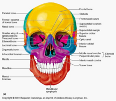
ocular traumas and what to look for when these patients first enter the trauma bay
Orbital bone fractures: Blow out fractures
EOM entrapment
Globe rupture
Eye penetration
Evaluate eyes early, as peri-orbital swelling occurs quickly and can obscure later exam
orbit is made of what bones?
frontal bone (roof/superior rim)
sphenoid bone & zygoma (lateral)
zygoma & maxilla (floor)
ethmoid & maxilla (medial)
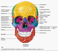
management of orbital bone fracture
CT localizes fractures
Consult placed to OMFS: Orbital floor "blow out" fractures usually need surgical repair
Avoid excessive eye pressure!
May apply ice packs to ease swelling
ocular muscle entrapment
- Occurs when eye muscles become trapped within a fracture (most commonly inferior rectus muscle)... requires urgent response
- Results in inability to actively move eye within all fields
warrants surgical exploration if there is no movement, let OMFS know
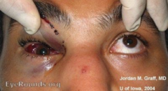
how does globe rupture present on PE
- decreased visual acuity
- pupillary defect
- teardrop pupil
- extrusion of vitreous fluid
- gross deformity of eye
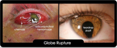
management of globe rupture
Requires STAT ophthalmology consult
Goal is to decrease intraocular pressure!
- elevate HOB, do not apply any pressure dressings
- avoid placing any medication or diagnostic eye drops into the eye
Place an eye shield over affected eye until evaluated by ophthalmology
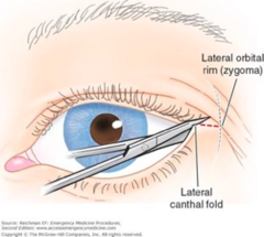
orbital hematoma
Causes loss of vision:
- Compartment syndrome of the eye
- May require lateral canthotomy to decompress orbit
eye penetration
- can cause?
- what to do with foreign bodies?
- imaging?
Can cause globe rupture
Any foreign bodies should be left in place, removal should be deferred to the ophthalmologist
Image with CT: Avoid MRI (may move metallic objects, takes more time than CT, more artifact movement)
Management of eye penetration
- Keep patient NPO
- Prophylactic antibiotics (Vanco & 3rd or 4th gen Cephalosporin) or Floroquinolone
- Tetanus prophylaxis
- Ophthalmology consult (may require transfer to tertiary center)
- Globe rupture and/or penetration will require surgical repair
nasal trauma includes
Nasal bone fractures
Cribriform fractures
Epistaxis
- Anterior
- Posterior
nasal bone fracture PE findings
may reveal nasal crepitus, deformity, edema
nasal bone fracture diagnostics
CT
management of nasal bone fracture
Ice & head elevation
Displaced fractures: ENT consult (may require closed reduction)
Nasal splint (may be internal or external to provide support)
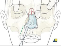
management of nasal bone fracture: septal hematoma
Septal hematoma may require drainage to prevent necrosis.
Drainage then requires nasal packing for 2-3 days (while edema goes down)
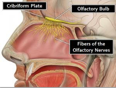
cribiform (ethmoid) fractures
- findings on PE
- diagnosed with?
- Exam may reveal CSF rhinorrhea
- Diagnosed via CT scan
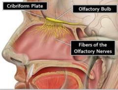
cribiform (ethmoid) fractures management
- these patients CANNOT have NG tubes placed (risk of tube entering brain)
- keep head of bead elevated 40-60 degrees to decrease ICP
- may result in loss of smell
**DO NOT discharge... concern for meningitis bc of disruption in bone at base of skull.
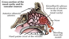
management of anterior epistaxis
pinch nose, wait 5-10 minutes minimum:
- if bleeding persists, consider nasal packing
- may also use cautery
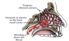
management of posterior epistaxis
consider consult to ENT or OMFS for placement of balloon catheter
Facial bone fractures
Majority of facial fractures do NOT require surgery
•Mandibular fractures
•LeForte fractures
•Sinus fractures
•Facial nerve injury
•Facial lacerations
which facial fractures require hospitalization and how are you managing them?
Nasoethmoid fracture: Monitor for CSF leak and risk of meningitis
Zygomatic arch fracture: Monitor for airway complications
LeFort fractures: Often require surgical repair
Facial fractures along with additional traumatic injuries
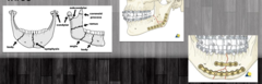
mandible fracture PE findings and imaging
- Malocclusion of jaw indicates mandible fx (can't close mouth)
- CT to evaluate extent of fracture
mandible fracture management
- Requires OMFS consultation
- Prophylactic antibiotics suggested (bc oral bacteria)
- Surgery requires ORIF mandible with plates and/or wires (2 screws above, 2 screws below, and not through)
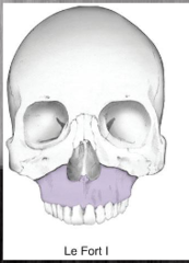
LeForte 1 fracture (definition and symptoms)
traverse fracture through maxilla above roots of teeth
symptoms: malocclusion
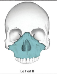
LeForte 2 fracture definition
fracture extends through maxilla, nasal
bridge, lacrimal bones, orbital floor and rim
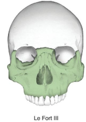
LeForte 3 fracture definition and symptoms
fracture than involves complete discontinuation of skull from the face
extends through nose and along medial wall of orbit, through floor of orbital and through lateral orbital wall and zygomatic arch, and through sphenoid
may involve CSF leak!!
management of sinus fracture
facial fx may extend into sinuses
require prophylactic antibiotic
sinus precautions (to minimize pressure w/in sinuses):
- Nothing inserted into nose
- Do not blow nose
- No drinking through straw
- Sneeze with mouth open
- No smoking
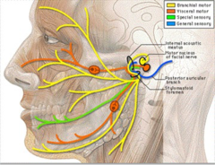
CN VII
motor branch of facial nerve
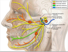
CN V
trigeminal nerve: sensation of facial nerve
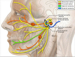
acute facial paralysis
- suggests?
- requires?
suggests facial nerve transection
requires immediate consultation with maxillofacial surgery
management: small simple facial lacerations
lacerations should be closed
management of large complex facial lacerations or avulsions
should be closed by plastic surgery, if available
what should you use for local anesthesia for facial laceration repair
Lidocaine 1%
DO NOT use epinephrine on facial structures (restricts the small capillaries)
eyelid lacerations (extending through eyebrow) require
closure by plastic surgery or ophthalmology
within 48 hours after injury!
steps to closing a laceration
1. Select your desired sutures, set up sterile field; you'll need:
- suture (0 is largest (dental floss), 60 is fine (like hair))
- needle driver
- pick-up forceps
- scissors
- needles and syringes to inject anesthetic
2. Irrigate the wound with saline/diluted hydrogen peroxide or betadine
3. Prep the area with antiseptic solution - open wounds should be cleaned and prepped with betadine solution (no alcohol solutions on the face or mucosal membranes)
4. Drape the area after prep dries
5. Inject local anesthesia into the lacerated edges to perform a field block
6. Debride any nonviable tissue
7. Start your closure - subcutaneous layer, if necessary or superficial closure
8. Apply antibiotic cream (Bacitracin) and apply desired dressings
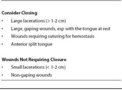
lip and tongue lacerations: management
simple small lacerations: can be primarily closed
complex lacerations traversing tongue: closed by OMFS
prophylactic antibiotics should be given due to introduction of oral flora
jaw dislocation may result from
opening the mouth too widely
once the condyle of the jaw is dislocated ______________ prevent self-reduction
muscles of mastication
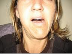
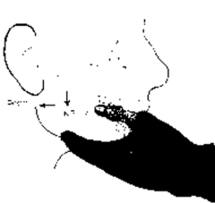
management of jaw dislocation
- Patient will require sedation
- Reduction achieved by placing thumbs overtop occlusal surface of teeth and pulling downward on the mandible then guiding posteriorly and superiorly back into temporal fossae
DO NOT attempt to reduce fractured mandibles
avulsed teeth and fractures are among ____________ in trauma patients
lowest priority
partially avulsed teeth should be
removed to prevent aspiration:
- only remove those easily done with gloved fingers
- DO NOT attempt to use any tools for removal
ellis 1 tooth fracture
involve enamel of tooth (least severe)
- painless
- evaluated outpatient
ellis 2 tooth fracture
yellow dentin exposed
- Pain
- Cover with calcium hydroxide & foil and refer outpatient
- Avoid solids
ellis 3 tooth fracture
Exposes dental pulp (most severe)
- Extremely painful
- Requires evaluation by dentist or orthodontist within 48 hr
alveolar ridge fractures results in
results in a group of teeth being dislodged
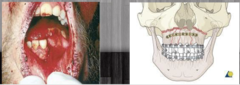
instability of the upper alveolar ridge can indicate
mid-face fracture
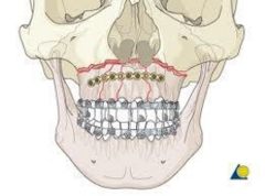
instability of the lower alveolar ridge can indicate
mandible
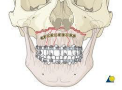
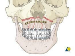
management of alveolar ridge fractures
- these fractures require stabilization with wires, arch bras or splinting by oral surgery
- DO NOT allow patient to eat or drink until fixation
- open fractures warrant prophylactic abx
laryngeal trauma PE
clinical triad:
- Hoarseness
- Subcutaneous emphysema
- Palpable fracture
management of laryngeal trauma
- Requires emergency intubation
- Can be diagnosed by CT if not obvious on exam
tympanic membrane rupture PE and treatment
- Use otoscope to examine TMs during secondary survey
Physical exam:
- TM perforated
- Hemotympanum
Treatment:
- Leave clots in place
- DO NOT irrigate
- ENT consultation
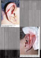
ear lacerations management
- Provide copious irrigation
- Debridement as necessary
- Closure to cover exposed cartilage and preserve cosmetic appearance (often plastic surg)
Avulsion, lacerations extending into ear canal, lacerations with middle or inner ear injury, or evidence of basilar skull fractures should be seen by plastic surgery and/or neurosurgery
primary survey of chest injury
Airway: ensure no obstructions
Breathing: assessing for bilateral breath sounds, equal chest expansion
Circulation: presence of pulses (central and peripheral)
Initiate ECG monitoring to assess for dysrhythmias
pneumothorax definition
Accumulation of air between the visceral and parietal pleura of the lungs
Can occur from blunt or penetrating trauma
pneumothorax results in
ventilation/perfusion defect:
- blood perfuses non-ventilated areas, resulting in un-oxygenated blood returning to the heart
- hypoxia results from poor perfusion
tension pneumothorax occurs when
a laceration in the lung creates a "one-way valve" for air to leak into the chest wall and accumulate under pressure
places pressure on to mediastinum
important to recognize early!
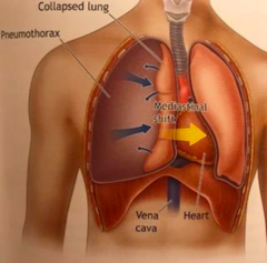
clinical diagnosis of tension pneumothorax
high index of suspicion based on symptoms to proceed with life-saving treatment
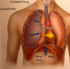
s/sx of tension pneumothorax
Symptoms include:
- Chest pain
- "Air hunger"
Signs include:
- Severe respiratory distress
- Tachycardia
- Hypotension (due to pressure placed on
mediastinum)
- Tracheal deviation AWAY from effected side
- Unilateral breath sounds
- Hyperresonance to percussion
- Neck vein distention (due to pressure
placed on mediastinum)
- Cyanosis
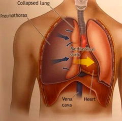
management of tension pneumothorax
DO NOT delay treatment for CXR!!!
Treatment: Needle Decompression
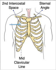
- Rapidly insert large bore (14g) needle into the 2nd ICS in the mid-clavicular line of the effected side (just inferior to clavicle)
- Keep catheter in place until a surgical chest tube has been inserted
- Once decompressed, then obtain CXR
cardiac tamponade definition
When a penetration or lac of the cardiac muscle allows blood to leak from the heart into the pericardial sac, causing pressure to build around the heart
Built up pressure prevents the heart from adequately filling & emptying
Decreased CO
Results in hypoxia
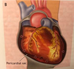
s/sx of cardiac tamponade
Symptoms:
- Chest pain
- SOB
Signs:
Beck's Triad:
- venous pressure elevation (blood can't get in)
- declined arterial pressure (blood can't get out)
- muffled heart tones
Kussmaul's sign: inc venous pressure with inspiration
dx and tx for cardiac tamponade
Dx:
- EKG
- FAST U/S
Tx:
• Pericardiocentesis: smaller effusion or no surgeon available
• Emergency thoracotomy: Large effusion with loss of vital signs
• Surgical repair of cardiac laceration: definitive treatment
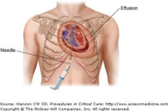
pericardiocentesis
- Needle drainage of the pericardial sac
- Use 16-18g long needle to drain sac
- Place drainage tube until patient can be transferred to definitive care
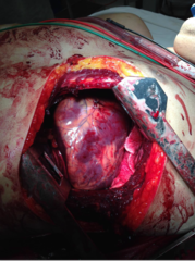
indications of emergent thoracotomy
May be performed only under certain situations
- Relief of large pericardial effusion
- Direct control of exsanguinating intra-thoracic hemorrhage
- Open cardiac massage (manual CPR)
- Allows cross-clamping of aorta to increase perfusion to heart and brain
ED thoracotomy followed by OR exploration of wounds
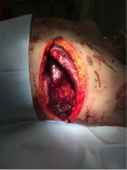
contraindications of emergent thoracotomy
- Absence of qualified surgeon
- Blunt chest trauma with pulseless electrical activity (low chance of survival)
- Penetrating wounds with no electrical activity on EKG
- No signs of life (reactive pupils, spontaneous movement, organized EKG)
secondary survey after cardiac tamponade
- CXR
- ABG
- EKG
- Bloodwork, including Troponin
- CT scan of the chest once patient has been determined to be hemodynamically stable
simple pneumothorax
- accumulation of air within pleura, described as a % of lung collapsed
- can convert to tension PTX under positive pressure ventilation
you should perform a CXR after surgery
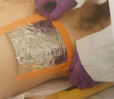
open pneumothorax
aka "sucking chest wound" (can see lac of chest and airflow)
requires placement of occlusive dressing, overtop of wound, secured on 3 sides
hemothorax
- Accumulation of blood within the pleural space
- Most commonly occurs due to lung laceration or laceration of intercostal vessel
- Can be associated with thoracic spine fractures
diagnostic studies for pneumothorax/hemothorax
CXR: A/P view
CT chest w/ IV contrast
s/sx of pneumothorax and hemothorax
Symptoms:
- SOB
- Chest pain
Signs:
Pneumothorax: decreased breath sounds, hyperresonance to percussion
Hemothorax: decreased breath sounds, dullness to percussion
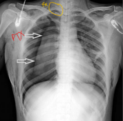
CXR pic of pneumothorax
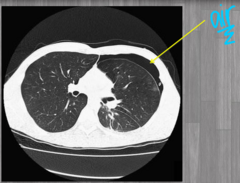
CT scan of pneumothorax
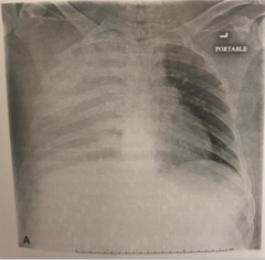
CXR of hemothorax picture
small hemothorax treatment
observation and serial CXR
large hemothorax treatment
Chest tube insertion to accurately monitor blood loss and need for OR exploration
OR criteria for hemothorax?
- 1500mL initial output when CT was placed
- Loss of 200mL of blood per hour x 2- 4 hours
- Requires multiple blood transfusions
treatment for pneumothorax (
often only detected on CT scan:
can be observed with serial CXR
treatment for pneumothorax (10-30% collapse)
call radiology to do IR drain
treatment for pneumothorax (>30% collapse)
requires insertion of surgical chest tube
treatment for a contained pneumothorax
consider interventional radiology to insert ultrasound guided chest tube (pigtail)
chest tubes/thoracostomy tubes
Placed into the 4th or 5th intercostal space, within the anterior mid-axillary line at the bedside with use of analgesics (IV pain medication and local)
Negative pressure contained system allows for lung re-inflation
Initially placed to wall suction then transitioned to "waterseal" prior to removal
If lung fails to reinflate with chest tube, may need OR and pleurodesis (glue lung tissue to chest cavity)down the road
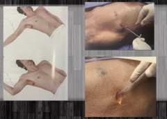
s/sx of lung contusion
Symptoms:
- chest pain
- SOB
Signs:
- Chest wall contusion
- Commonly occurs with rib fx
- SpO2 less than 90% on room air
lung contusion diagnosis and treatment
Diagnosis:
- serial CXR
- Chest CT (will show difference between contusion and hemothorax)
Treatment:
- Serial CXR
- SpO2 monitoring
- Patients with low SpO2 and chronic lung conditions may require intubation and ventilator support
blunt cardiac injury/cardiac contusion diagnosis and treatment
can range from clinically silent injury, transient arrhythmias, to cardiac wall rupture
Dx:
- EKG
- FAST exam (US)
- Troponin
- Echocardiogram
Tx:
- Management of arrhythmias
- Evaluation by cardiac surgeon
- Management of MI
s/sx of sternal and rib fractures
Symptoms:
- severe chest pain
- back pain
- SOB (worse with deep breaths from rubbing)
Signs:
- Crepitus on chest wall palpation
- Chest wall contusion
- Low SpO2
flail chest (definition and what it causes)
fracture of 2 or more adjacent ribs in 2 or more places
causes:
- paradoxical movement
- inadequate ventilation
- hypoxia
sternal and rib fractures diagnosis and treatment
Dx: CXR and CT chest
Tx:
- Analgesia/Pain Meds! so they breathe and don't dvlp PNA!
- Repeat CXR
- Incentive Spirometry
- Intubation & ventilatory support for patients with multiple rib fx/lung disease
OR:
- Open reduction of rib fractures (rib plating)
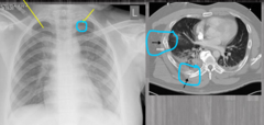
signs of tracheal/bronchiole injuries
- Hemoptysis
- Chest wall subcutaneous emphysema (disruption to trachea)
- Tension PTX (chest tube and lung still doesn't re-inflate)
- Failure of lung to re-expand after chest tube placement
HIGHLY FATAL
tracheal/bronchiole injuries diagnosis and treatment
Dx: Bronchoscopy
Tx:
- Intubate (opposite main stem... only oxygenate one side)
- Operative repair trachea
aortic injury notes
- Highly fatal injury, cause of many DOA on scene
- Those who survive typically have an incomplete tear with a contained hematoma
- No specific s/sx
- High index of suspicion for those with decelerating forces (high falls)
aortic injury diganosis and treatment
Dx: Helical CT chest with contrast
Tx: primary repair of torn segment or endovascular repair by vascular surgeon
major vessel injuries management
- may require emergent thoracotomy
- cross clamping of vessel
- emergent OR exploration with repair of lacerated vessel