Audition - Cochlear Anatomy
 Call Kai
 Learn
Learn Practice Test
 Spaced Repetition
 Match
Match Flashcards
 Knowt Play
1/103

There's no tags or description
Looks like no tags are added yet.
Name | Mastery | Learn | Test | Matching | Spaced | Call with Kai | Chat |
|---|
No analytics yet
Send a link to your students to track their progress
104 Terms
What are the two main "shells" of the inner ear?
Bony labyrinth and membranous labyrinth
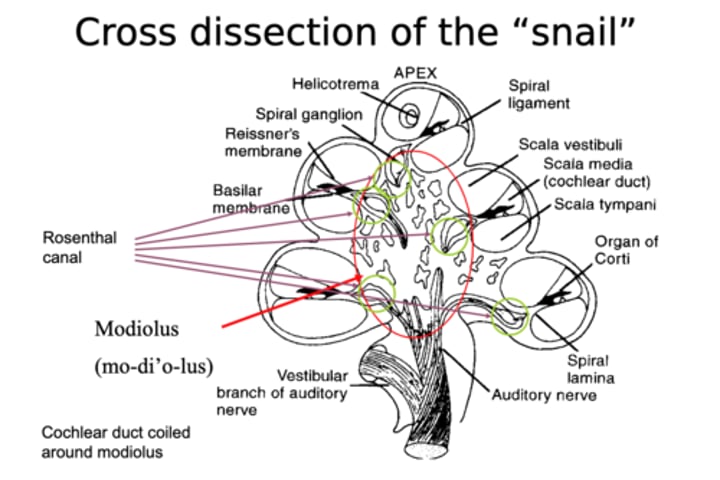
Cross dissection of cochlea
What are the two main parts of the inner ear?
The cochlea and the vestibular apparatus (for balance)
What does the cochlea do?
processes acoustic information, very sensitive to sound
How does sound get into the inner ear?
through the oval window
What parts make the vestibular apparatus?
semicircular canals and the vestibule
Where is the cochlea located?
petrous of the temporal bone
What is frequency selectivity?
at each location along BM, neurons are sensitive to certain sound frequencies
Which structure that is related to sound conduction is also closely located to the vestibular apparatus in the cochlea?
the oval window
What does receptor specificity mean?
It refers to the affinity a certain neurotransmitter has for a particular receptor. Remember from Dr. Wang's lecture that the receptor cells and neurons aren't much different between the cochlea and the vestibule. It is the accessory structures that make the cochlea much more sensitive to sound.
What determines receptor specificity?
Receptor specificity is determined by accessory structure, not the nature of the receptor cells.
What is coiled around the modiolous?
the Rosenthal canal
On the "snail"/cochlea, what frequencies are at the apex and what are at the base?
Low frequencies are at the apex, high frequencies at the base.
Where do high frequency spiral ganglion neurons send their axons? Where do low frequency fibers go?
The high frequency SGNs send their axon more peripherally in the trunk of the auditory nerve.
Low frequencies fibres are closer to the center of the trunk.
What can scales be analogous to?
chambers/compartments
How many scales does the cochlea have?
3
What are the scalas?
Scala tympani, scala media, scala vestibuli
What are the scalas separated by?
The basilar membrane and Reissner's membrane
What is the pathway between SV and ST?
helicotrema
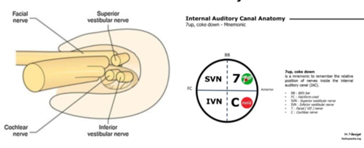
What is the 7UP and Coke mnemonic?
- this mnemonic tells us the relative position of nerves inside the internal auditory canal
- the internal auditory canal is essentially like a hole in the internal auditory meatus that carries all the important neural fibres in and out of the brain
- the top left is the superior vestibular nerve and bottom left is inferior vestibular nerve
- top right is CN7 (so 7UP)
- bottom right is cochlear nerve (so COKE down)
What is the helicotrema? (Basically, how can the location be described)
A hole at the apex of the cochlea, and it connects the scala vestibuli and the scala tympani
How could you describe the spiral ganglion neurons?
The spiral ganglion neurons innervate the hair cells of the Organ of Corti (they project to HCs laterally). The high frequency SGNs send their axon more peripherally in the trunk of the auditory nerve. Closer to the center of the truck are more low frequency fibers.
Where are the spiral ganglia neurons housed?
Inside Rosenthal's canal
What are the two ways the 8th nerve is organized?
1) frequency configuration
2) auditory vs. vestibular
Where can we see the relationship across the facial, vestibular and cochlear nerve?
inside the internal auditory meatus
What else goes to the inner ear via the internal auditory meatus?
the blood vessels
What is the habenula perforata?
small holes through which the auditory nerve is projected out from the SGNs
What can affect the spiral ganglia neuron count in the Rosenthal Canal?
ototoxic drugs
What connects the perilymph space and the CSF?
cochlear aqueduct
What is the endolymphatic sac? What does it do?
- small space
- balances solution
- important for metabolism
- responsible for reabsorption of endolymph
What does the size of the BM impact?
frequency
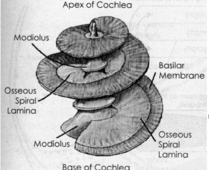
The BM becomes ____ from basal to apical turn
wider
The vibration of the BM stimulates _____
sensory cells
What does the tectorial membrane do?
structure that bends the stereocilia (located above the HCs)
The spiral plate becomes ____ from basal to apical turn
narrower
Why does the BM vibrate more?
because its soft
What are the five main parts of the membrane labyrinth (once the bony shell is removed)?
Scala vestibuli, scala tympani, scala media, basilar membrane and Reissner's membrane
What is on the lateral wall of the SM?
stria vascular (soft tissue) and spiral ligament
What is the pathway of sound?
vestibule, cochlea (scala vestibuli and then scala tympani), partially released from round window
Why do we hear sound in the cochlea if it goes to the vestibule first?
accessory structures
the cochlea is built for hearing
Which parts have perilymph and which have endolymph?
SV and ST - perilymph
SM - endolymph
What is perilymph similar to?
What is endolymph similar to?
extracellular fluid
intracelullar fluid
Perilymph is similar to extracellular fluid and _____
shares a similar chemical composition to CSF (the perilymph space and CSF are connected through the cochlear aqueduct)
What does the lateral wall of the scala media contain?
stria vascularis and the spiral ligament
Geometrically, where does sound go before hitting the cochlea?
The sound gets into the vestibule before the cochlea
Why do we hear sound through the cochlea?
In general, we hear sound through the cochlea because the cochlea possesses all the necessary structures for hearing. The vestibule is not "built" for hearing. It's all about the accessory structures!
What is the endolymphatic sac responsible for?
reabsorption of endolymph
What is an associated condition of the endolymphatic sac?
Meniere's disease (also called endolymphatic hydrops)
What is the perilymph space connected with? How (ie via which part)?
The perilymph space is connected with CSF (the fluid in the brain) via the cochlear aqueduct.
On the coiled turns of the cochlea, describe how the relationship of the osseous spiral lamina and basilar membrane changes from the base to the apex?
The basilar membrane is narrower at the base, with its width increasing toward the apex. The osseous spiral lamina is wider at the base, and narrower at the apex.
Describe the impedance components and its relation to the frequency of sound in relation to the width of the basilar membrane. Note that the organ of Corti is small.
There are different resonant frequencies throughout the cochlea due to gradients of the BM. The mass and stiffness components change with the width of the BM.
The shifting of the BM width is important for frequency analysis. The narrower the BM, the better response to higher frequencies (i.e., at the base). The apex is responsible for low frequency sound (wider).
Which volume is larger, the scala vestibuli or the scala tympani? What is one reason for this?
The volume of the SV is slightly larger than that of the ST, partially because it is connected to the vestibule. (represented on slide 25)
What is another way to describe the 'up and down' movement of the basilar membrane? What does this do?
It's the effective vibration of the BM. It helps balance pressure.
What happens to acoustic force as it goes into the cochlea at the oval window?
It causes the BM to vibrate, and it becomes partially released through the round window.
What causes the pressures imbalance?
When the sound (acoustic energy) enters the oval window, it goes through the oval window to the SV to the ST via the helicotrema and eventually to the round window. It doesn't fully get released at the round window due to the different dimensions between the SV and the ST as well as the different flexibilities between the OW and RW (the RW is more flexible. Since it doesn't fully get released, there is a pressure imbalance. (The BM vibrates to balance it.)
How does the vibration of the basilar membrane compare to that of the bone vibration and osseous spiral lamina in sound conduction?
The BM vibrates more than the bone vibration as well as the osseous spiral lamina.
Where is the Organ of Corti located?
on the BM
Describe the resonant frequencies of the cochlea? What is the reason?
The resonant frequencies of the cochlea change along the BM. At the base, there is a greater response to higher frequencies because the BM is narrower here. At the apex, there is a greater response to lower frequencies because the BM is wider here.
What are the two main (big) categories of cochlear cells?
Hair cells and supporting cells.
What is the perilymph space connected with? How (ie via which part)
CSF, through cochlear aqueduct
What are four examples of the supporting cells?
Pillar cells, Deiter cells, sulcus cells and Henson's cells
What are the two other cells?
Claudius C, fibrocytos
What cells are also called the rod of Corti?
Pillar cells
What did researchers used to think occurred at the rod of Corti?
it used to be thought of as the source of cochlear resonance
What is the connection between the tectorial membrane and inner hair cell stereocilia?
The stereocilia on the OHCs are "connected" to the tectorial membrane (the stereocilia are physically embedded in the tectorial membrane, at least for OHCs). For the IHCs, the stereocilia are bent by Henson's strip and hydraulic coupling.
What is the supporting cell at the top and bottom of the outer hair cell?
Deiter cells.
Where do hair cells contact the fluid? Which fluid?
Only the top surface of the HCs contact the endolymph. The bodies of the HCs are in fluid very similar to perilymph (Corti fluid).
Where are tight junctions located in the organ of corti?
Around the tops of the HCs and supporting cells.
What is the primary role of tight junctions?
to separate endolymph and perilymph
Where are tight junctions formed?
at the level of the reticular lamina
What is the basilar membrane permeable to?
perilymph
What structure is located above the hair cells?
the tectorial membrane
What kind of structure is the tectorial membrane (composition)?
gel protein and fibrous structure
How does the tectorial membrane relate to the outer hair cell and inner hair cell?
The stereocilia of the OHCs are physically embedded in the tectorial membrane, but the stereocilia of the IHCs are not.
What direction do the connections of the tectorial membrane make the stereocilia move?
In a radial direction (from the modiolus to the lateral wall).
When do the stereocilia move in a radial direction?
when the BM moves up and down
What are the two ways the IHC stereocilia bend?
The potential mechanisms are Henson's stripe and hydraulic coupling (vibrations of water—this one is more important).
Which hair cells are supported by the Deiter cells?
OHCs
Describe what the "fingers" of the Deiter cells do?
the fingers go from the bottom of one OHC to the top of another OHC
What is the freedom of movement like for the outer hair cells?
The larger portion of the OHC bodies are free of contact with supporting cells. I.e., the bodies are free to move.
What is the take home message about Deiter cells in relation to their connection with OHCs?
Deiter's cells don't attach to the OHC bodies. It is the fingers of the DCs that go from the bottom of one OHC to the top of another.
What is meant by the mosaic shape as it relates to the hair cells?
The mosaic configuration refers to the fact that each HC in isolated from other HCs. Each is surrounded by supporting cells. It is due to developmental genetic reasons: The SCs and HCs from the same stem cells. In basic terms, the idea is that the HCs let the cells around them know that they can't also be hair cells
What happens if hair cells die?
Hearing loss - if HCs die, they cannot be regenerated
How do we number the outer hair cells from the inside out?
1, 2, 3 from the inside out.
What supporting cells participate in making this mosaic shape?
The Deiter cells and the outer pillar cells.
Where is stereocilia?
On the top surface of the Organ of Corti (i.e. on the reticular lamina)
What is the difference in configuration of stereocilia for the OHC and IHC?
OHC—W-shape. IHC—straight line.
How does the size of the stereocilia change?
the most lateral row is the tallest
How do the OHC and IHC differ in their contact with the sulcus cells?
IHCs—fully contacted with SCs.
OHCs—largely free of contact with SCs.
Why is the difference of contact between OHC and IHC to sulcus cells important?
The OHCs are responsible for the amplification of sound, which they (partially) do by changing shape. Therefore, they need to move freely. (But remember they are still restricted at the top and bottom. Only the length changes (shape change))
What is the shape of the OHC? IHC?
OHC - cylinder
IHC - vase
Is the OHC lateral wall thick or thin? What is the function of this?
OHC lateral wall is thick, which is what allows them to stand up without support of supporting cells.
What is at the top and bottom of OHCs?
Contact with supporting cells at the top and bottom. Tight junction at the top and the nuclei at the bottom.
What type of neurons innervate the OHC? And which nerve?
Afferent: Type II SGNs. Efferent: Medial efferent. Nerve: Auditory nerve.
What is the difference between the cuticular plate and the reticular lamina?
Cuticular plate is on top of each HC. Reticular lamina is the plate structure formed by the tops of HCs and supporting cells.
Is the IHC lateral wall thick or thin?
thin
Where are the IHC nuclei?
in the middle of the cell
Which neurons innervate the IHC?
Afferent: Type I SGNs. Efferent: Lateral efferent
Which innervation as it relates to the hair cell (IHC or OHC) is the main ascending info pathway? What about the other one?
IHCs- Type 1 SGN is the main ascending information pathway from the cochlea to the brain. The OHCs only synapse with type II SGNs, which we know very little about their functions. The major role of the OHCs is to not provide neuronal information pathway, but to provide mechanical amplification.
1. What is the difference (in general) between an efferent and afferent pathway?
Afferent pathway: carries information to the brain. Efferent: carries information away from the brain.