Old Material - US Safety, CEU, Panoramic, Elastography, TDI, Speckle Tracking, & Strain, Harmonics, Signal Processing, Doppler Signal Processing
 Call Kai
 Learn
Learn Practice Test
 Spaced Repetition
 Match
Match Flashcards
 Knowt Play
1/149

There's no tags or description
Looks like no tags are added yet.
Name | Mastery | Learn | Test | Matching | Spaced | Call with Kai | Chat |
|---|
No analytics yet
Send a link to your students to track their progress
150 Terms
Hand Hygiene
Most effective method for preventing the spread of microorganisms
Cleaning
Removal of visible soil
Soap & water
Essential before HLD and sterilization
Low-Level Disinfection (LLD)
Destruction of most bacteria and some viruses and fungi
Does not inactivate tuberculosis or bacterial spores
High-Level Disinfection (HLD)
Destruction of all micro-organisms, except bacterial spores
Sterilization
Destroys all forms of microbial life
Uses chemical sterilants or germicides, steam under pressure, dry heat, ethylene oxide (EtO) gas, hydrogen peroxide gas plasma, and liquid chemicals
Ergonomics
The science of fitting your work environment to you rather than fitting you to your working environment
Probably applicable to hobbies and leisure activities also
Manufacturer Responsibilities
Ultrasound system - machine, monitor, transducers, exam table, chairs
Employer Responsibilities
Risk management
Protocols and scheduling
Examination area
Computer workstation
Sonographer Responsibilities
Neutral posture
Safe patient handling
Practicing ALARA
Increase overall gain
Decrease power
Avoid high TI and MI values
Minimize scanning time
Avoid using Doppler on early embryos
Power
Rate at which energy is transmitted
Intensity
Power/Energy ÷ Area
Bioeffects
Adverse effects that ultrasound causes on biological tissues
Thermal Index (TI)
Power that raises temperature in tissues
TIS - soft tissue
TIB - bone
TIC - cranial bone
Thermal Index (TI) Intensities
B-mode < color Doppler < spectral Doppler
Mechanical Index (MI)
Pressure amplitude estimation related to cavitation - potentially forms/bursts gas bubbles
≤ 1.9
Spatial Peak (SP)
Intensity at the center of the beam
Spatial Average (SA)
Intensity averaged across the entire beam
Temporal Peak (TP)
Maximum intensity in the pulse - measured when pulse is on
Temporal Average (TA)
Average intensity over one beam cycle - from beginning of one push to the beginning of the next
Pulse Average (PA)
Average intensity over a single pulse
Beam Intensities in PW
TP > PA > TA
Beam Intensities in CW
TP = TA
Good Patient Care Skills
Utilize patient safety techniques for transfers
Make sure the patient is comfortable & informed
Maintain cleanliness
Listen
Contrast Imaging
Utilization of an agent to enhance the contrast of structures or fluids in the human body
Microbubbles
Designed to remain within vasculature
Suspended in fluid solution
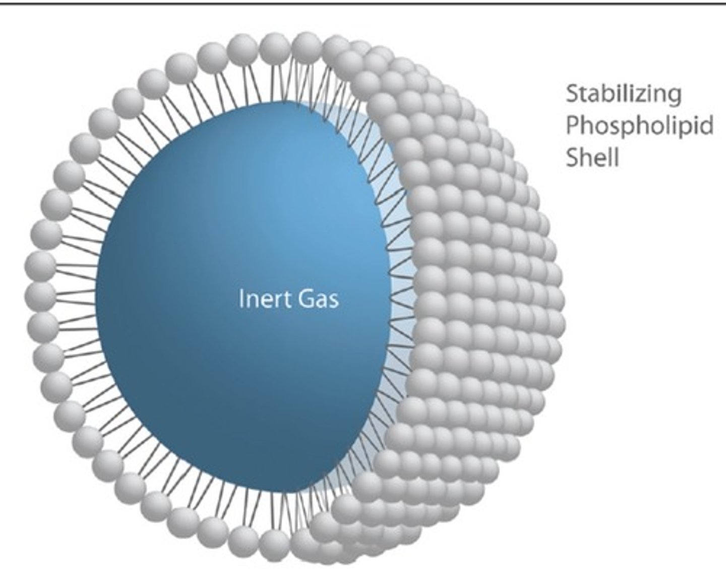
Inner Component of Microbubble
Gas bubble
Degrade at capillary level - clear from bloodstream via lungs
Outer Component of Microbubble
Phospholipid shell
Broken down by macrophages in liver & spleen
Microbubble Longevity
Determined by chemical composition and equipment settings
Longer longevity = better image with fewer injections
Echoes Created by Microbubbles
Linear reflection - acoustic mismatch
Non-linear scatter - bubble size
Non-linear harmonics - expansion/contraction
Behavior of Microbubbles
Compressible - allows bubbles to vibrate, expand, and contract to produce echoes at harmonic frequencies
Microbubbles with Low Pressures & MI
Greater linear reflection and backscatter
Microbubbles with High Pressures & MI
Greater non-linear backscatter and harmonics
Microbubbles with Very High Pressures & MI
Bubble destruction
Equipment Parameters for Contrast Imaging
Sensitivity
Specificity
Resolution
Dual Display
Assist in determining if lesions are benign or malignant
Recognizing vascular phases, hypoenhancement, & hyperenhancement
Current CEUS Uses
Cardiac - endocardial boarders
Abdomen - liver & kidneys
Vascular - thrombus
Future CEUS Uses
Targeted microbubbles - more sensitive & earlier detection
Delivery of targeted therapies
CEUS Risks
Hypersensitivity reactions - rash
Allergic reactions - anaphylaxis
Contraindications for CEUS
Known intracardiac shunt
Pregnancy
Sensitivity/allergy
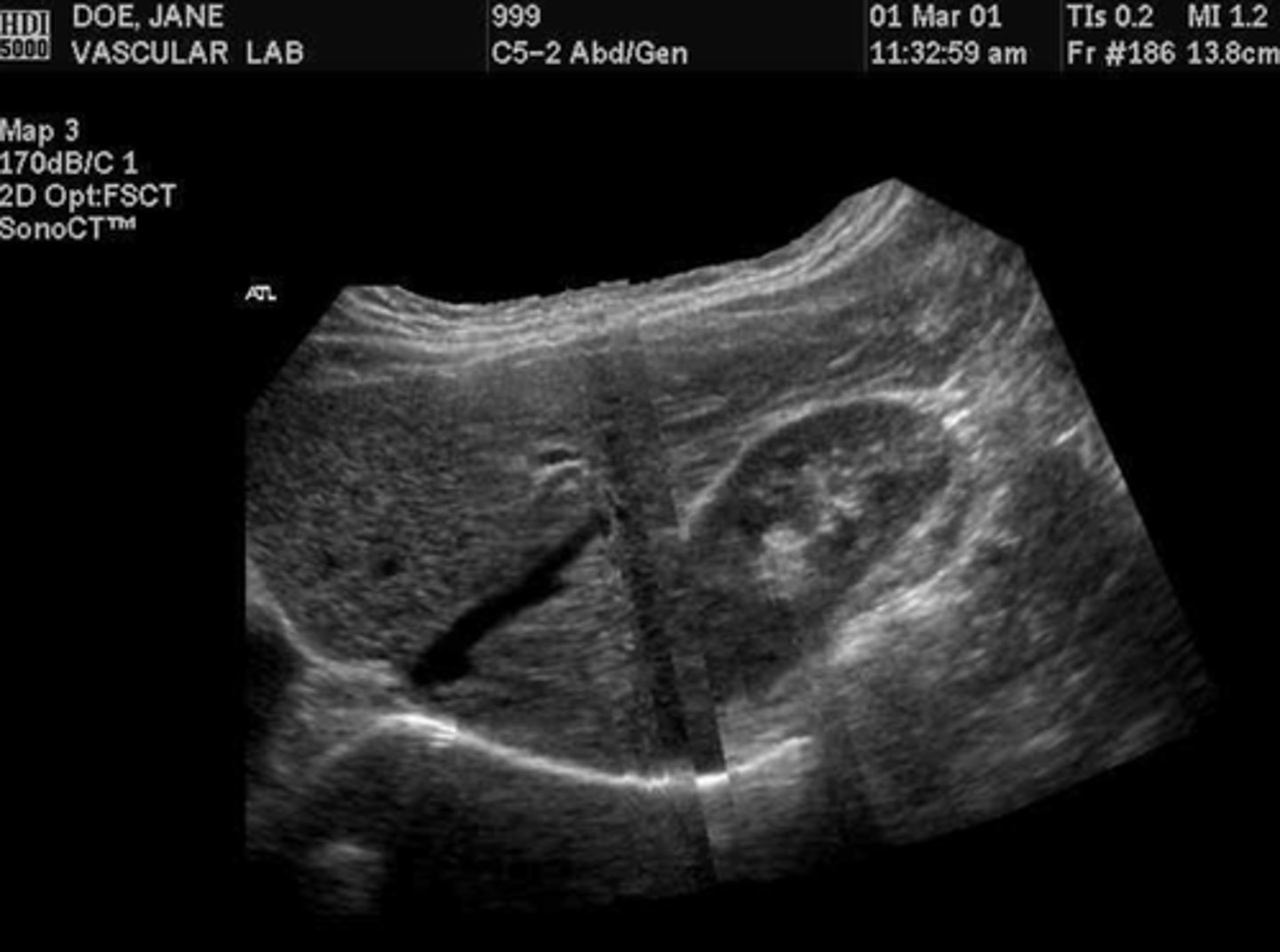
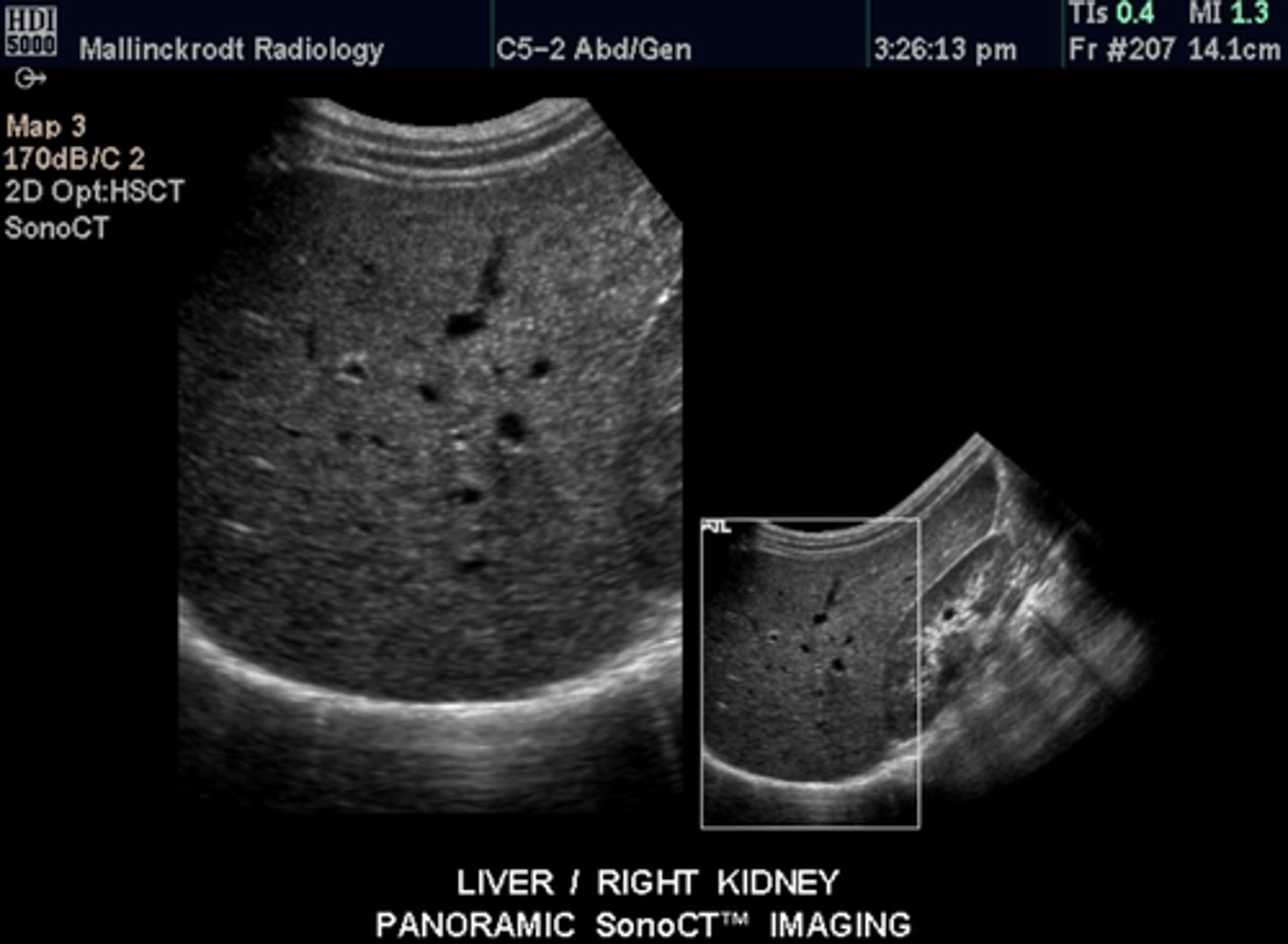
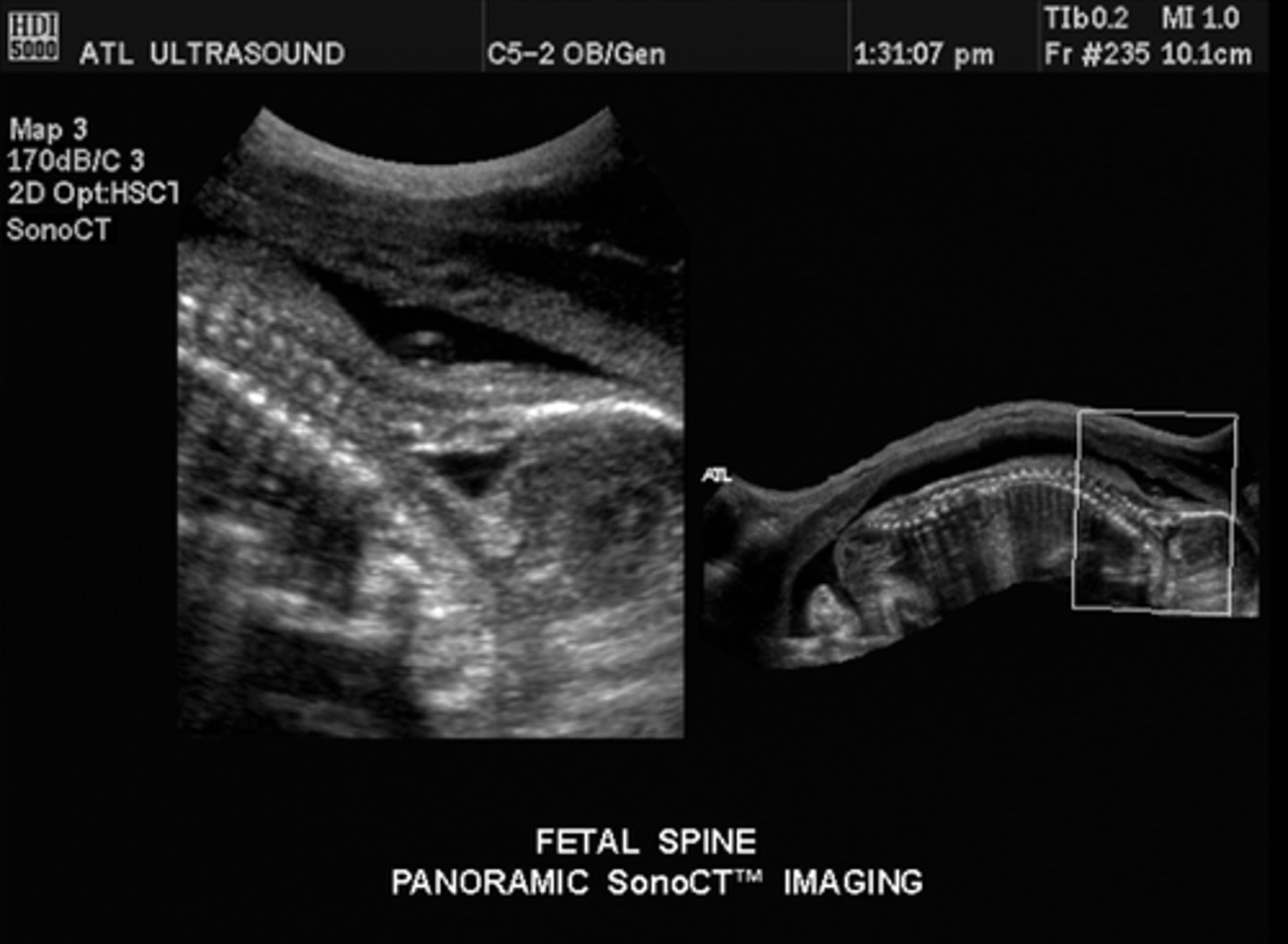
EFOV/Panoramic Imaging
Multiple frames are acquired while detecting scan line position & angle - 15-20/second
Increased contrast resolution, SNR, and tissue differentiation
Decreased speckle artifact
Frame rate is not affected
EFOV/Panoramic Imaging Transducers
Linear
Curvilinear
Real-Time EFOV/Panoramic Imaging
Current anatomy is shown as the transducer is moved
Live feed until the image is frozen
Static EFOV/Panoramic Imaging
Previously scanned anatomy is shown - image grows larger as the transducer is moved
Machine continuously takes still images and combines them into a larger image
Applications of EFOV/Panoramic Imaging
Tracking/following a structure
Showing anatomical relationships & orientation
Showing dimensions of larger pathologies
EFOV/Panoramic Imaging Artifacts
Smearing - mistakes in sweep speed
Stacking/Misregistration - scan head is not perpendicular to skin/rotated/off-axis, will see a jagged edge in frames
EFOV/Panoramic Imaging Optimization Techniques
Pre-scan
Consistent speed
Transducer perpendicular to skin
Have patient hold breath/stay still
Elasticity
Tendency of tissue to resist deformation with applied force or resume its original shape after removal of force
Longitudinal Waves
Motion is parallel to direction of sound beam
Shear Waves
Motion is perpendicular to direction of sound beam
Elastography
Evaluates tissue stiffness/elasticity
Form of strain imaging - does not replace 2D imaging
Disadvantages of Elastography
Normal values depend on vendor and are not interchangeable
Cannot compare values to MRI elastography
Strain Imaging
Measures physical tissue displacement parallel to the applied normal stress
Strain Elastography (SE)
Measures tissue displacement from manual compression or internal physiologic motion (heart)
Stress is parallel to direction of sound beam
Measured by radiofrequency echo correlation-based tracking and Doppler processing
Acoustic Radiation Force Impulse Strain Imaging (ARFI)
Measures tissue displacement from stress applied by high-intensity acoustic radiation force (pushing pulse) from the transducer
Stress is parallel to direction of sound beam
Measured by radiofrequency echo correlation-based tracking and Doppler processing
Shear Wave Imaging
Uses dynamic stress to generate shear waves (can be parallel or perpendicular)
Wave speed provides estimates of tissue elasticity
1D Transient Elastography (TE)
Non-imaging method
A-mode images
Utilizes transducer and mechanical vibrating device to apply external stress
2D Shear Wave Elastography (SWE)
ARFI utilized at a single point to induce tissue displacement
Measures speed of shear waves perpendicular to direction of stress
Used on B-Mode
Point Shear Wave Elastography (pSWE/ARFI quantification)
ARFI utilized with multiple points to induce stress
Real-time visualization of elastogram
Used on B-Mode
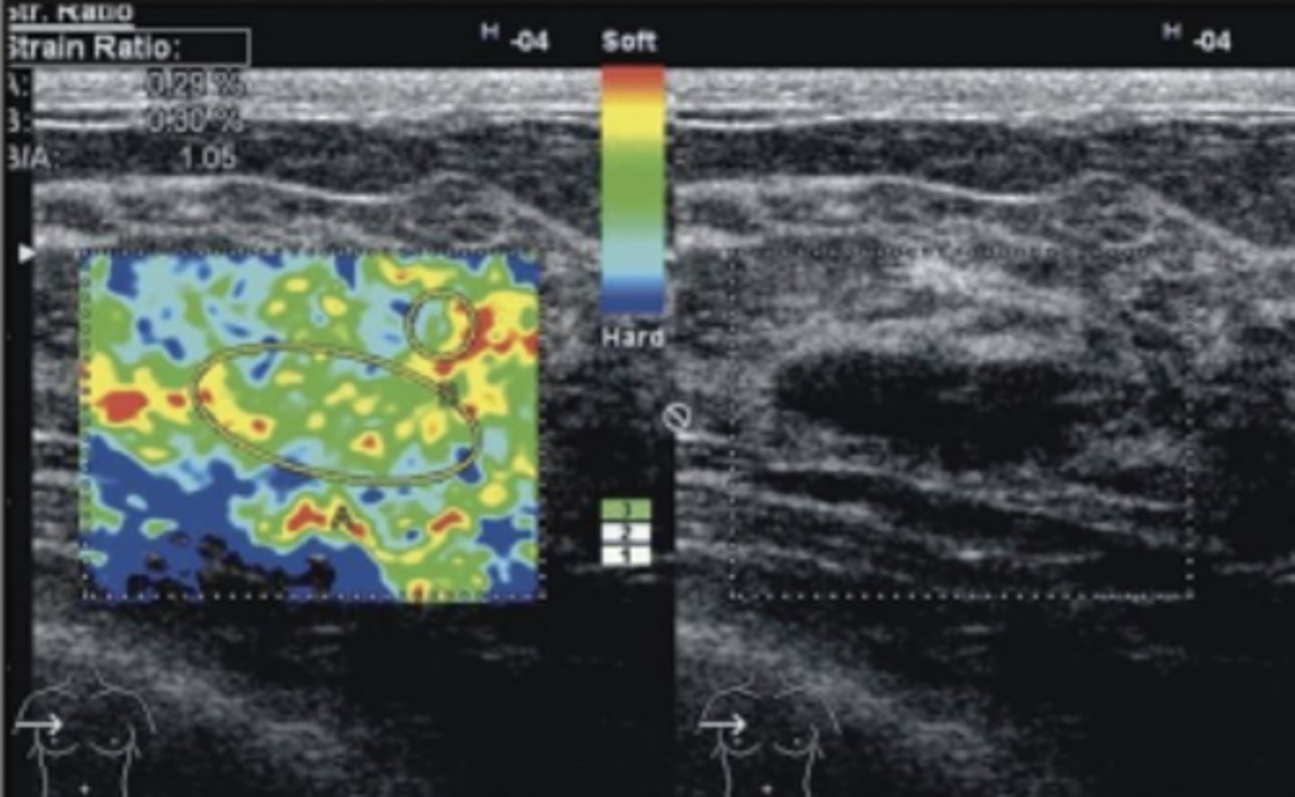
Most common color coding on Elastography
Blue = Increased stiffness/hard
Red = Decreased stiffness/soft
List the clinical applications for Elastography
Liver pathologies/diseases - fibrosis, hepatitis, cirrhosis, alcoholic or autoimmune disease
Characterization of lesions - breast, prostate, thyroid, kidney, lymph nodes
Tissue Doppler Imaging (TDI)
Uses Doppler/frequency shifts to detect movement in myocardium
Best when motion is parallel to sound beam
Cannot distinguish between passive and active motion of muscle fibers
Used with color and PW Doppler
Tissues
Low velocity
High amplitude
Blood
High velocity
Low amplitude
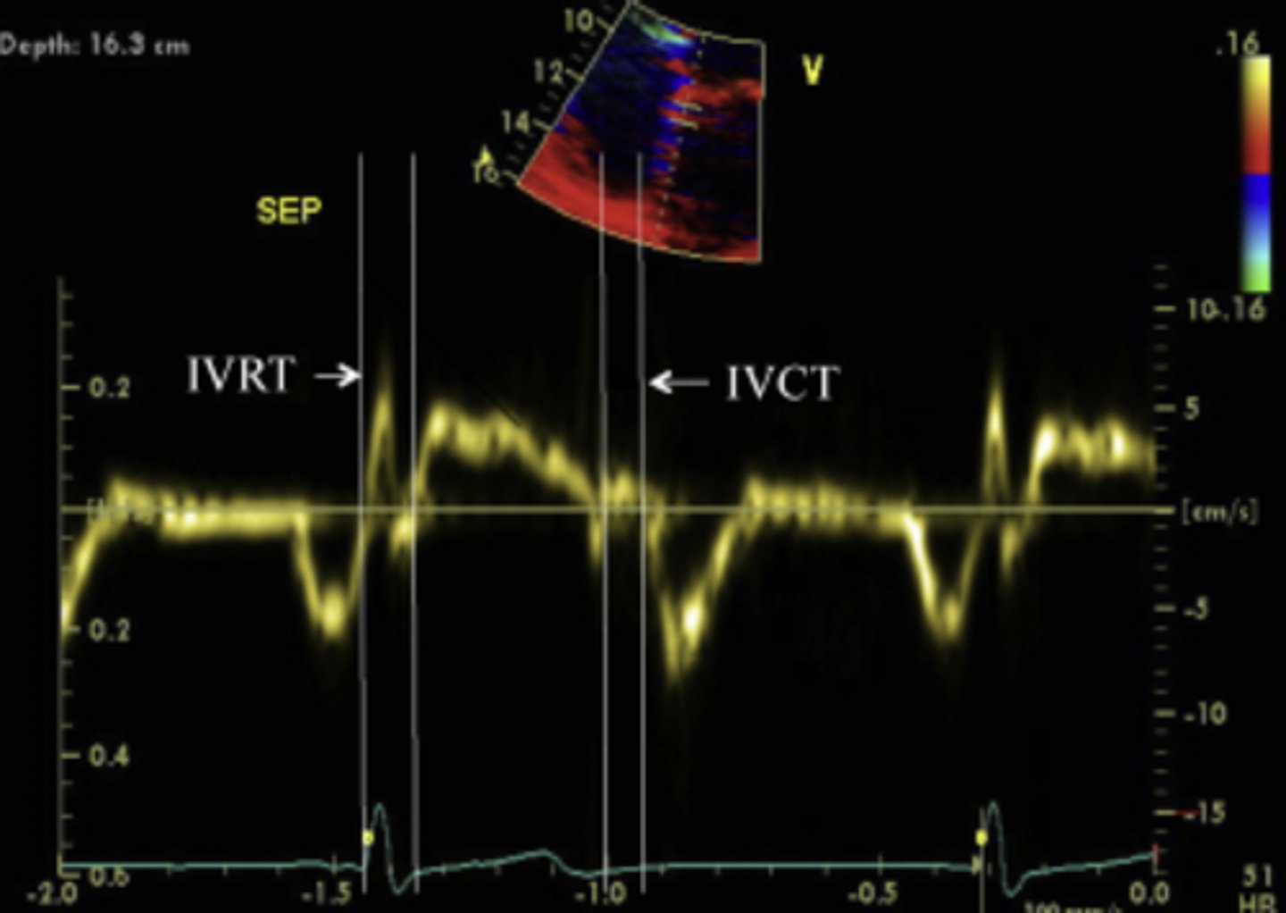
PW TDI
Measures peak & mean velocities in systole (Sa/Sm), early diastole (Ea/Em), and end diastole (Aa/Am).
Systole - Annulus descending
Early diastole - annulus ascends (away from apex)
End diastole - atrial contraction
Advantages of PW TDI
High temporal resolution
Can compare to MV inflow Doppler
Disadvantages of PW TDI
Cannot assess multiple segments simultaneously
Color TDI
Myocardial motion & velocities represented by color on 2D image
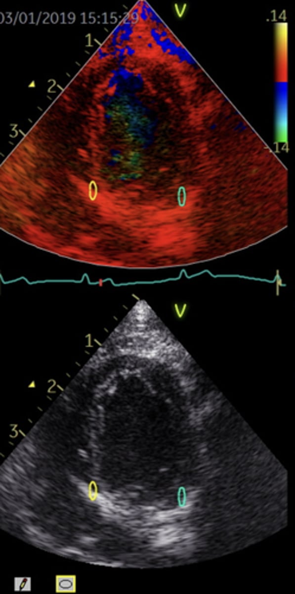
Advantages of Color TDI
Better spatial resolution
Able to assess multiple segments at one time
Speckle Tracking
Uses strain rate to detect LV wall motion and myocardial thickening
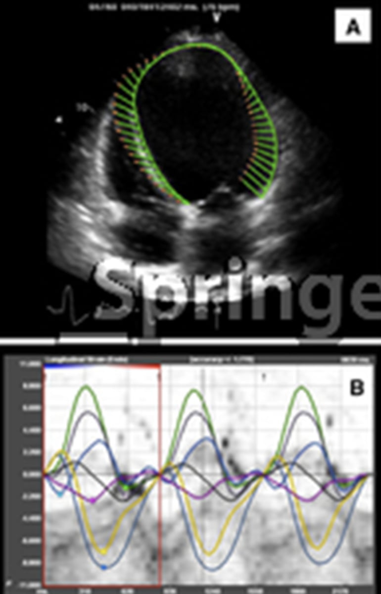
Advantages of Speckle Tracking
Easy to use
Only need one cardiac cycle
Can assess more than on plane in a single image
Independent angle of insonation
Can detect disease early
Reproducible
Better interpretation of ventricular pacing than TDI
Disadvantages of Speckle Tracking
Needs clear image of boarders to work
Needs high temporal resolution
Difficult to use on large LV
Uses for Speckle Tracking
Coronary Artery Disease
Cardiomyopathy
Valvular Disease
LVH
Dyssynchrony
Heart Failure
EF
Ablation
Right Ventricular Function
Tissue Tracking
Must recognize end-diastole & end-systole
Must define a region of interest
Strain
Deformation of an object from its original shape due to applied stress
Dimensionless - percentage of deformation
Change in length of an object to its original length
Strain Formula
(Change in length - original length) ÷ original length
Strain Measures...
Regional LV function
Peak systolic strain
Average deformation within a myocardial segment
Local shortening, thickening, & lengthening of myocardium
Strain Timing
Based on QRS complex
Positive Strain
Elongation (diastole)
Negative Strain
Shortening (systole)
Advantage of Strain Imaging
Detects local myocardial dysfunction before global ventricular impairment
Uses of Strain Imaging
Systemic Hypertension
Duchenne Muscular Dystrophy
Hypertrophic Cardiomyopathy
Myocardial Infarction
Dyskinesis (Disorganized movement)
Severe pulmonary regurgitation (after repair of Tetralogy of Fallot)
Ischemic heart disease
Diabetic heart disease
Amyloidosis (Protein build up in tissue)
Hypertrophy
Follow up other treatment plans
Spatial Correlation Method of Strain
Uses 2-dimensional pattern-matching
Uses before and after deformation images
Shows longitudinal and lateral displacement
Long processing time
Phase-Shift Tracking Method of Strain
Based on autocorrelation
Can rapidly & precisely determine longitudinal tissue motion
Cannot measure large displacements with aliasing
Does not compensate for movements
Combined Autocorrelation Method of Strain
Uses phase-domain processing
No aliasing
Rapid detection of longitudinal displacement
Compensates for movements
Strain Rate
Speed of deformation of an object from its original shape
Change in velocity related to distance between segments
Rate of change in strain per second
Strain Rate Formula
(V2 - V1) ÷ Distance
Strain Rate Measures...
Rate/speed of deformation of myocardial walls
Apical Strain Rate
Rate of lengthening
PSAX Strain Rate
Rate of thickening
2 objects can have the same strain, but different strain rates
History of Harmonics
1990's - harmonic frequencies can be generated in native tissues
Harmonics
Frequencies that are integer multiple of the fundamental frequency
Original/Fundamental Frequency
One frequency
Sine wave
Attenuation begins as soon as pulse enters body
Harmonic Frequency
Multiples of the original frequency - often integer multiples of 2
Non-linear wave/non-linear propagation
Generated at center of beam where intensity is the highest
Begins after skin line, strengthens with depth, then looses intensity
Returning harmonic signals is less intense than the original frequency
Linear Sound Propagation
Sine wave
Small acoustic pressure
Tissue shows elasticity
No new harmonic frequencies created