23 Vestibular System: function and dysfunction
 Call Kai
 Learn
Learn Practice Test
 Spaced Repetition
 Match
Match Flashcards
 Knowt Play
1/49

There's no tags or description
Looks like no tags are added yet.
Name | Mastery | Learn | Test | Matching | Spaced | Call with Kai | Chat |
|---|
No analytics yet
Send a link to your students to track their progress
50 Terms
VERTICAL: macula of saccule
HORIZONTAL: macula of utricle
ROTATIONAL: cristae of semicircular canal
What is the difference between the vertical, rotational, and horizontal parts of the vestibular system? Where are they?
True
(stimulation of one side causes inhibition of the other)
TRUE OR FALSE: the vestibule of the vestibular system provides continuous tonic input
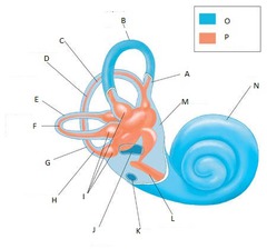
Bony perilymph in blue, membranous endolymph in orange
What are the main parts of the vestibular system labyrinth?
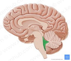
Medial longitudinal fasciculus
This structure associated with the vestibular system is the pathway from vestibular nuclei to LMN in motor nuclei of cranial nerves III, IV, and VI to coordinate and conjugate eye movement as the head changes position
VIII (vestibulocochlear)
Which cranial nerve is largely associated with the vestibular system?
in the 4th ventricle
Where is the vestibular system located?
1. spinal cord
2. rostral brainstem
3. cerebellum
Projections from the vestibular nuclei have 3 main targets. What are they?
Cerebellum
What connection to the vestibular system helps maintain position of the head, trunk, and limbs when body moves, controlling rate, range, and force?
Continuous inhibitory impulses
What kind of impulses does the cerebellum project back to the vestibular nuclei?
Vestibulospinal tract
What is the major descending tract from the vestibular system that travels to ALL segments of the ipsilateral spinal cord?
1. facilitation of ipsilateral extensor muscles
2. inhibition of ipsilateral flexor muscles
3. inhibition of contralateral extensor muscles
What is the point of the vestibulospinal tract, what does it cause?
1. *facilitation* of ipsilateral extensor muscles
2. *inhibition* of ipsilateral flexor muscles
3. *inhibition* of contralateral extensor muscles
The vestibulospinal tract causes:
1. *facilitation or inhibition* of ipsilateral extensor muscles
2. *facilitation or inhibition* of ipsilateral flexor muscles
3. *facilitation or inhibition* of contralateral extensor muscles
Because you're falling to the left and the body wants to stop you from falling left: Increase extensor tone on the left, decrease flexor tone on the left AND dec. extensor tone on the right.
I push you on the right side. What does the vestibular system do to react?
Stabilizes images on the retina
How is the vestibular system related to eye movements?
Opposite
Movement of the head induces compensatory eye movements in the *same/opposite* direction from the vestibular system.
Motor nuclei of CN III (oculomotor), CN IV (trochlear nerve), and CV VI (abducens)
Vestibular neurons communicate with the motor nuclei of WHAT cranial nerves for compensatory eye movements?
Turning head slowly and focus eyes on something: slow phase (L lateral rectus muscle and R medial rectus muscles working to keep eye trained in front of me)
Letting my eyes move to where my head is: fast phase
I turn my head to the right slowly while focusing my eye on something in front of me. What is it called when my head turns and my eyes can focus on that thing, versus when I let my eyes snap to where my head it going.
Fast phase of eye movement = controlled by higher centers NOT vestibular
Which phase of nystagmus movement is NOT controlled by the vestibular system?
Fast phase to the right for a left inner ear problem, but head tilt to the left (side of lesion)
note: the fast phase normally points AWAY from the lesion
If I have a lesion in my left inner ear, where will the fast phase for nystagmus be towards? Where will the head tilt be?
Clinical signs for problems with the vestibular system
Head tilt, pathologic nystagmus, positional strabismus, and vestibular ataxia are all examples of:
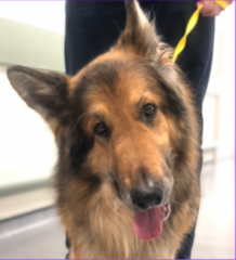
Head will be tilted and the ventrally deviated ear (lower ear) is the side of the lesion
EX: the dog in the picture likely has a right side lesion
How do you know which side the lesion is on with a vestibular tract lesion and head tilt?
Pathologic nystagmus
This is a "jerky nystagmus" with distinct fast and slow phase, described by the direction of the fast phase
By the direction of the fast phase. The fast phase points AWAY from the lesion.
(so if fast phase is to the left, the lesion is on the right)
How do you define a pathologic nystagmus? How do you know where the lesion is?
Horizontal, Rotary, Vertical
What are the 3 different types of pathologic nystagmus in animals?
On the right side
An animal has a right head tilt with a horizontal nystagmus fast phase to the left. Where is the lesion?
Pendular
Which type of nystagmus commonly occurs in siamese or Himalayan cats but is NOT a sign of vestibular dysfunction?
Strabismus
This is a ventrally or ventrolaterally deviated globe when the head is extended, commonly on the same side as the lesion
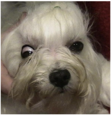
There is loss of CN III (oculomotor), CN IV (trochlear nerve), and CV VI (abducens) innervation
What does strabismus (the eyes not looking in the same direction), tell you about cranial nerves?
Vestibular ataxia
This common vestibular system abnormality sign refers to wide based stance, leaning, listing, tight turning circles, falling or rolling
a. Proprioceptive deficits: usually NOT present with peripheral, usually present with central
b. Deficits other than CN VII or VIII: not with peripheral, but possible with central
c. Vertical Nystagmus: does not happen with peripheral, but does with central
d. Change in direction of fast phase of nystagmus: does not occur with peripheral, but does with central
e. Speed of nystagmus: the faster the nystagmus, the more likely it is peripheral
What are the major differences between peripheral and central vestibular disease in terms of the following categories:
a. proprioceptive deficits
b. Deficits other than CN VII or VIII
c. Vertical Nystagmus
d. Change in direction of fast phase of nystagmus
e. Speed of nystagmus
Postural deficits
If central disease regionally affected the ascending general proprioception, what will you see?
Hemiparesis
If central disease regionally affected the descending UMN, what will you see?
Depressed consciousness
If central disease regionally affected the ARAS, what would you see?
a. head tilt and nystagmus are OPPOSITE to true side of lesion
b. ataxia is OPPOSITE to true side of lesion
c. postural reaction deficits are on SAME SIDE as lesion
(basically everything that normally happens is the exact opposite, hence the paradox)
What is the problem with PARADOXICAL vestibular disease in terms of:
a. head tilt and nystagmus
b. ataxia
c. postural reaction deficit
Paradoxical vestibular disease because the cerebellum sends INHIBITORY impulses to the vestibular nuclei at all times
If disease of the cerebellum happens (flocculonodular, caudal peduncles, fastigial nucleus), what might happen and why?
Cats
Bilateral vestibular disease is more common in *cats or dogs*
Head tilt and pathologic AND physiologic nystagmus MISSING
INSTEAD you see animal crouched low to ground with wide lateral head excursions (because they don't know where the earth is)
What classic vestibular sign is often missing with bilateral vestibular disease? What will you see instead.
Peripheral
Otitis media/interna, idiopathic, and inflammatory polyps are examples of things that cause *central or peripheral* vestibular dysfunction
Otitis interna/media
What is the MOST COMMON CAUSE of peripheral vestibular dysfunction
CN VII (facial) deficits and Horner's
Otitis interna/media is the most common cause of peripheral vestibular dysfunction. What things commonly are seen concurrent with this condition?
Otoscopy, bulla radiographs
How do you diagnose otitis interna/media?
Peripheral vestibular disease, head tilt to side of lesion
What will this cause?
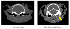
There is beam hardening = distorted because the petrous portion of the temporal bone is one of the densest in the body
What is one of the problems seen with using CT of the skull to see the brain?
Canine idiopathic vestibular disease (geriatric)
What is the second most common cause of peripheral vestibular dysfunction in dogs?
Otitis media/interna NOT idiopathic disease
Is CN VII deficit and Horner's commonly seen concurrently with *canine idiopathic vestibular disease* or *Otitis media/interna*?
Normal
Dogs with idiopathic vestibular disease have *normal or abnormal* proprioception
Canines: OLDER, geriatric disease
Cats: any age
What is the difference between canine and feline idiopathic vestibular peripheral disease?
Nasopharyngeal polyps
These are growths off the epithelia or tympanic bullae that commonly cause noticeable stertor and are a common cause of vestibular signs in young cats
Central
Hypothyroidism, neoplasia, meningoencephalitis, cerebrovascular accidents, and metronidazole toxicity are common causes of *central or peripheral* vestibular dysfunction.
1. head tilt and nystagmus suggests: vestibular or balance problem
2. what is responsible for other cranial nerve abnormalities described: CN VII (facial) for the drooping of the face. menace involves CN II (vision) and CN VII (blinking). palpebral involves CN V (facial sensation) and CN VII (blinking). So, because the animal can track the cotton ball but cannot blink, we can deduce there is a CN VII facial nerve problem.
3. based on all neuro findings: peripheral is more likely
WHY?: postural reactions are normal. deficits are within CN VII and VIII. smell history is important for otitis interna/media.
4. neurolocalization: right side peripheral vestibular system and right facial nerve
5. how do we explain facial nerve problems: facial nerve is commonly affected with labyrinthe disease and concurrent with otitis media/interna.
CASE: GET OUT SOME PAPER:
SIGNALMENT: 8 yo. female spayed cocker spaniel
HISTORY: owners woke up and found dog with severe head tilt and she couldn't maintain her balance
PE: moist malodorous ears. face asymmetrical but hard to tell with head tilt. bright alert responsive.
NEURO EXAM: right side head tilt. moderate vestibular ataxia. listing and circling tight to the right. horizontal nystagmus with left fast phase.
REFLEXES: absent menace on right. absent palpebral on right. drooping of right lip. spinal and postural reactions all normal. can follow cotton ball with the eyes.
ANSWER THE FOLLOWING QUESTIONS:
1. head tilt and nystagmus suggests what type of problem?
2. what is responsible for other cranial nerve abnormalities described?
3. based on all neuro findings, is central or peripheral disease more likely?
4. neurolocalization?
5. how do we explain facial nerve problems?