14. A1.2 Nucleic Acids
 Call Kai
 Learn
Learn Practice Test
 Spaced Repetition
 Match
Match Flashcards
 Knowt Play
1/32

There's no tags or description
Looks like no tags are added yet.
Name | Mastery | Learn | Test | Matching | Spaced | Call with Kai | Chat |
|---|
No analytics yet
Send a link to your students to track their progress
33 Terms
State the 2 primary functions of nucleic acids
Hereditary material by replicating and transferring genetic information to the next generation
Code for protein production in the process of gene expression
State the two types of nucleic acids used in cells
DNA - deoxyribonucleic acid
Passes heredity information between generations of cells
Codes for making RNA during transcription
RNA - ribonucleic acid
Codes for making proteins during translation
mRNA, rRNA, and tRNA are the three main types of RNA involved in protein synthesis
Outline the meaning and implication of DNA being the genetic material of all living organisms
Evidence of universal common ancestry of life
State why RNA viruses do not falsify the claim that all living things use DNA as the genetic material
Some viruses use RNA as their genetic material.
However, because viruses are not made of cells, they are not considered to be living.
List the 3 components of a nucleotide.
nucleotides (monomer), nucleic acid (polymer)
Nucleotides are composed of:
nitrogenous base
Adenine (A)
Thymine (T)
Cytosine (C)
Guanine (G)
Uracil (U)
a five-carbon "pentose" sugar (ribose or deoxyribose)
A negatively charged phosphate group
Draw the basic structure of a single nucleotide (using circle, pentagon and rectangle), labelling the carbons of the pentose sugar.
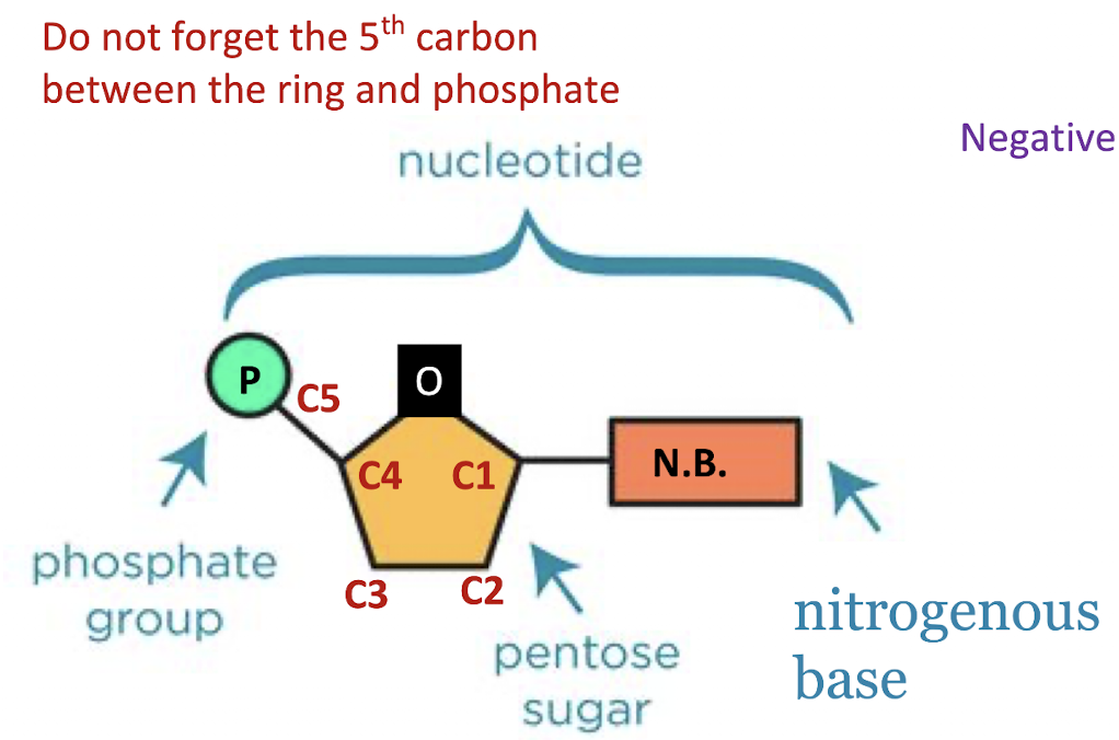

nitrogenous base connecting to carbon-1
carbon-5 branches out of the ring
phosphate group connecting to carbon-5
Define “backbone” as related to nucleic acid structure
Phosphodiester bond:
Each phosphate group is covalently bonded to the 3rd carbon of the pentose sugar before it and to the 5th carbon of the next sugar
Nucleotides connect to form a strong sugar-phosphate backbone, from 5’ to 3’
5’ terminal = the end where the last phosphate is
3’ terminal = the end where a phosphate could link to the 3rd carbon
DNA: double helix, 2 sugar phosphate backbones that run antiparallel
RNA: one sugar-phosphate backbone
Function: provides structural support
The sharing of electrons in the covalent bond between sugar and phosphate provides strength to the structure.
The strength maintains the nucleotides in their specific sequence, (for storing, replication and expression of genetic information)

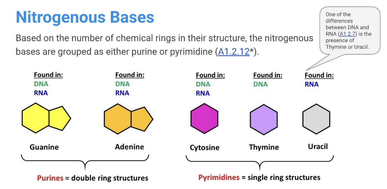
State a similarity and a difference between the nitrogenous bases
Outline how the sequence of bases in a nucleic acid serves as a ‘code’.
CODON = a set of three bases that codes for an amino acid
Define gene
A gene is a specific sequence of nitrogenous bases in DNA nucleotides that codes for the making of a protein.
Describe the structure of a DNA double helix
The two strands of DNA is antiparallel
= they are parallel but run in opposite directionsAt each end of the double helix, one strand is 5’ and the other is 3’
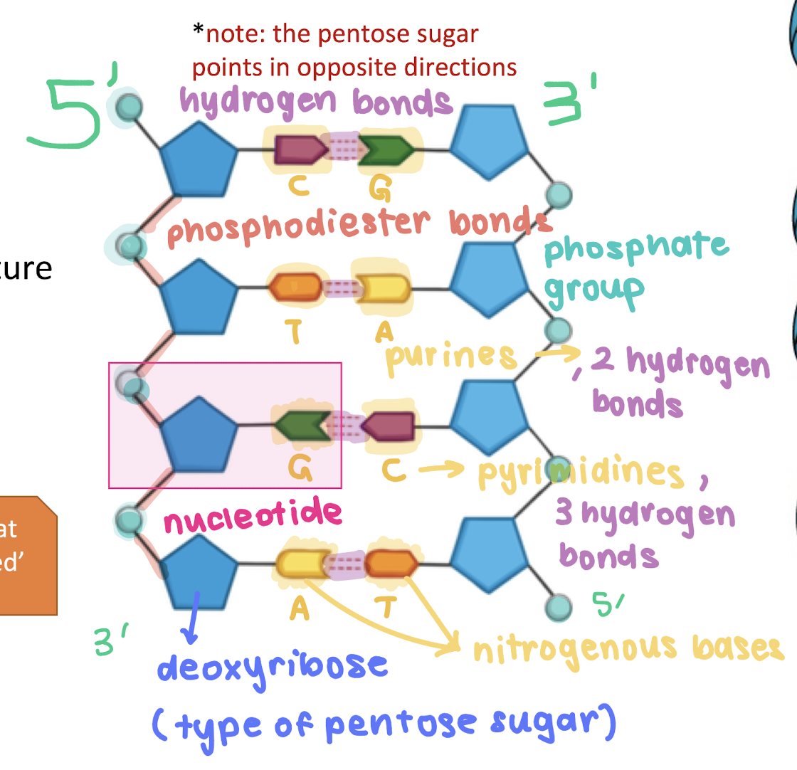
The two strands ‘twist’ to form a double-helix structure
The strands are held together by hydrogen bond between the complementary base pairings.
Adenine – Thymine ( 2 H-bonds)
Guanine – Cytosine (3 H-bonds)
Compare and contrast the structures of DNA and RNA
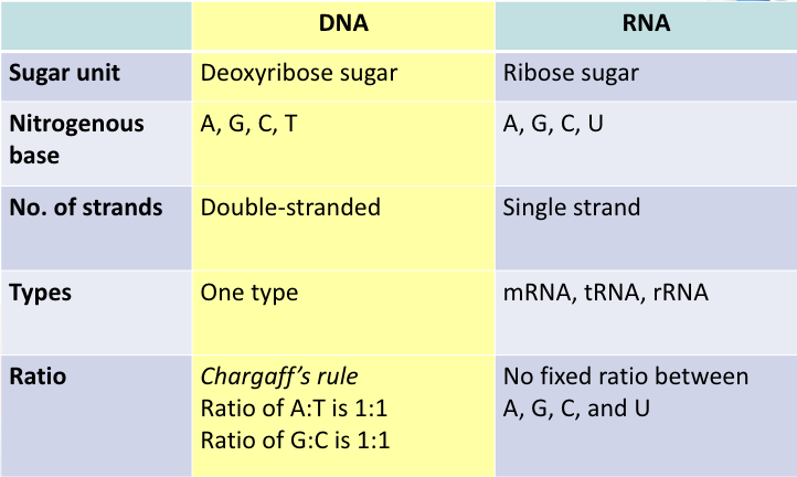

Compare and contrast the functions of DNA and RNA

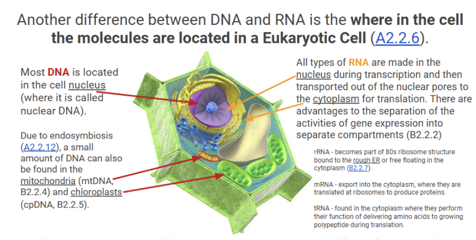
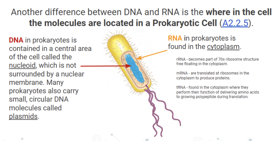
Compare and contrast the location of DNA and RNA in prokaryotic and eukaryotic cells
Outline the role of complementary base pairing in maintaining the DNA sequence during DNA replication
DNA replication occurs before cell division.
DNA replication is "semi-conservative"
each of the original DNA strands serves as a template for the creation of a new strand.
DNA polymerase III builds the new strand by "reading" the template and adding the complementary DNA nucleotide.
This results in the newly built strand having the same sequence of bases as the other template strand.
In this way, replication builds two identical DNA molecules, each with one original and one new strand.

Understanding translation and transciption in DNA
Outline the role of complementary base pairing in transmitting the genetic code in transcription
Transcription is the synthesis of RNA using a DNA template.
The enzyme RNA polymerase builds an RNA strand by "reading" the DNA template and adding the complementary RNA nucleotide.
RNA strand having the complementary sequence of bases as the DNA, thereby maintaining the information stored in the sequence of nucleotides of the code.
Outline the role of complementary base pairing in transmitting the genetic code in translation
Translation is the synthesis of a polypeptide from mRNA.
ribosome builds a polypeptide by reading the mRNA template and binding the coded amino acid to the polypeptide chain
amino acids are brought to the ribosome by a tRNA.
tRNA forms a temporary bond to the mRNA using complementary base pairing
Outline why there is a limitless diversity of DNA base sequences
4 bases = This can code for 20 different amino acids
Polypeptide length can range from 20-2000 amino acids
Average length of a polypeptide is around 300 = 20300 = ~ 1.4… x 10390 possible polypeptide or ~1.24 x 10723 different base sequence
The arrangement of any base sequence is possible and a gene can also be of any length
And can code for a limitless possibility of polypeptide with difference amino acid sequences which leads to a very diverse range of genes
There is an almost limitless capacity of DNA for storing a wide range of data with great economy as it only needs 4 different bases, can be store within such a small structure of a cell and can be easily replicated
Outline why conservation of the genetic code across all forms of life is evidence of common ancestry
All organisms using the same genetic material – DNA (excluding virus)
All organisms uses the complementary base pairing (prokaryote uses uracil in their DNA instead of thymine)
And all organisms replicate their DNA, transcribed and translate their DNA into protein
Most using the same 3-base codon to code for amino acid sequence
Suggesting a universal common ancestry (despite the hypothesis that RNA is the first genetic material to evolve)
*In very limited organisms, some codons have different meaning, which raised some hypothesis that genetic code might not be universal, or that codons have occurred after the origin of life
Universal definition in this context: The genetic code is universal, which means that all living organisms use this same genetic code. Even viruses use this same code
Directionality of RNA & DNA
DNA strands are antiparallel and DNA & RNA strands are produced from the 5’ to 3’ direction
Meaning that DNA nucleotides & RNA nucleotides can only be added to the 3’ end of a growing strand
This is because the condensation reaction to form the sugar phosphate bone occurs between the hydroxyl group of phosphate (attached to the 5’ carbon) in the new nucleotide and the hydroxyl group attached to the 3’ carbon
The addition of the nucleotide during RNA/DNA synthesis involve the cleavage of two phosphate group from the dNTP (deoxyribose nucleoside triphosphate) which releases energy to form the phosphodiester bond
Identify and label the 5’ and 3’ ends of the daughter DNA strands on a diagram of the DNA replication fork
Since both strands of DNA are used as a template during DNA replication, there are differences in how replication occurs on each strand
Leading strand: the new DNA polymer
3' end is at the opening of the replication forkLagging strand: the new DNA polymer
3' end is opposite the opening of the replication fork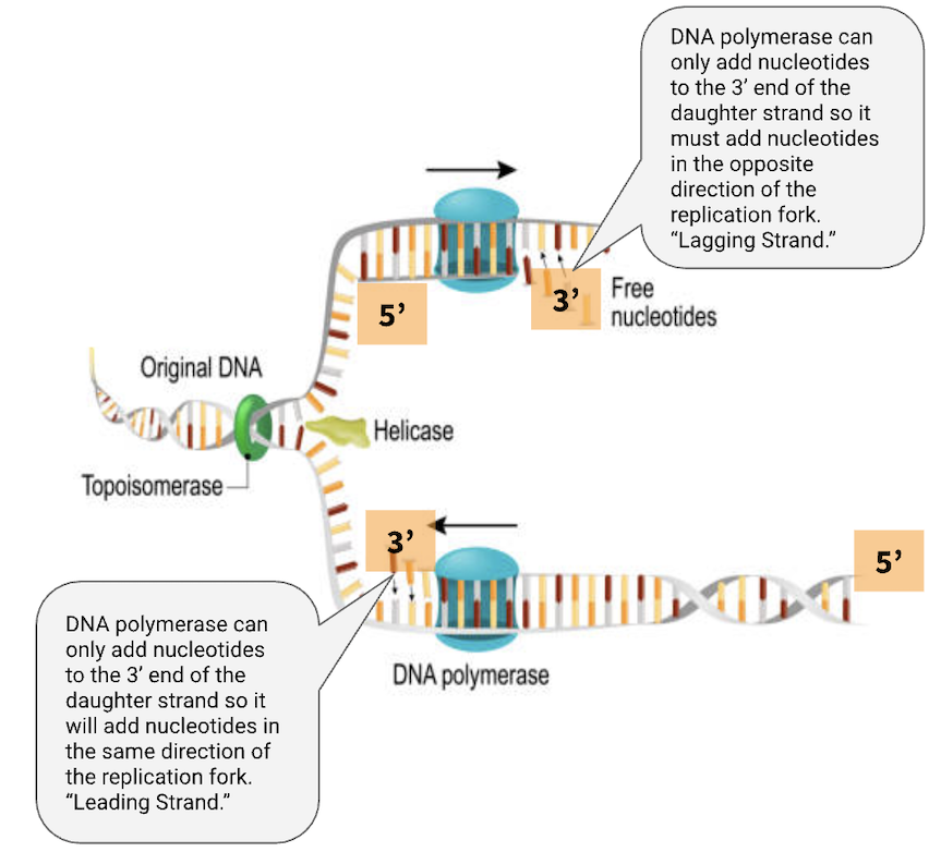

Identify and label the 5’ and 3’ ends of RNA on a diagram of the transcription bubble

Outline the impact of DNA directionality on translation

Compare and contrast the structures of purines and pyrimidines
Compare: All have nitrogen
Contrast: Purines = double ring structures, Pyrimidines = single ring structures
Purine to pyrimidine bonding
A can only pair with T & C can only pair with G
The bases pair upside down with each other
The complementary base pairs have
equal length between the two strands
State two consequences of purine-to-pyrimidine bonding on the structure of DNA
When a pyrimidine is paired with a purine, the width dimension of both pairs is identical. This means that the DNA sugar-phosphate backbones have a consistent diameter throughout the entire molecule.
The hydrogen bonding between the double helix helps to greatly stabilise the structure.
Function of nucleosomes
Eukaryotic Cells: have histones to help package DNA into nucleosomes
The nucleosome contributes to supercoiling of DNA and is an adaptation that allows the packing of large genome within a small space in eukaryotes
It also protects the DNA from damage and allows chromosomes to move during mitosis & meiosis without entanglement.
Structure of nucleosomes
A nucleosome is made up of
eight histone proteins (octamer) with DNA coiled around it
A linker DNA connects one nucleosome to the other
Another histone protein H1, binds the DNA to the core particle
Forming a beads-on-a-string structure (chromatosomes) – genes can still be activated

Draw and label the structure of a nucleosome, including the H1 protein, the octamer core proteins, linker DNA and two wraps of DNA

DNA packaging
DNA packing with nucleosomes occurs only in eukaryotic cells.
Prokaryotic cell DNA remains "naked" meaning it does not wrap around histone proteins.

The H1 proteins will interact and form a ring of 6 nucleosomes which will coil and stack upon one another together to form a fibre which is 30nm in diameter (also called a solenoid) - like a hollow tube
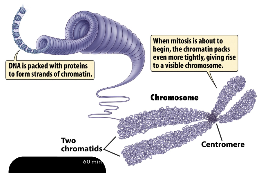
This 30nm fibre form further loops that coils around each other and condense into chromatins (700nm) – part of the chromosome
During prophase of mitosis and meiosis, DNA condenses even more by supercoiling into chromosomes (D2.1.6). Condensing DNA into chromosomes prevents DNA tangling and damage during cell division.

Outline the procedure of the Hershey and Chase experiment
1940s – scientists were unsure whether protein or DNA was the hereditary material
Proteins were more favoured as it can produce a great variety due to 20 naturally occurring subunits instead of only 4 nucleotide subunits
Experiment
It was known then that virus:
Binds to host cell and inject their genetic material into cells
Its non-genetic material remain outside of cell
The infected cell will then produce large numbers of virus until it bursts and releases the viruses into the environment
So the substance injected inside of the virus must contain the hereditary material for producing new viruses
Virus has a protein coat, and a DNA core
They choose to investigate T2 bacteriophage that is a very simple virus that consists only of a protein coat (capsid) and a strand of DNA
Grew one batch of virus with radioactive sulfur (35S) --> label proteins since protein only have sulfur and DNA does not have.
Grow another batch of virus with radioactive phosphorus (32P) —> label the DNA but not the protein
Test: if the radioactive material is inside the bacteria or was left outside.
They then infect the bacteria with radioactively-labelled virus (separately)
Separate the bacteria and virus through centrifugation as the
Heavier bacteria sink to the bottom to form a pellet
The rest of the virus remains in supernatant (top layer liquid)
If protein was the genetic material, then they should find more 35S protein in the bacteria (pellet) & no radioactive in the supernatant
If DNA is the genetic material, then they should find more 35P DNA in the bacteria (pellet) & no radioactive protein in the supernatant.
Results:
35S remain in supernatant
32P in pellet – meaning that DNA is in the bacteria

Describe implications of Chargaff’s data that showed a 1:1 ratio of purine to pyrimidine in a sample of DNA
Levene was a scientist who discovered nucleotides and proposed the structure of nucleic acid – Tetranucleotide Hypothesis
Another scientist – Chargaff decided to calculate the different amount of purine and pyrimidines in different organisms
He found that the 4 bases was not equal in amount as proposed by the tetranucleotide hypothesis
Instead, he found that the amount of
adenine = thymine & cytosine = guanine
Although Chargaff did not figure out the structure of DNA,
Chargaff’s data falsified the tetranucleotide hypothesis that there was a repeating sequence of the four bases of DNA
