Generation of diversity in B and T cell receptors-part 1
 Call Kai
 Learn
Learn Practice Test
 Spaced Repetition
 Match
Match Flashcards
 Knowt Play
1/30

There's no tags or description
Looks like no tags are added yet.
Name | Mastery | Learn | Test | Matching | Spaced | Call with Kai | Chat |
|---|
No analytics yet
Send a link to your students to track their progress
31 Terms
What are the four key features of the adaptive immune system mentioned in the lecture?
The four features are:
Diversity: A vast number of B cells and T cells can recognize an enormous range of antigens.
Clonal selection: Clonal selection is the process where, within a pool of lymphocytes, one specific clone that binds a foreign antigen is selected to proliferate and expand, creating an army of identical lymphocytes to eliminate the antigen. It is based on the fact that every lymphocyte is antigen-specific.
Immunologic memory: Immunologic memory means that when a foreign antigen returns to the body a second time, the response is faster and stronger because a clone of lymphocytes that has seen the antigen before is already present. This is the principle behind vaccination.
Self vs. non-self recognition: "Self" refers to the body's own proteins and structures; "non-self" refers to foreign antigens. Since BCRs and TCRs are generated randomly by genetic recombination, some may recognize self-structures (autoreactive cells). During development, these autoreactive cells are eliminated, and only functional cells are released into the periphery. This is called education.
What types of antigens do B cells recognize, and how?
B cells recognize native antigens by their shape. The antigen can be in solution (e.g., toxins) or on cells (e.g., cancer cells, bacteria, viruses). B cells do not require cell-to-cell contact.

What types of antigens do T cells recognize, and how?
T cells recognize antigenic fragments (peptides) derived from antigens that are presented on the surface of other cells by MHC molecules. T-cell function depends on cell-to-cell contact.
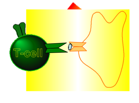
What are BCR and TCR, and what is their key characteristic?
BCR (B cell receptor) is the surface-bound antibody on B cells, and TCR (T cell receptor) is on T cells. They are highly variable, meaning different B and T cells have unique receptors that can differentiate between a vast array of molecules. They are created by genetic recombination during development.

What is the difference between a BCR and an antibody?
The BCR is the membrane-bound form of immunoglobulin on the surface of a B cell. When secreted, it is called an antibody (soluble form).
Describe the basic structural composition of an antibody.
An antibody consists of two identical heavy chains and two identical light chains. The heavy chains are paired to each other by disulfide bonds, and each light chain is paired to a heavy chain by a disulfide bond.

What are the two functional regions of an antibody, and what are their roles?
The variable region (N-terminus, includes both heavy and light chains) binds to the antigen and varies between antibodiesand there are two variants of the light chain constant region: lambda (λ) and kappa (κ) – not important for antibody function. The constant region determines the antibody's function and is found in both heavy and light chains; it has nothing to do with antigen specificity.

What are immunoglobulin (Ig) domains, and what are CDRs?
Ig domains are repeating homologous globular units in the heavy and light chains. Within the variable region, there are complementarity determining regions (CDRs), also called hypervariable regions, which create the antigen-binding surface. The amino acids in these regions are highly variable between antibodies.

hat are the five antibody isotypes, and what distinguishes them?
The five isotypes are IgD, IgM, IgG, IgA, and IgE. They differ in the length of their constant region, their hinge region, and their glycosylation patterns. The constant region of the heavy chain is important for antibody function.
Some of the antibodies that are secreted from a B cell can form complex. For example IgM can stick together with the help of j-chain or joining chain so they can bring five IgM (Pentameric) together and this strengthens the interaction between the antibody and the antigen. And another example is IgA that can be found in monomer or a dimer (dimeric) form in the blood.

Which is the first antibody the B cell produce during an immune response? and give 2 example of where can you find isotypes in the body?
The B cells usually first produce IgM and IgD, but later on, they can switch to other isotypes like IgG, LgA or IgE, depending upon the antigen and the type of immune response or if it is bacterial infection or parasite.
IgM is found in the blood, while dimeric LgA are found in the respiratory mucosa, intestinal mucosa and milk.
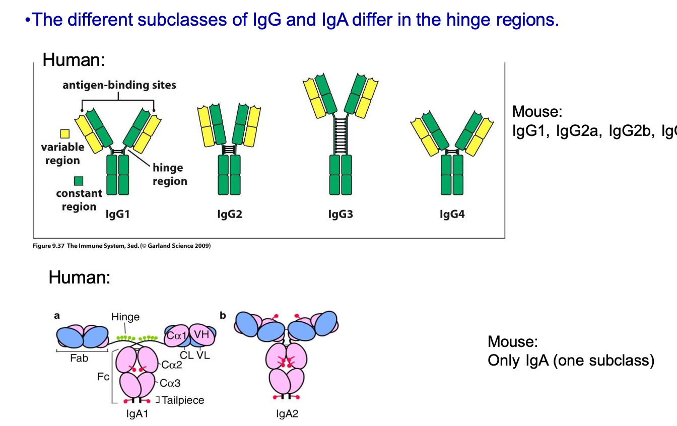
What are the different subclasses that IgG and IgA has?
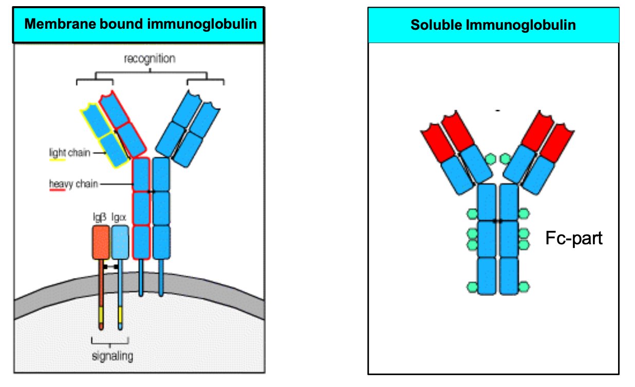
What are the key structural and functional differences between membrane-bound immunoglobulins (BCRs) and soluble immunoglobulins (antibodies) regarding their attachment, signaling, and regions?
Location & Attachment:
BCR: Anchored to the B-cell membrane via a transmembrane domain.
Soluble Antibody: Lacks a transmembrane domain; circulates freely in blood, body fluids, and tissues.
Intracellular Signaling:
BCR: Has no intracellular domain of its own. It relies on accessory molecules Ig\(\alpha \) and Ig\(\beta \) to start the intracellular signaling cascade when an antigen binds.
Soluble Antibody: Does not perform direct transmembrane cell signaling.
The Fc Part (Fragment of Crystallization):
Origin: Named because it crystallizes easily when cleaved from the antigen-binding arms.
Function: Binds to Fc receptors on other immune cells to trigger effector functions.
Give me an overview of B and T lymphocyte development:
1. Commitment of stem cell to B or T cell lineage
2. Rearrangement (= recombination) of BCR and TCR receptor genes
3. Checkpoints at several stages – selection of cells that express functional
and non-harmful receptors
4. Differentiation to functionally and phenotypically distinct subpopulations of B and T cells.
- B cells: Follicular, marginal zone, B1 B cells
- T cells: CD4+ helper and CD8+ cytotoxic αβ T cells, γδ T cells, NKT cells
• The developing B and T cells proliferate/spread/grow at several stages during the early developmental process, providing a large pool of cells which can form useful lymphocytes.
• Sites for development: Fetal liver, bone marrow (B cells), thymus (T cells)
What gene segments make up the light chain loci and the heavy chain loci? And where are the antibody gene loci located in humans?
The light chain loci are made of V (variable), J (joining), and C (constant) gene segments.
The heavy chain loci are made of V, D (diversity), J, and C gene segments.
The lambda light chain locus is on chromosome 22, the kappa light chain locus is on chromosome 2, and the heavy chain locus is on chromosome 14.

Describe the step-by-step process of the recombination of antibodies:
Heavy Chain Recombination
Germline DNA: Multiple un-rearranged V, D, J, and C segments exist in the progenitor cell.
First Recombination Step: One D segment joins with one J segment.
Second Recombination Step: The joined D-J unit joins with one V segment.
Recombination Complete: Somatic recombination for the heavy chain finishes here.
Transcription: The rearranged DNA is transcribed into a primary transcript RNA.
Splicing: RNA splicing occurs to generate functional mRNA.
Translation: The mRNA is translated to form the heavy chain polypeptide.
Region Mapping: The blue C segment forms the constant region, while VDJ forms the variable region.
Light Chain Recombination:
Timing: The heavy chain rearranges first in B cells, followed by the light chains.
Germline DNA: The light chain starts un-rearranged with several V, J, and C segments.
No D Segments: Light chains completely lack D segments.
Somatic Recombination: One V segment directly recombines with one J segment.
Transcription: The rearranged segments are transcribed into primary transcript RNA.
Splicing: Introns are removed to create the functional mRNA.
Translation: The mRNA is translated to form the final light chain polypeptide.
Region Mapping: The blue C region forms the constant region, while VJ forms the variable region.

What is junctional diversity, and what processes contribute to it?
Junctional diversity occurs during recombination when V, D, and J segments are joined. Nucleotides between segments are removed (nibbling), and nucleotides are added: P nucleotides (palindromic, template-dependent) and N nucleotides (non-template-dependent). This is random and creates diversity, but can also cause frame shifts leading to non-functional receptors.
Which CDR region has the highest diversity, and why is it important?
CDR3 has the highest diversity because it has the highest amino acid variation between antibodies. It is the most important region for antigen specificity.
What is the calculated potential receptor repertoire, and why is the actual number lower?
The calculated potential repertoire is around 10^11. The actual number in an individual is lower (around 10^7) because the random processes can yield non-functional receptors (due to frame shifts) and autoreactive cells, which are eliminated.
What is the VDJ recombinase (RAG1/RAG2 complex), and what does it do?
RAG (Recombination Activating Gene) 1/2 complex is the VDJ recombinase, expressed only in developing B and T cells. It recognizes Recombination Signal Sequences (RSS) at the ends of V, D, and J segments, initiates somatic recombination by causing DNA breaks and hairpin loop formation.
What is the role of terminal deoxynucleotidyl transferase (Tdt)?
Tdt is expressed only in developing B and T cells. It adds non-templated N nucleotides to the junctions between gene segments, creating junctional diversity.
What is the role of DNA double-stranded break repair (DSBR) enzymes?
DSBR enzymes are expressed in all cell types. They join together and ligate the junctions between cut gene segments, completing somatic recombination.
What happens in RAG knockout mice, and what condition does this model?
RAG knockout mice lack B cells and T cells because no recombination of gene segments occurs. This results in severe combined immunodeficiency (SCID). In humans, SCID is caused by a recessive mutation in the RAG1/2 complex.
What are the stages of B cell development in the bone marrow?
1. Pluripotent Stem Cell Stage
Commitment: The cell commits to the B cell lineage.
Status: No heavy or light immunoglobulin chains are made yet.
2. Pro-B Cell Stage
Heavy Chain Rearrangement: The genetic segments of the heavy chain begin fusing.
Step 1: The D and J segments fuse together first.
Step 2: The combined DJ segment then fuses with a V segment.
Two Chances: The cell has two alleles (one from each chromosome), giving it two separate chances to successfully rearrange a functional heavy chain.
Light Chain Status: The light chain is completely untouched during this stage.
3. Large Pre-B Cell Stage
Pre-BCR Expression: The newly formed heavy chain pairs with a temporary, non-variable surrogate light chain (made of VpreB and 𝜆5 polypeptides).
First Checkpoint: This temporary pair forms the pre-B cell receptor (pre-BCR) on the cell surface to test if the heavy chain works.
Intracellular Signaling: Successful expression sends a signal into the cell nucleus via Ig-alpha and Ig-beta .
Allelic Exclusion: The signal immediately halts heavy chain rearrangement on the second chromosome allele, ensuring the B cell only produces one type of heavy chain.
Proliferation: The cell undergoes rapid division, creating multiple pre-B cells sharing the exact same successful heavy chain.
What are the stages of B cell development in the bone marrow? part 2
4. Small Pre-B Cell Stage
Light Chain Rearrangement: The successful pre-BCR signaling prompts the cell to stop dividing and start rearranging its light chain genes.
Four Chances: The cell has four total chances to build a working light chain across its genetic loci.
First Preference: It tries the kappa light chain alleles first.
Backup Option: If both kappa alleles fail, it attempts to rearrange the lambda light chain alleles.
Light Chain Allelic Exclusion: Once a successful light chain is produced, further light chain rearrangement stops, and the surrogate light chain is permanently shut down.
5. Immature to Mature B Cell Stage
BCR Assembly: The newly minted light chain pairs with the existing heavy chain.
Surface IgM: This complete antibody is expressed on the surface as a fully functional IgM receptor.
Final Maturation: The cell leaves the bone marrow and finishes maturing, expressing both IgM and IgD on its surface to protect the body against pathogens.
What are the two main checkpoints during lymphocyte development?
The first checkpoint checks if the cell expresses a functional pre-antigen receptor (pre-BCR for B cells: Rearranged heavy chain + surrogate light chain (invariant), pre-TCR for T cells: Rearranged Beta-chain + surrogate Alpha-chain (invariant)).
The second checkpoint ensures the cell expresses a functional, complete antigen receptor. In T cells, this is called positive selection (checking if the TCR recognizes MHC-peptide complexes).
What is negative selection, and what happens to autoreactive immature B cells?
Negative selection eliminates B and T cells that recognize self-antigens (autoreactive cells). However, self-reactive immature B cells are given one more chance: they can edit their receptors by changing gene segments, a process called receptor editing. This does not happen in T cells.
What is somatic hypermutation, and when does it occur?
Somatic hypermutation is a process unique to B cells where random mutations occur in the variable region when B cells encounter antigen. It happens in the presence of antigen, usually in lymphoid organs. Its purpose is to increase the affinity of the BCR towards the antigen; B cells with higher affinity are selected during the immune response.
What are the three main subtypes of B cells?
The three subtypes are: B1 cells, follicular B2 cells, and marginal zone B2 cells.
Describe the characteristics of B1 cells. (less variable receptors B cell)
• Best defined in rodents.
• Are located in the peritoneum and mucosa.
• Formed in fetal liver and self-renew in periphery.
• Mainly produce poly-specific antibodies to polysaccharide and lipid antigens (”natural antibodies”).
Describe the characteristics of marginal zone B2 cells. (less variable receptors B cell)
• Are mainly located in the spleen (mice and humans) and lymph nodes (humans).
• Formed in bone marrow and differentiate in spleen.
• Mainly produce low-avidity IgM antibodies to polysaccharide antigens (”natural antibodies”).
Describe the characteristics of follicular B2 cells.
Follicular B2 cells are the most common B cells in lymphoid organs. They are developed in the bone marrow and differentiate further in the spleen. They have more variable receptors compered to B1 and marginal B cells.