Pathology Flashcards by Robbins and Cotran
0.0(0)
Studied by 3 people0%Exam Mastery
Build your Mastery score
 AP Practice
Supplemental Materials Call Kai
Call KaiCard Sorting
1/4

Earn XP
Last updated 2:13 PM on 8/31/23
Name | Mastery | Learn | Test | Matching | Spaced | Call with Kai | Chat |
|---|
No analytics yet
Send a link to your students to track their progress
5 Terms
1
New cards
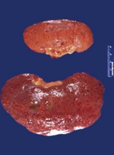
A 70-year-old woman has had a blood pressure of 160/105 mm Hg for many years. Abdominal ultrasound shows the decreased size of one kidney. What gross morphologic description applies to the abnormal kidney? What is the likely cause of this finding? How could this affect the patient's renal function? What cellular organelle plays a major role in this process? What cellular protein processing pathway is involved?
ANS: The smaller kidney is atrophic.
ANS: Diminished blood supply (renal artery stenosis as a result of atherosclerosis) with chronic ischemia likely caused this. Although remaining individual cells in the parenchyma are smaller ( cellular atrophy ), most of the organ shrinkage is attributable to cell dropout because of ischemic injury.
ANS: Diminished blood supply leads to increased renin secretion by the hypoperfused kidney, which causes hypertension.
ANS: Lysosomes play a major role in cellular atrophy through autophagy.
ANS: Cellular atrophy is mediated through increased protein degradation by the ubiquitin-proteasome pathway.
ANS: Diminished blood supply (renal artery stenosis as a result of atherosclerosis) with chronic ischemia likely caused this. Although remaining individual cells in the parenchyma are smaller ( cellular atrophy ), most of the organ shrinkage is attributable to cell dropout because of ischemic injury.
ANS: Diminished blood supply leads to increased renin secretion by the hypoperfused kidney, which causes hypertension.
ANS: Lysosomes play a major role in cellular atrophy through autophagy.
ANS: Cellular atrophy is mediated through increased protein degradation by the ubiquitin-proteasome pathway.
2
New cards
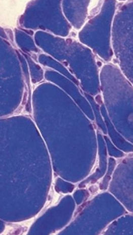
A 55-year-old man has repeated trauma to his upper arms from operating a jackhammer. He now has hand and forearm weakness. A skeletal muscle biopsy specimen reveals the pattern shown at the right. What is the microscopic description of these myocytes? What features support the diagnosis? Why are smaller angulated fibers grouped together?
ANS: Myocyte cellular atrophy is present as a result of lysosomal autophagy and increased proteasomal degradation.
ANS: Small angulated fibers with occasional central nuclei are grouped together.
ANS: Skeletal muscle fibers in a motor unit are randomly enervated; nerve injury initially leads to scattered myocyte atrophy within any given motor unit. After one nerve is injured, however, an adjacent neuron can branch and reinnervate denervated myocytes. If that neuron is now injured, the result is group atrophy of myocytes
ANS: Small angulated fibers with occasional central nuclei are grouped together.
ANS: Skeletal muscle fibers in a motor unit are randomly enervated; nerve injury initially leads to scattered myocyte atrophy within any given motor unit. After one nerve is injured, however, an adjacent neuron can branch and reinnervate denervated myocytes. If that neuron is now injured, the result is group atrophy of myocytes
3
New cards
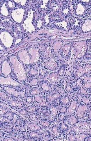
At 40 weeks' gestation, a 20-year-old woman delivers a healthy infant and begins nursing postpartum. Her breasts show bilateral enlargement. The microscopic appearance of her breast tissue is shown. What is the morphologic description? What is the substance represented by the pink-staining material within the lobules? What hormonal stimuli account for these findings?
ANS: The female breast lobules have increased cellularity. This is consistent with physiologic hyperplasia from elevated estrogen and progesterone levels during pregnancy.
ANS: The pink secretions within the lobules are milk. The breast is a modified apocrine gland; lipid-rich secretions are formed through the budding of apical portions of epithelial cytoplasm.
ANS: Estrogen and progesterone bind to breast epithelium steroid hormone receptors to cause specific nuclear mRNA transcription. This results in protein translation that promotes cellular proliferation and gain of function.
ANS: The pink secretions within the lobules are milk. The breast is a modified apocrine gland; lipid-rich secretions are formed through the budding of apical portions of epithelial cytoplasm.
ANS: Estrogen and progesterone bind to breast epithelium steroid hormone receptors to cause specific nuclear mRNA transcription. This results in protein translation that promotes cellular proliferation and gain of function.
4
New cards
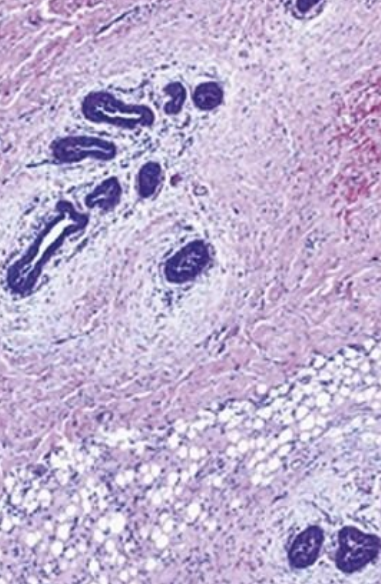
A 22-year-old man has had bilateral breast enlargement for the past 5 years. The microscopic appearance of his breast tissue is shown. This lesion and the tissue shown on Side A represent estrogen effects on breast epithelium. What is the difference between the effects seen in male breasts versus female breasts? Why might this man have elevated estrogen levels?
ANS: As opposed to physiologic lobular hyperplasia in the female breast, the male breast lacks lobules, and elevated estrogen levels lead primarily to ductular hyperplasia with associated increases in extracellular matrix. This leads to hyperplastic male breast tissue, which is called gynecomastia.
ANS: Gynecomastia can suggest estrogen-secreting tumors (e.g., adrenal, testicular), exogenous estrogen administration, drug effects (e.g., mimicking estrogen, causing increased endogenous production), or abnormal estrogen metabolism (e.g., as a result of cirrhosis)
\
ANS: Gynecomastia can suggest estrogen-secreting tumors (e.g., adrenal, testicular), exogenous estrogen administration, drug effects (e.g., mimicking estrogen, causing increased endogenous production), or abnormal estrogen metabolism (e.g., as a result of cirrhosis)
\
5
New cards