M1: Defects of Enamel and Dentin
 Call Kai
 Learn
Learn Practice Test
 Spaced Repetition
 Match
Match Flashcards
 Knowt Play
1/18

Earn XP
Description and Tags
Name | Mastery | Learn | Test | Matching | Spaced | Call with Kai | Chat |
|---|
No analytics yet
Send a link to your students to track their progress
19 Terms
Hypoplastic Amelogenesis Imperfecta
Radiographic features: Enamel reduced in bulk, shows thin layer over occlusal.
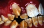
Hypoplastic Amelogenesis Imperfecta
Treatment: No treatment is necessary
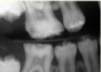
Hypomaturation Amelogenesis Imperfecta
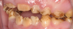
Hypomaturation Amelogenesis Imperfecta
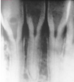
Radiographic features: Affected enamel exhibits radiodensity similar to dentin
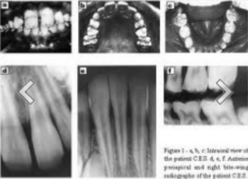
Hypocalcified Amelogenesis Imperfecta
Radiographic features: Density of enamel and dentin are similar.
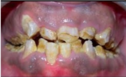
Dentinogenesis Imperfecta
Affects both primary and permanent dentition.
Have blue brown discoloration
With distinctive translucence
Enamel frequently separates easily from underlying defective dentin.
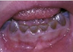
DI Type I
Radiographic features: Partial or total obliteration of pulp chamber and root canals
DI Type II
Radiographic features: Partial or total obliteration of pulp chambers and root canals.
DI Type III
Radiographic features: Periapical radiolucencies
DI Type I
Clinical features: Primary teeth are more severely affected than permanent teeth.
DI Type II
Clinical features: Only have dentin abnormalities and no bone disease.
DI Type III
Clinical features: “bradwine type” multiple pulp exposures in deciduous, which is not seen in type I and II
Dentin Dysplasia
Is also known as “rootless teeth”
Rare disturbances of dentin formation, which had an abnormal pulpal morphology
DD Type I Radicular
Clinical features: Premature tooth loss may occur because of short roots or periapical inflammatory lesions.
Type I (Radicular type)
Radiographic features: roots are extremely short, pulps are almost completely obliterated
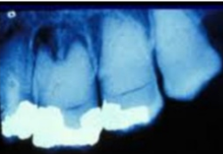
DD Type II Coronal
Clinical features: Color of the primary dentition is opalescent while in permanent dentition is normal.
Type II (Coronal type)
Radiographic features: Deciduous - roots are extremely short, pulps are almost completely obliterated.
Permanent dentition - abnormally large pulp chambers in the coronal portion of the teeth.
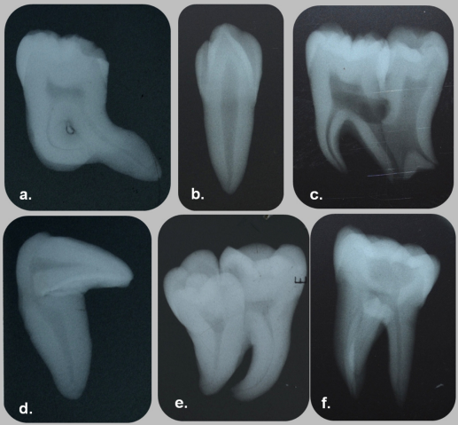
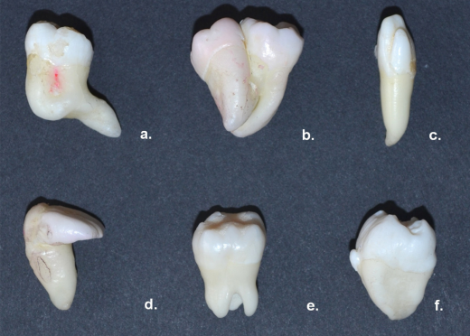
a. Root dilaceration of mandibular molar; b. Concrescence between mandibular first and second molar; c. Talons’ cusp on the labial aspect of maxillary lateral incisor; d. Crown dilaceration of maxillary central incisor; e. Taurodontism in maxillary second molar; f. Enamel pearl on buccal aspect of mandibular third molar
a. Root dilaceration of mandibular molar; b. Talons’ cusp lined by enamel containing core of dentin and pulp in maxillary lateral incisor; c. Fusion of root between mandibular second and third molar; d. Crown dilaceration of maxillary central incisor; e. Concrescence between mandibular first and second molar with dens evaginatus; f. Enamel pearl seen as radio-opaque foci near the furcation area