Lecture 13: The Reproductive System I
1/53
There's no tags or description
Looks like no tags are added yet.
Name | Mastery | Learn | Test | Matching | Spaced |
|---|
No study sessions yet.
54 Terms
What is the pelvic girdle? WHat does it connect?
pelvic girdle = a ring of bones that links your upper body to your legs.
Main Job: Connect your spine (axial skeleton) to your legs (appendicular skeleton).

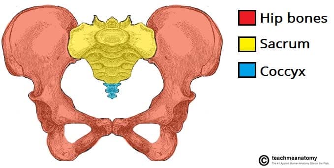
What are the 3 main bones that make up the pelvic girdle?
Hip Bones (Right & Left) – Each hip bone is like one half of the bowl that makes up your pelvis.
Sacrum – This is the big triangle-shaped bone at the bottom of your spine.
Coccyx – Your tailbone, the tiny pointy part under the sacrum.
What are the 2 points of attachment in the pelvic girdle?
2 Points of attachment**
The back of the ring = sacrum + hip bones - sacroiliac joints(x2)
The front of the ring = pubic symphysis

pelvic inlet vs pelvic outlet?
The pelvic inlet is the top entrance to the tunnel.
The pelvic outlet is the bottom exit of the tunnel.
So, anything coming into the pelvis (like a baby during birth or digestive organs) passes through the inlet, and anything going out (like a baby during vaginal delivery) goes through the outlet.

in relation to the inlet and outlet - what is the true pelvis? false pelvis?
FALSE PELVIS:
Located above the pelvic brim/inlet
It's part of the abdomen and helps guide organs into the pelvis.
TRUE PELVIS:
Located below the pelvic brim/outlet
This is the area where reproductive organs sit and where childbirth happens.


WHat are 5 key differences in the female pelvis and their significance?


Because during pregnancy, a fetus grows inside the uterus, which is in the pelvis. The bones need to make room and not block the baby from coming out.
That’s why:
The inlet is wide to allow the baby to enter the birth canal.
The outlet is round and large to help the baby pass through during birth.
The sacrum and coccyx are less curved so they don’t get in the way.
What is the pelvic floor?
Imagine your pelvis is a bowl with an open bottom (called the pelvic outlet). If we just left it open, all your organs (like your bladder, uterus, rectum) would fall down!
So… the body covers the bottom of the bowl with a group of muscles called the pelvic floor,

what is the function of the pelvic floor?
Why Do We Need the Pelvic Floor?
Your pelvic floor muscles do 3 big jobs:
Hold up organs (like your bladder, uterus, and rectum) — kind of like a trampoline under them
Control peeing and pooping — helps you "hold it in" until you're ready 🚽
Deal with pressure — like when you cough, sneeze, jump, or push (the muscles need to tighten so things don’t leak or fall out)
This is why Kegels (those pelvic exercises) are a thing — to strengthen these muscles!
What are the 4 major muscle that make up the pelvic floor? what is the must group called?
LEVATOR ANI MUSCLES:
Puborectalis
Pubococcygeus
Iliococcygeus
Coccygeus
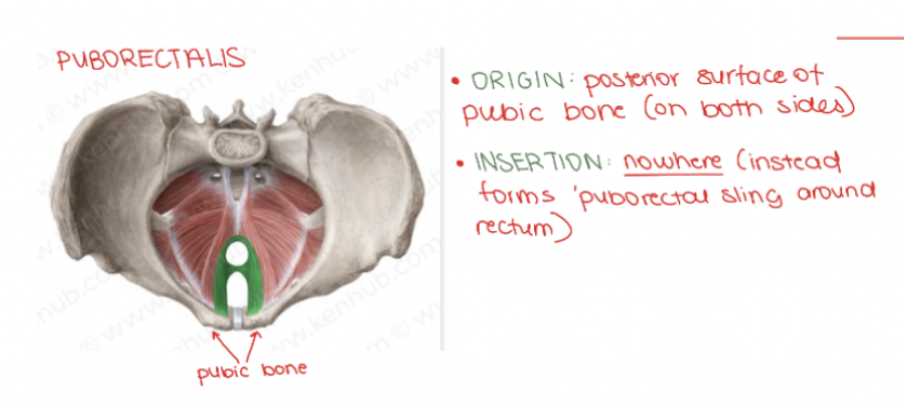
What does PUBORECTALIS do?
makes a sling around the rectum
hold in poop
originates in the anterior pelvis (tendinous arch)
no insertion point! just wraps around to make a sling
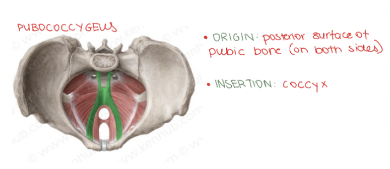
What does PUBOCOCCYGEUS do?
connects the pubic bone to the coccyx (tailbone)
pubo = front to coccygeus = back
originates from the anterior pelvis (tendinous arch)
inserts at the back (coccyx)
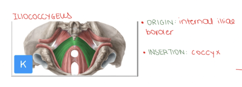
What does ILIOCOCCYGEUS do?
connects the ilium to the coccyx
connects side of the hip to the tailbone
originates from the internal surface of the illiac border (tendinous arch)
inserts at the back (coccyx)
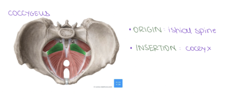
What does COCCYGEUS do?
connects the ischial spine to the coccyx
helps close off the pelvic girdle
originates from the ischial spine
inserts at the coccyx
What is the common origin point for all the levator ani muscles?
COMMON ORIGIN = TENDINOUS ARCH

What are the 2 main openings in the pelvic floor? What do they allow for?
Rectal Hiatus - opening for rectum/anus (poo exits)
Urogenital Hiatus - opening for urethra and vagina**

What are other muscles/features you see but are NOT part of the pelvic floor
**Think of piriformis and obturator internus as background helpers. They're not floor muscles, but they're nearby, and help define the walls of the pelvis.
Piriformis - back wall of pelvis, goes out to the butt
Obturator Internus - side wall of pelvis
**Tendinous Arch - thick fascia over obturator internus (the levators attach here!!!)


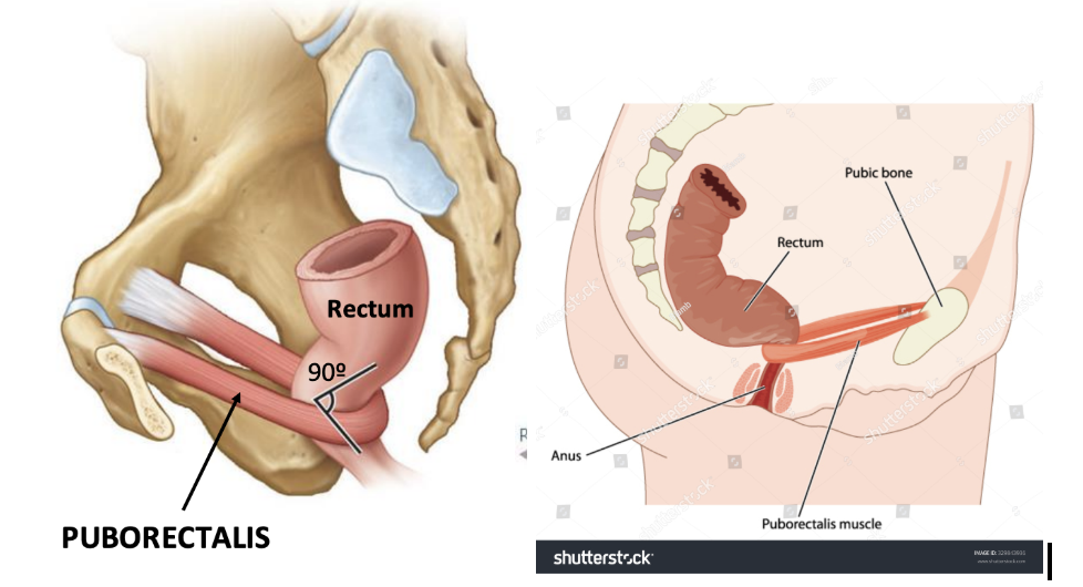
What is the positioning of the puborectalis in relation to the rectum?
It's shaped like a sling or a loop that wraps behind your rectum.
It literally pulls the rectum forward into a sharp angle (about 90°), kind of like bending a straw or hose.
What is tonic contraction?
Tonic contraction = the puborectalis is always a little tight, even when you're not thinking about it. This keeps you from pooping your pants randomly.
WHat happens to the puborectalis when you actually need to poo
When you actually need to poop: You relax puborectalis, the kink disappears, and now poop can pass easily.
Why is squattig better for pooing in comparison to sitting?
SITTING
rectum stays at a 90 degree angke
poop has to work around the bend
SQUATTING
puborectalis relaxes
rectum straightnes
easier smoother pooping

What is the FIRST step/structure poop will encounter on its way out the body (out of four)
RECTUM
Rectum = the end of your large intestine
Like the "waiting room" for poop.
Inside the rectum are 3 folds:
Superior, Middle, and Inferior Rectal Folds
These folds slow down poop and trigger your brain to feel like "I need to go!"

What is the SECOND step/structure poop will encounter on its way out the body (out of four)
RECTAL AMPULLA
Rectal Ampulla = the poop storage pouch
"Ampulla" just means a bulge or swollen area.
When it fills, you feel the urge to poop
What is the THIRD step/structure poop will encounter on its way out the body (out of four)
ANORECTAL JUNCTION
Anorectal Junction = the change from rectum ➝ anal canal
A narrowing point where the final control area begins.
What is the FOURTH step/structure poop will encounter on its way out the body (out of four)
THE ANAL CANAL
3–5 cm long
This is the exit tunnel for poop.
It contains:
Anal Valves – these help secrete mucus to lubricate the area and make pooping easier.
Internal Anal Sphincter – automatic/involuntary muscle that helps keep the anus closed.
External Anal Sphincter – somatic/voluntary muscle, so you control when to poop.
Perianal Skin - skin around the anus; thin and moist which helps with friction and the passage of poo
What are the 3 mechanisms by which poo is controlled to leave the body or stay in the body at a given time?
Internal Anal Sphincter
automatic/involuntary muscle that helps keep the anus closed.
you cannot control it
once the body senses poo it beginning to fill up - it automatically opens
External Anal Sphincter
somatic/voluntary muscle, so you control when to poop
you can control it
you can hold in your poop until it’s appropriate to release
Puborectalis
also helps here by keeping everything bent and closed

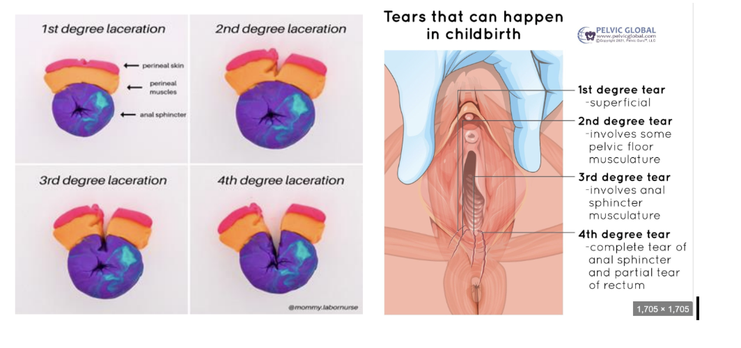
Clinical corelate: What can cause tearing of the pelvic floor muscleS?
When giving birth vaginally, the baby passes through the pelvic floor muscles. This can cause tears (lacerations).
Clinical corelate: What are the 4 degrees of this tearing?

1st/2nd degree = can be stiched up easily
3rd/4th degree = serious - may need surgery (**4th = straight to operating room!)
Clinical correlate: What is an episiotomy?
Sometimes, a doctor may choose to cut the area intentionally to prevent a worse tear.
Median = Cut straight down
Mediolateral = Cut sideways at an angle – safer, less likely to extend into anus
Doctors try to avoid episiotomies unless really needed because natural tearing often heals better.

Clinical correlate: What is sometimes a result of this tearing in female?
a weakened pelvic floor
this can cause vaginal prolapse
After birth, especially if tearing happens, the pelvic floor can weaken. That means it can’t hold up the organs well anymore.
This leads to prolapse = things start to fall out of place, like:
Bladder
Uterus
Vagina
They can bulge out of the vaginal opening.

What is pelvic viscera?
"Viscera" = internal organs
"Pelvic viscera" = internal organs inside your pelvis
Some pelvic viscera are in everyone (like the bladder and rectum), and others are sex-specific, depending on if someone is assigned male or female at birth.
What are the SHARED pelvic viscera? (2)
These are found in all people, regardless of sex:

What are the pelvic viscera found in people ASSIGNED MALE AT BIRTH - AMAB (2 additional ones)

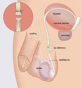
in addition to the bladder + rectum, men have the prostate + penis/scrotum/testes

What are the pelvic viscera found in people ASSIGNED FEMALE AT BIRTH - AMAB (2 additional ones)

in addition to the bladder + rectum, women have the uterus + vagina

**WHAt is a temporary organ found in individuals assigned female at birth?
The Placenta!
Grows during pregnancy (in the uterus)
Is considered a temporary organs
Supports the fetus by giving oxygen/nutrients
Leaves the body after birth
Note: The peritonium form the abdomen does not continue into the pelvis - rather drapes over the pelvic organs and creates pouches within the pelvis. What are the pouches called in men and where are they located? in women?
ASSIGNED MALE AT BIRTH (AMAB)
Rectovesical pouch = space between the rectum and the bladder

ASSIGNED FEMALE AT BIRTH (AFAB)
Vesicouterine pouch: Between bladder & uterus
Rectouterine pouch (a.k.a. pouch of Douglas): Between uterus & rectum

SUMMARY;

VISCERA FOR INDIVIDUALS ASSIGNED MALE AT BIRTH - What is the 9 step pathway of sperm? List first 5 here before it enters in the pelvic girdle
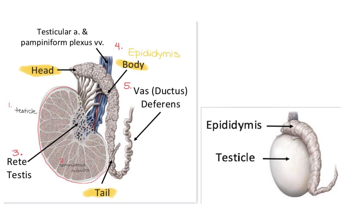
Testicle (Testis)
This is where sperm is made.
Located in the scrotum (outside the body) because the body is too hot for sperm to develop properly.
The scrotum keeps the testicles cooler (around 35°C instead of 37°C).
Seminiferous tubules
Tiny tubes called seminiferous tubules make sperm.
Rete Testis
Like a hallway inside the testicle that collects sperm.
Epididymis
A long, coiled tube that sits on top of the testicle.
Sperm travels through it for 1–2 days to mature (learn to swim and survive).
Has three parts:
Head = entrance
Body = where sperm matures
Tail = storage area before ejaculation
Vas (Ductus) Deferens
A thick tube that carries sperm during ejaculation.
Travels up from the epididymis, into the pelvis via the inguinal canal.
Loops behind the bladder to meet with other reproductive structures
VISCERA FOR INDIVIDUALS ASSIGNED MALE AT BIRTH - What is the 9 step pathway of sperm? List last 4 here after it enters in the pelvic girdle
Seminal Vesicle:
Vas Deferens meets with the Seminal Vesicle
A gland that adds fluid to sperm to make semen.
Fluid is rich in sugar (fructose) to feed sperm.
So now we have sperm + seminal fluid = semen
**The merging of the Vas Deferens + Seminal Vesicle happens POSTERIORLY (behind the bladder)
The Prostate Gland
Located under the bladder
The ejaculatory ducts pass through it: Adds more fluid to semen — contains enzymes and activates sperm so they start swimming
Also has openings into the urethra for prostatic fluid
Bulbourethral Glands (Cowper's Glands)
Located below the prostate, near the membranous urethra
Adds pre-ejaculate fluid to clean and neutralize the urethra.
This happens before ejaculation to protect sperm from acid.
What does this fluid do?
Neutralizes acidity in the urethra (leftover from urine)
Lubricates the urethra for smooth passage of semen
Protects sperm when they come through
Urethra
Carries both urine and semen, but not at the same time.
internal urethral sphincter closes during ejaculation so pee doesn’t mix with semen

SUMMARY;

VISCERA FOR INDIVIDUALS ASSIGNED MALE AT BIRTH - Why is this complex setup nessecary?
Because sperm are delicate, and need:
The right temperature
The right nutrients
The right pH (not too acidic)
The right environment to survive and swim
Each part of the male reproductive tract helps with that.
VISCERA FOR INDIVIDUALS ASSIGNED FEMALE AT BIRTH - What are the structures of the female system from the outside in (7)

What is the cervix?
It's the "gate" between the uterus and vagina.
What does the cervix look like in someone who is nulliparous vs. parous
Normally it’s tight and round like a donut.
Nulliparous = Person who has never had a vaginal birth → cervix is small and round
Parous = Person who has had a vaginal birth → cervix is wider and more oval due to stretching

in what 2 ways does the cervix change during labor
Dilation - How open the cervix is (1–10 cm)
Effacement - How thin the cervix is (0–100%)
Fully ready to push: 10 cm dilated AND 100% effaced

What are the main parts of the uterus (3) What are the 3 layers?
Uterus
Fundus: Top rounded part of the uterus (above where the fallopian tubes connect)
Body: Main central portion of the uterus (where a fertilized egg implants and the fetus grows)
Isthmus: Narrow bottom part of the uterus, just above the cervix (transitions into the cervix)
Layers of the Uterus (Body)
Perimetrium: Outer layer (like skin)
Myometrium: Middle muscle layer (contracts in labor!)
Endometrium: Inner lining that sheds during your period if no pregnancy happens
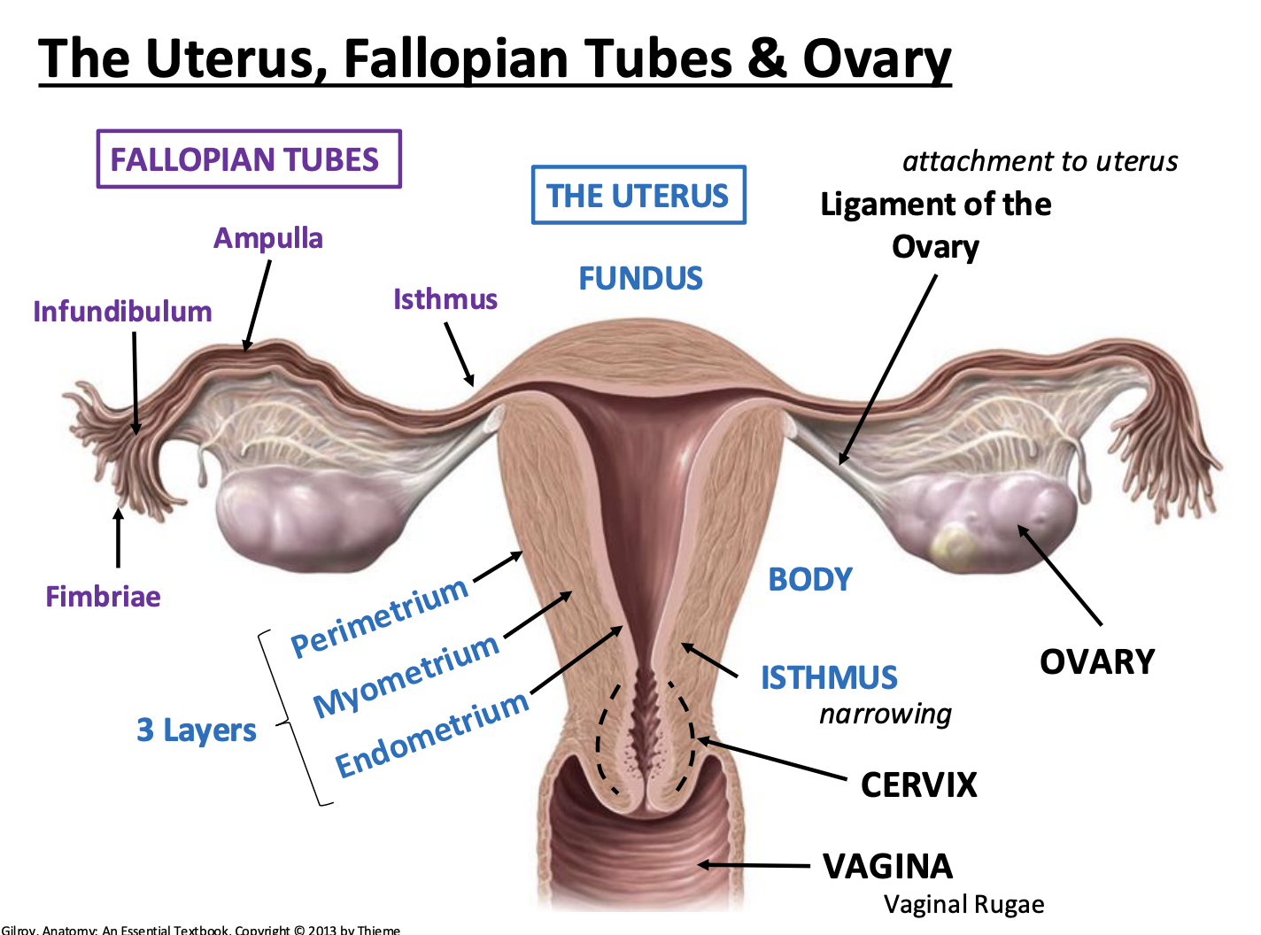
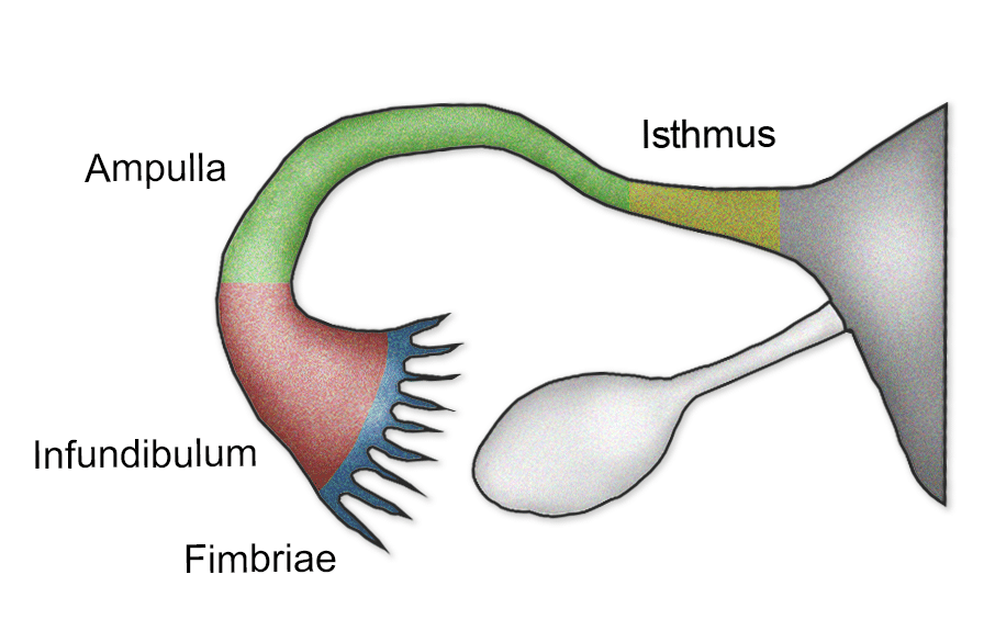
What are the main parts of the fallopian tubes? (4)
Fallopian tubes (Uterine tubes)
These are the pathways for the egg. Each has 4 parts:
Fimbriae: Finger-like structures that "grab" the egg
Infundibulum: Funnel-shaped opening near the ovary
Ampulla: Most common place for fertilization 💥
Isthmus : Narrow part attached to the uterus
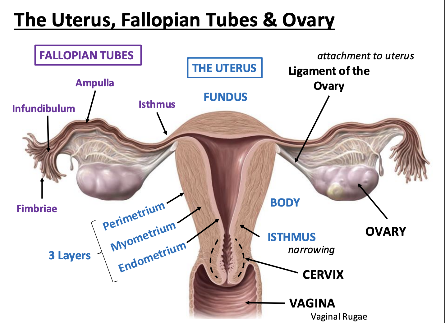
What is the ovaries?
The ovary is a female reproductive organ that produces eggs (ova) and releases hormones like estrogen and progesterone. It plays a key role in the menstrual cycle, ovulation, and hormone regulation.
The ovary is NOT physically attached to the tube or uterus - so how does the egg in the ovary get fertilized in the tubes
The egg is released from the ovary during ovulation.
It does NOT go directly into the fallopian (uterine) tube right away because The ovary and fallopian tube are NOT directly connected - There is a small gap between them
The egg enters the fimbriae — the finger-like ends of the fallopian tube.
These fimbriae wave and create a current (like little arms waving "come here!").
This movement guides the egg from the ovary into the opening of the fallopian tube.
****So the egg is released into the open abdominal cavity, and then the fimbriae scoop it up into the tube.
What is a specialized feature of the vagina? where else do we see this?
Rugae are folds in the internal lining of certain organs that allow them to expand.
They are found in the stomach, vagina, and urinary bladder.
What is Anti version? Anti flexion?
Version: Angle between vagina and cervix
Flexion: Angle between cervix and uterus
“Version = vaginal angle, Flexion = uterine bend”
Normal Position:
Anteverted (tilted forward)
Anteflexed (bent forward over bladder)
Most common uterine position

What are some abnormal uterine bending? significance on fertility?


Can affect fertility by making it harder for sperm to reach the egg — may be corrected with surgery.
What are the 4 stages of uterine growth during pregnancy in comparison to fruit?

Then baby drops into the pelvis just before birth (40 weeks) = “lightening”
Clinical Correlate: What is an etopic pregancy?
Normal: Egg is fertilized in the fallopian tube and implants in the uterus.
Ectopic = fertilized egg implants outside the uterus
Where can the egg implant? why can this happen?

Why is it possible?
Because the fimbriae and ovary are open to the peritoneal cavity.
The egg or sperm can wander and implant in the wrong place.
These pregnancies are not viable and can be life-threatening if not caught early (especially tubal — can rupture the tube).