UNC BIOL 252 Lab Exam 1
 Call Kai
 Learn
Learn Practice Test
 Spaced Repetition
 Match
Match Flashcards
 Knowt Play
1/282

There's no tags or description
Looks like no tags are added yet.
Name | Mastery | Learn | Test | Matching | Spaced | Call with Kai | Chat |
|---|
No analytics yet
Send a link to your students to track their progress
283 Terms
vertebral foramen
canal through which spinal cord passes

vertebral canal
formed by vertebral column and contains spinal cord and the beginnings of spinal nerves

cervical enlargement
supplies nerves to the shoulder and upper limbs

lumbar enlargement
nerves to pelvic region and lower limbs

spinal nerves
31 pairs of nerves arising from the spinal cord
conus medularis
The cone-shaped termination of the spinal cord

cauda equina
collection of lumbar sacral, and coccygeal spinal nerves below the end of the spinal cord

filum terminale
extension of pia mater that anchors the spinal cord to the coccyx

C1-C8 nerves are named according to vertebrae (inferior or superior) to the nerve
inferior
For all spinal nerves outside of C1-C8, they are named according to the the (superior or inferior) vertebrae
superior
The end of the spinal cord only extends to which vertebra?
L1
dermatome
Area of skin supplied by a single pair of spinal nerves
Meninges
cover and protect the brain and spinal cord

dura mater
thick, outermost layer of the meninges surrounding and protecting the brain and spinal cord

epidural space
fat-filled space surrounding the dura mater that is the common site of injection for anesthetic

arachnoid mater
middle layer of the meninges

subarachnoid space
a space in the meninges beneath the arachnoid membrane and above the pia mater that contains the cerebrospinal fluid
pia mater
Innermost layer of the meninges

denticulate ligaments
20 pairs of lateral projections that anchor the spinal cord to the dura mater and limit the movement of the spinal cord

filum terminale
the continuation of the pia mater past the conus medularis that anchors the end of the spinal cord to the sacrum
central canal
the canal of the spinal cord that runs through the center and connects the ventricles of the brain; filled with cerebrospinal fluid
white matter
outer region of the spinal cord that contains myelinated axons
gray matter
the cell bodies of neurons and their synapses with other neurons
somatic neurons have terminals in the:
skin, skeletal muscles, or bones
visceral neurons have terminals in:
smooth muscles, cardiac muscles, glands, or other viscera within body cavities
the two categories of peripheral nervous system neurons
motor or sensory
motor neurons bring signals from ________ to the __________
spinal cord; muscles and glands of the body
sensory neurons bring information into the ___________ from the ________
spinal cord; skin and other organs
dorsal horn
the cell bodies here are interneurons that receive connections from sensory neurons
the dorsal-most region of the dorsal horn receives ____________________ neurons from the _________________
somatic sensory; skin, skeletal muscles, and bones
the region of the dorsal horn closest to the center of the spinal cord contains interneurons that receive connections from _______________________ neurons from __________________
visceral sensory; organs within body cavities
lateral horn
contains cell bodies for visceral motor neurons
the visceral motor neurons within the lateral horn send signals to ___________________
smooth muscle, cardiac muscle, and glands
ventral horn
contains cell bodies for somatic motor neurons
somatic motor neurons send signals to:
skeletal muscle
descending pathways
the systems of neurons that bring information from the brain down to the appropriate muscle or gland of the body
corticospinal tract
information travels from the cortex to the spinal cord via two neurons
the upper motor neuron of the corticospinal tract travels within the __________ and is an _________________ of the CNS
corticospinal tract; interneuron
the lower motor neuron of the corticospinal tract travels with the ____________ and eventually reaches its _________________
spinal nerve; skeletal muscle target
ascending pathways
The systems of neurons that bring information up to the brain. Signals travel along three neurons. An example is the spinothalamic tract.
1st order neuron
the sensory neuron extends from the source of the stimulus to the dorsal horn where it synapses with a 2nd order neuron
2nd order neuron
this interneuron crosses to the other side of the spinal cord and then its axon enters the white matter. from the white matter, the axon occupies the spinothalamic tract until reaching its snaps (sensory nerve action potential) in the thalamus
3rd order neuron
this next interneuron relays the signal from the 2nd order neuron up to the cerebral cortex, allowing conscious awareness of sensation
spinal nerve
a peripheral nerve attached to the spinal cord

dorsal root
contains only sensory neurons that are entering the dorsal horn of the spinal cord

dorsal root ganglion
contains cell bodies of sensory neurons

sensory neurons are called ____________ because of the single process that stems from the cell body
unipolar
ventral root
contains only axons of motor neurons
spinal reflexes
involuntary and stereotyped reactions
polysynaptic reflex
the stimulus travels along a sensory neuron that synapses with one of several interneurons in the dorsal horn then several sets of motor neurons are stimulated to exact a motor response
monosynaptic reflex
sensory neuron synapses directly onto motor neuron in the ventral horn
the withdrawal reflex is an example of a _______________
polysynaptic reflex
the patellar reflex is an example of a _________________
monosynaptic reflex
rami
branches of spinal nerves
dorsal and ventral ramus
carry somatic motor and somatic sensory neurons to the skin, muscle, and bones
communicating rami
carry visceral motor and visceral sensory neurons to the smooth muscle, cardiac muscle, and glands of the body

sympathetic ganglia
contain a second cell body for the sympathetic nervous system (the first is in the lateral horn spinal segments T1-L2)

sympathetic train/trunk
runs parallel to the spinal cord and distributes innervation to the viscera
visceral sensory neurons travel from ___________ through the ___________ and into the ____________ before reaching the ____________
their visceral targets; communicating rami; dorsal root; dorsal horn
referred pain
pain that is felt in a location other than where the pain originates
The pain of a heart attack may be felt in the left shoulder; this is an example of
referred pain
intercostal nerves
supply muscles of the ribs, anterolateral thorax, and abdominal wall

plexus
combine somatic motor and sensory neurons from as many as 6 ventral rami
brachial plexus
C5-T1 spinal nerve rami supplies the upper limb

lumbosacral plexus
Formed of L2-S2 rami. Supplies the lower limb.

central pattern generators
neural mechanisms in the spinal cord that generate rhythmic patterns of motor output for locomotion
What explains why the cervical and lumbar enlargements are larger than the spinal cord between them?
A.There are more neurons in these regions that receive information from the limbs
B.The neurons between the enlargements are smaller, but approximately equal in number to those in the enlargements
C.There are more neurons in these regions that send information to the limbs
D.The spinal nerves have to narrow to pass through the intervertebral foramen
A and D
Why are the lumbar, sacral, and coccygeal spinal nerves present within the lumbar vertebral canal?
A. The spinal cord grows more slowly, but will reach the lower parts of the vertebral canal in adulthood.
B. Spinal nerves pass through the thoracic vertebral canal on their way to the appropriate intervertebral foramen in the sacrum and coccyx.
C. The spinal cord becomes the filum terminale, so there's nothing left but spinal nerves in this region.
D. The spinal cord no longer resides in the these regions but the spinal nerves still pass through on their way to the approbpriate intervertebral foramen.
D
Which of the following is NOT found anywhere within the dorsal root, ventral root, or spinal nerve?
A. Motor axons
B. Interneuron axons
C. Sensory axons
D. Sensory neuron cell bodies
B
Match the functional grouping of the peripheral nervous system with the central nervous system structure with which it begins or ends.
A. Dorsal horn
B. Lateral horn
C. Ventral horn
1. Somatic motor
2. Somatic sensory
3. Visceral motor
4. Visceral sensory
1. C
2. A
3. B
4. A
A monosynaptic reflex involves a total of _____ neurons.
2
For most of the spinal cord, a given spinal segment is located more _________ than the corresponding spinal nerve's exit from the vertebral canal.
A. posteriorly
B. superiorly
C. laterally
D. medially
E. anteriorly
F. inferiorly
B
If it is part of an ascending pathway, a neuron that has its cell body in the dorsal horn will...
A. have its axon traveling with a spinal nerve
B. synapse in the cerebral cortex
C. be a sensory neuron
D. have its axon in a white matter column
E. synapse in the thalamus
D and E
An upper motor neuron...
A. synapses in the dorsal horn
B. is an interneuron
C. is a motor neuron
D. may have its cell body in the primary motor cortex
E. may have its cell body in the thalamus
F. synapses in the ventral horn
B,D, and F
The sympathetic chain contains a group of cell bodies called the sympathetic chain ___________ . This structure contains a second cell body for the sympathetic nervous system, the first in the series being found in the _______ horn of spinal segments T1-L2. The purpose of the ___________ rami is to carry visceral motor and visceral sensory neurons to and from this sympathetic chain.
ganglia, lateral, communicating
Check all that apply: Which structures contain motor neurons?
A. Ventral root
B. Dorsal ramus
C. Ventral ramus
D. Dorsal root ganglion
E. Dorsal root
A, B, and C
The region of the spinal cord that gives off the L1 spinal nerves is located ___.
A. At the first lumbar vertebra
B. Superior to the first lumbar vertebra
C. Inferior to the first lumbar vertebra
B
Match the functional grouping with the appropriate part of the spinal cord.
A. Lateral horn
B. Dorsal horn
C. Ventral horn
1. Somatic motor neurons originate here
2. Visceral motor neurons originate here
3. Sensory neurons terminate here
1. C
2. A
3. B
A fractured vertebra damages the left ventral root of a single spinal nerve. What is the impact to the patient's left side?
A. No motor control in the region supplied by that spinal nerve.
B. No sensation in the region supplied by that spinal nerve.
C. Neither motor control nor sensation to the region supplied by that spinal nerve.
A
Match the cell bodies of neurons with their location.
A. Dorsal root ganglia
B. Ventral horn of the spinal cord
C. Sympathetic chain ganglia
D. Dorsal horn of the spinal cord
1. Sensory neuron cell bodies
2. Motor neuron cell bodies
3. Interneuron cell bodies
4. Sympathetic neuron cell bodies
1. D
2. B
3. A
4. C
The grouping of ventral rami that travel to innervate the upper limb is called the " _________________ plexus."
brachial
Four weeks following fertilization the forerunner to the brain and spinal cord is formed, the _________________
neural tube
6 major divisions of the brain
cerebrum, diencephalon, midbrain, pons, medulla oblongata, cerebellum
nuclei
the central gray matter of the brain
ventricles
fluid filled cavity of the brain
cortex
superficial region of gray matter
white matter of the brain
contains myelinated axons traveling to and from the brain's nuclio and cortex
cerebrospinal fluid
a fluid that nourishes and protects the brain; produced from blood at the choroid plexuses in the walls of the ventricles
lateral ventricles
A set of paired ventricles lying within the cerebral hemispheres.

septum pellucidum
thin membrane that separates lateral ventricles

3rd ventricle of brain
connected to the lateral ventricles by the interventricular foramen
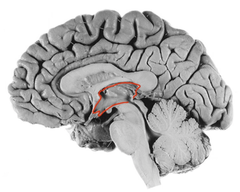
interventricular foramen
connects lateral ventricles to third ventricle
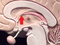
cerebral aqueduct
a narrow tube interconnecting the third and fourth ventricles of the brain, located in the center of the mesencephalon

4th ventricle
ventricle between pons and cerebellum

central canal of spinal cord
4th ventricle narrows to become this

dural venous sinuses
large veins in the dura mater that drain blood away from the brain and scalp
arachnoid granulations
Extensions of the arachnoid mater that allow excess CSF to be absorbed by the dural sinuses
falx cerebri
located within the longitudinal hemisphere and separates the two cerebral hemispheres

superior sagittal sinus
A venous sinus located in the midline just dorsal to the corpus callosum, between the two cerebral hemispheres.

tentorium cerebelli
located within the transverse fissure that separates cerebrum from cerebellum
