Chapter 13 - Central Nervous System Overview: Key Terms and Concepts
1/82
There's no tags or description
Looks like no tags are added yet.
Name | Mastery | Learn | Test | Matching | Spaced |
|---|
No study sessions yet.
83 Terms
Rostral
toward the nose/snout
Caudal
toward the tail
Cerebrum
largest part of the brain...left and right hemispheres
Diencephalon
gives rise to the thalamus, hypothalamus, and epithalamus
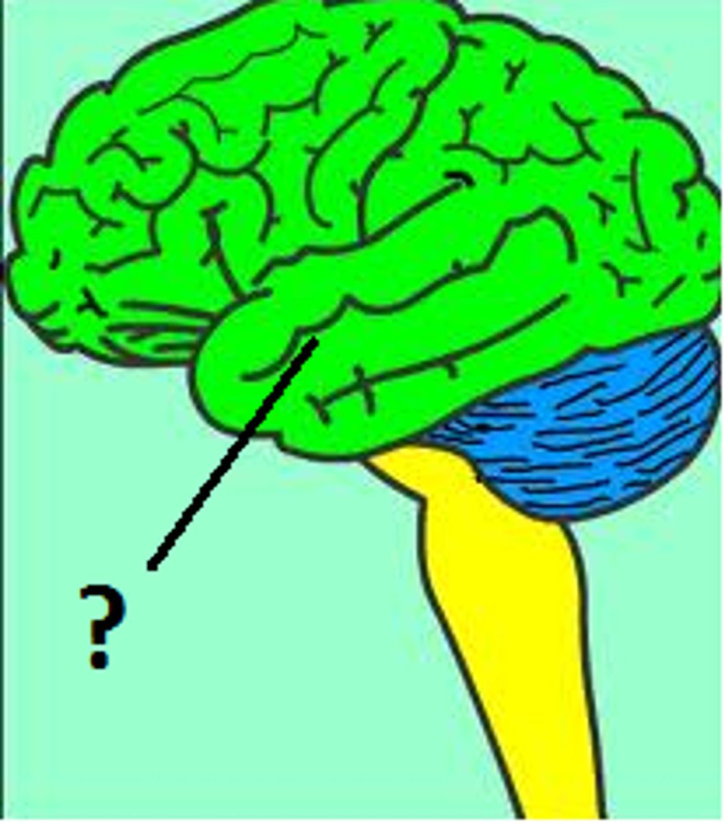
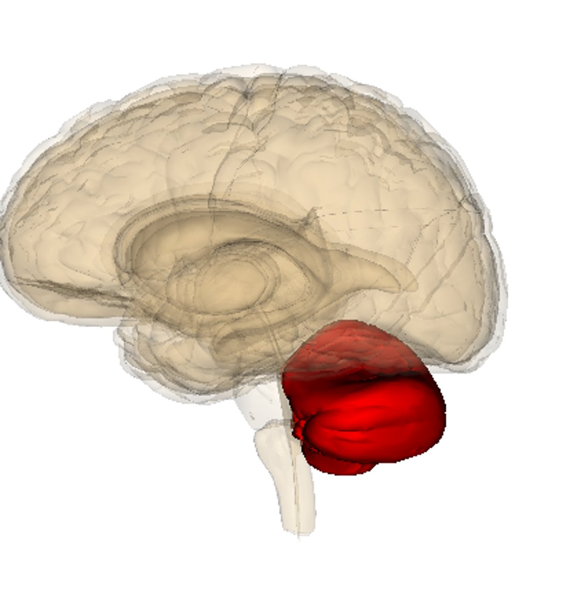
Cerebellum
second largest part of the brain
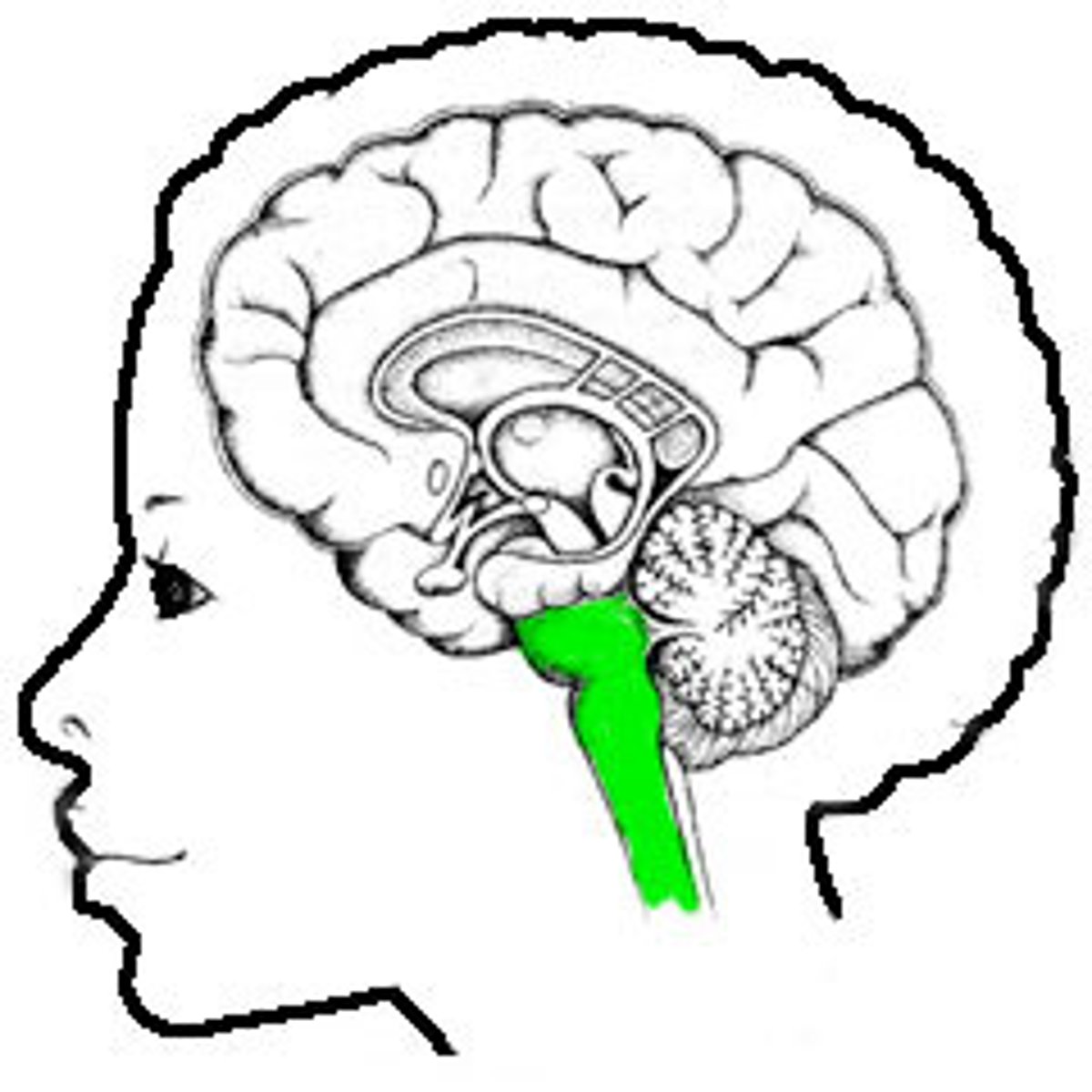
brain stem
rostral continuation of the spinal cord; consists of the medulla oblongata, pons, and midbrain
lateral ventricles
cerebrum
third ventricle
diencephalon
cerebral aqueduct
brain stem: midbrain
fourth ventricle
pons, cerebellum, medulla obolongata
central canal
spinal cord
Brain ventricles
cerebrospinal fluid filled cavities within the brain
Continuous with each other and central canal
Lined with ciliated ependymal cells
provide CSF to nearby brain regions
What does CSF provide?
cushion, nutrients, buoyancy
What is the function of external wrinkles on cerebrum?
increase surface area
Fissure
deep groove
Sulcus
where groove is
Gyrus
wrinkles
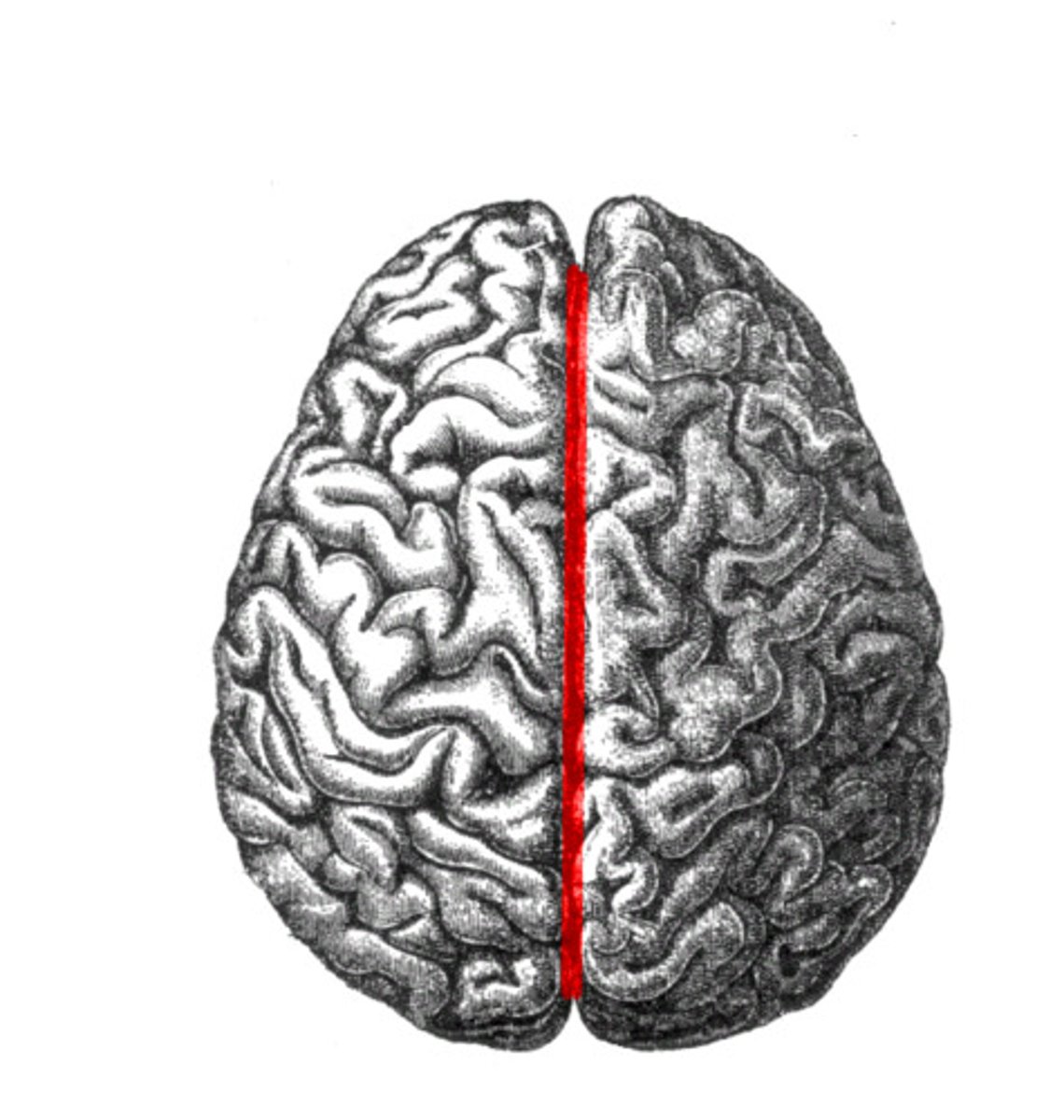
Longitudinal Fissure
separates left and right cerebral hemispheres
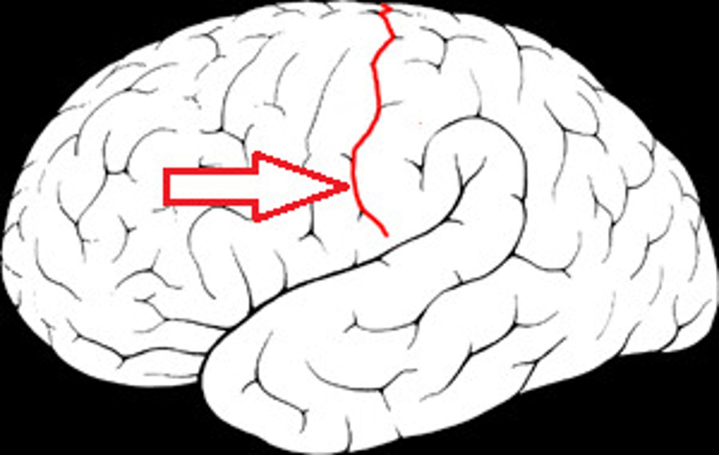
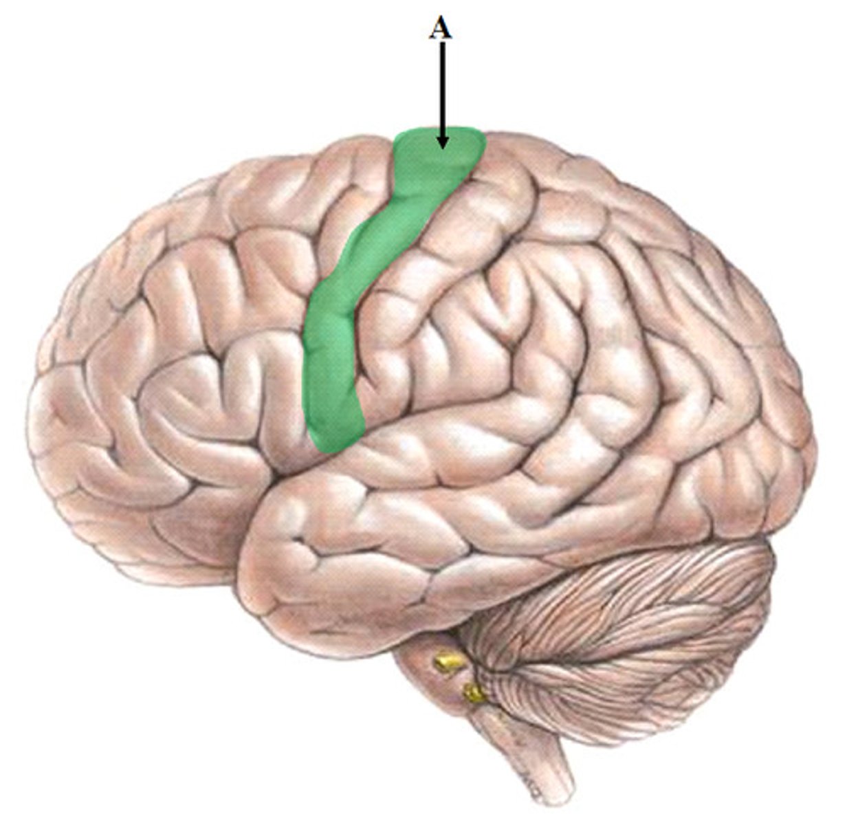
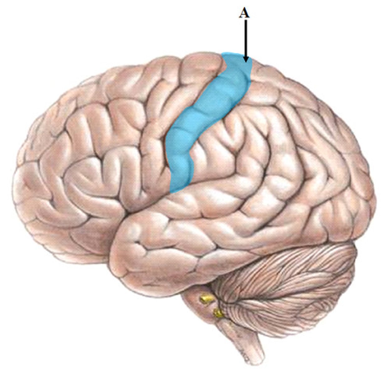
Central sulcus
separates frontal and parietal lobes
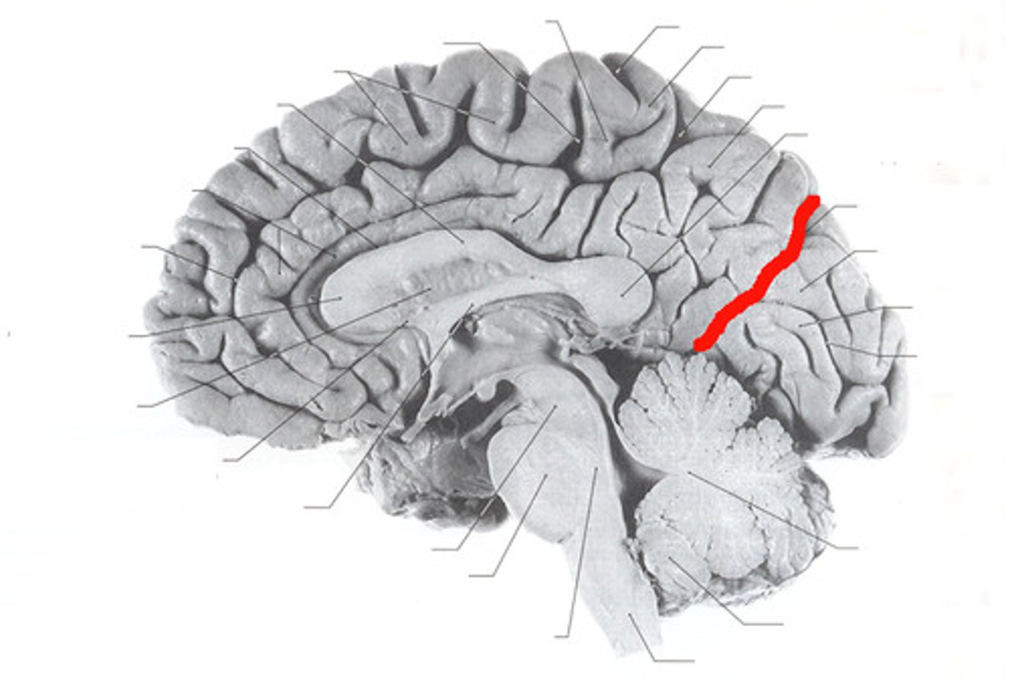
Parieto-occipital sulcus
seperates parietal and occipital
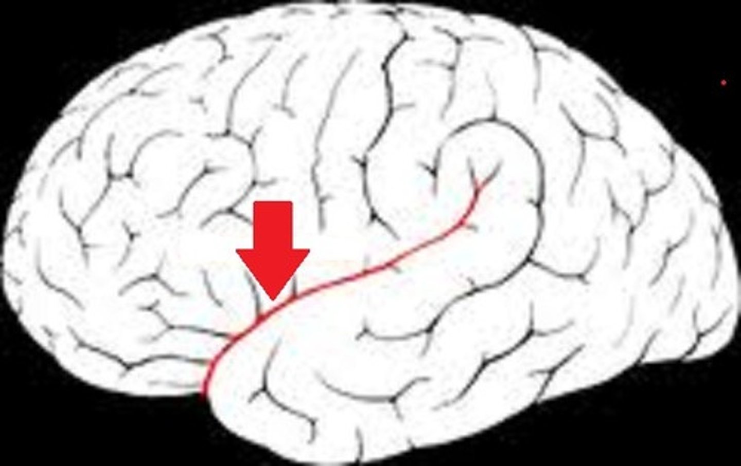
lateral sulcus
Separates temporal lobe from parietal and frontal lobes
precentral gyrus
before central sulcus; in frontal lobe
postcentral gyrus
after central sulcus; in parietal lobe
function of precentral gyrus
primary motor cortex
function of postcentral gyrus
primary somatosensory cortex
function of temporal lobe
primary auditory cortex
function of occipital lobe
primary visual cortex
What determines amount of cortex used for a body part?
amount of cortex devoted to a given body region is proportional to either the number of muscles and motor units there or the number of receptors for a particular body part
three fibers of cerebral white matter
commissural fibers, associational fibers, projection fibers
commissural fibers
connect right hemisphere to left
Largest= corpus callosum
associational fibers
Intrahemispheric
Long or short
projection fibers
connects cortex to lower CNS areas
vertical fibers
Whats in cerebral deep grey matter?
basal nuclei (ganglia)
Basal nuclei( ganglia)
initiates and terminates body movements
suppresses unwanted movements
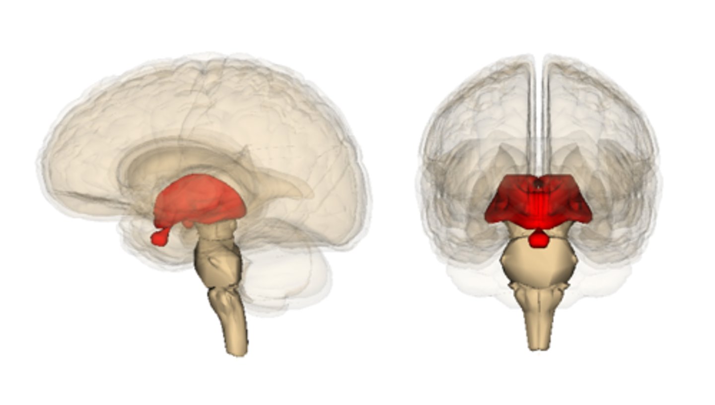
Thalamus: shape and how many?
Egg shaped, one for each hemisphere
hypothalamus: location
below the thalamus
Epithalamus (pineal gland) function
secretes melatonin
Thalamus function
"gateway to the cerebral cortex"; major relay station for most sensory inputs; filters info on its way to cerebral cortex
Ventral posterolateral nucleus function
sensory
medial geniculate body
auditory
lateral geniculate body
visual
function of hypothalamus
Maintains Homeostasis by regulating body temp, hunger, thirst
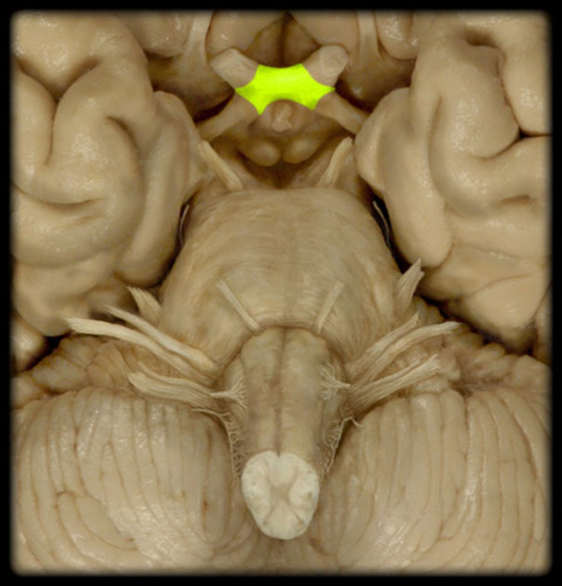
Optic chiasm
crossover point for optic nerve
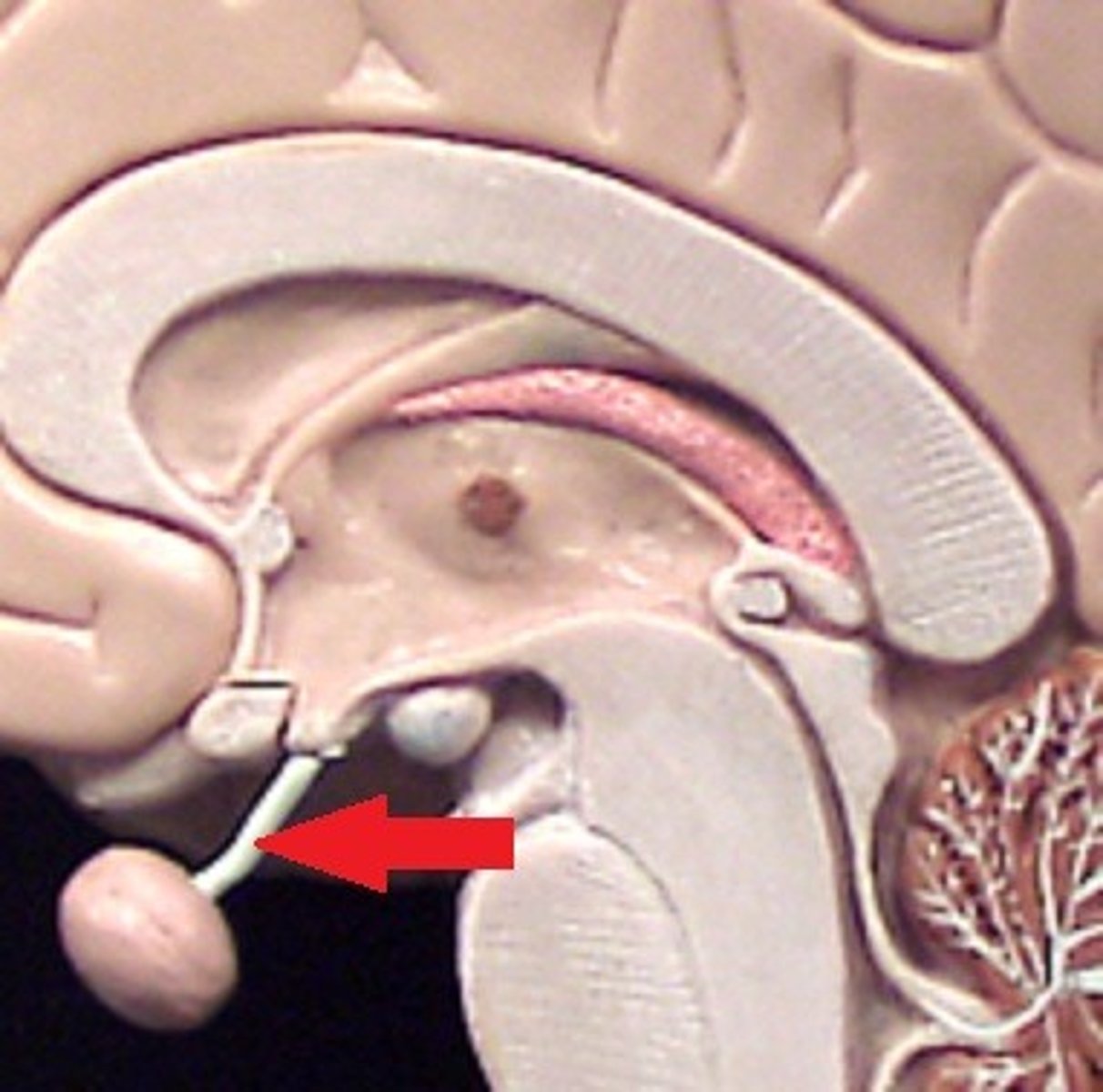
infundibulum
connects hypothalamus to pituitary
interthalamic adhesion
connects thalamus
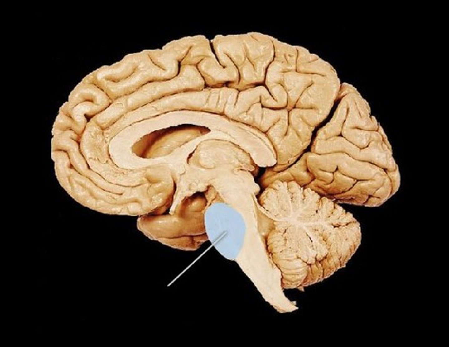
cerebral peduncles
composed mostly of motor axons from the cortex to the cerebellum and spinal cord
looks like columns/pillars supporting the cerebrum
located ventrally
cerebral aqueduct
passes through the center of midbrain; 3rd ventricle to fourth
corpora quadrigemina
nuclei that form 4 bumps on the dorsal midbrain
Superior colliculi: visual reflexes
Inferior colliculi: auditory reflexes
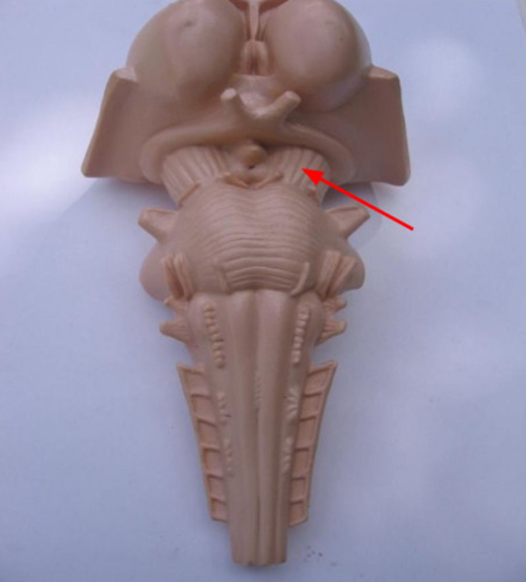
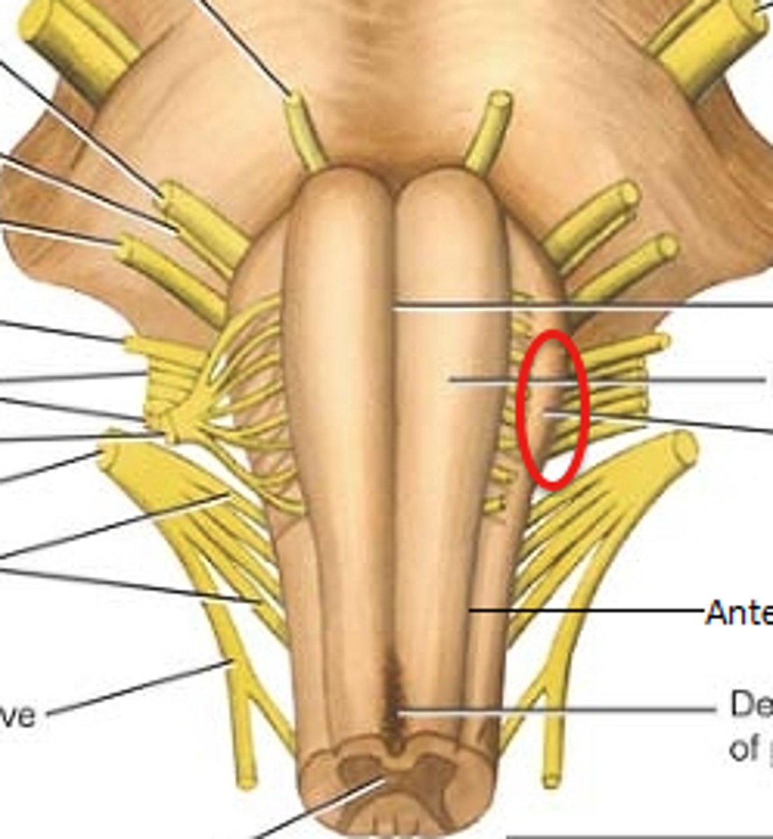
Pons location
between midbrain and medulla oblongata; functions as bridge between brain stem and cerebellum
viewed ventrally as superficial, transverse fibers
Two structures on medulla oblongata?
continuous with superior aspect of the spinal cord
pyramids and olives
Pyramids
motor tracts on anterior surface of medulla
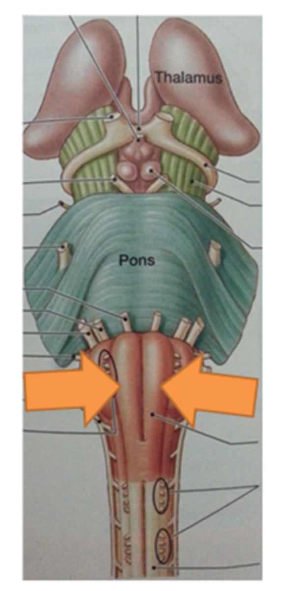
Olives
nuclei that are lateral to pyramids; sensory relay station
Cerebellum structure
two hemispheres
cortex- grey matter with many folds called folia
white matter-tracts called arbor vitae
deep grey matter
What connects two hemispheres of cerebellum?
vermis
anterior and posterior lobe of cerebellum function
motor coordination of trunk and limbs balance
flocculonodular lobe of cerebellum
equillibrium via head/eye motor coordination
What are the protective coverings of the CNS?
skull, vertebrae, meninges
Meninges
connective tissue sheaths that surround the brain and spinal cord
Three layers of meninges
Dura mater: most superfifical
Arachnoid matter: middle
Pia mater: deepest
two layers of dura mater
periosteal layer and meningeal layer; two layers continuous unless sinus
arachnoid granulations
allow CSF to drain into sinus
Is CSF only found inside the brain?
No, inside and outside
Where is CSF deposited
dural venous sinuses around the brain
Falx Cerebri
separates the two cerebral hemispheres
tentorium cerebelli
separates cerebrum from cerebellum
falx cerebelli
separates the two cerebellar hemispheres
Spinal cord: position relative to brain and function
Inferior to head
sensory and motor innervation of the whole body
two-way conduction pathway between the brain and body
major integration center for reflexes
Most caudal part of CNS?
conus medullaris
Does spinal cord extend to end of spinal column?
No
Location of spinal cord segments?
typically superior to respective vertebrae
Where does the spinal cord end?
L1 or a little beyond; 31 pairs of spinal nerves
Are spinal nerves apart of the CNS?
No
Grey matter of spinal cord
neuron cell bodies and axons with little myelin
white matter of spinal cord
abundantly myelinated axons
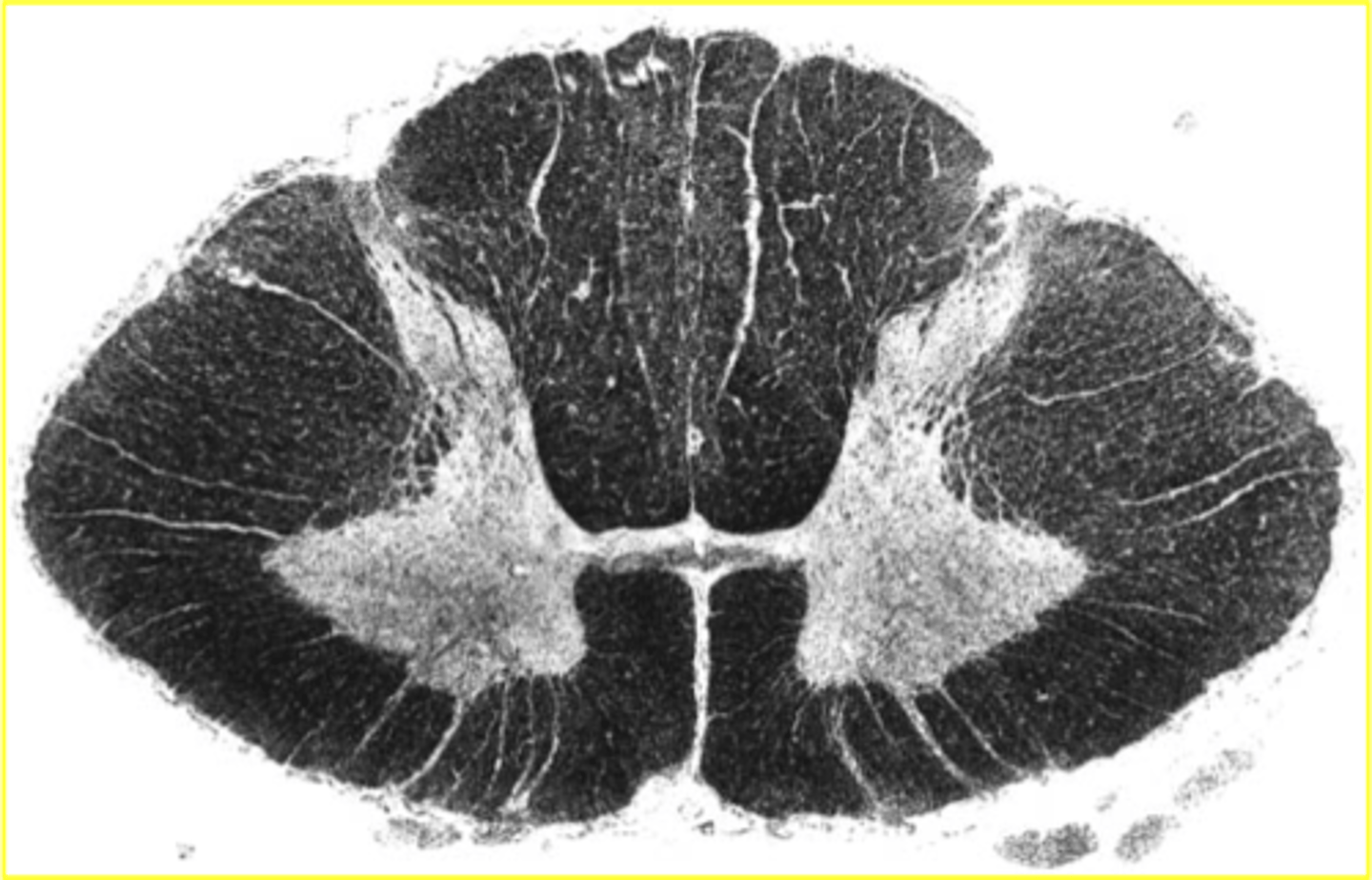
Cervical spinal cord segments
large amounts of white matter; anterior grey horn is large
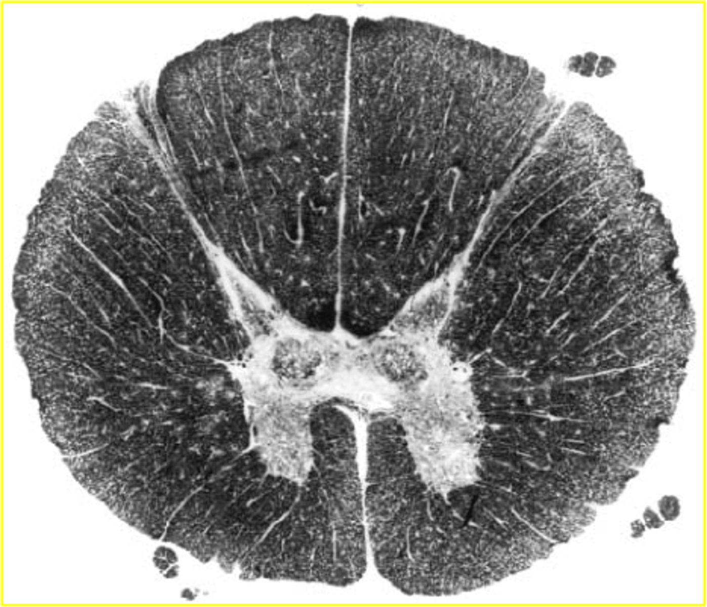
thoracic spinal cord segment
very small amount of grey matter
lumbar spinal cord segment
large anterior/posterior grey horns; considerably less white matter compared to cervical
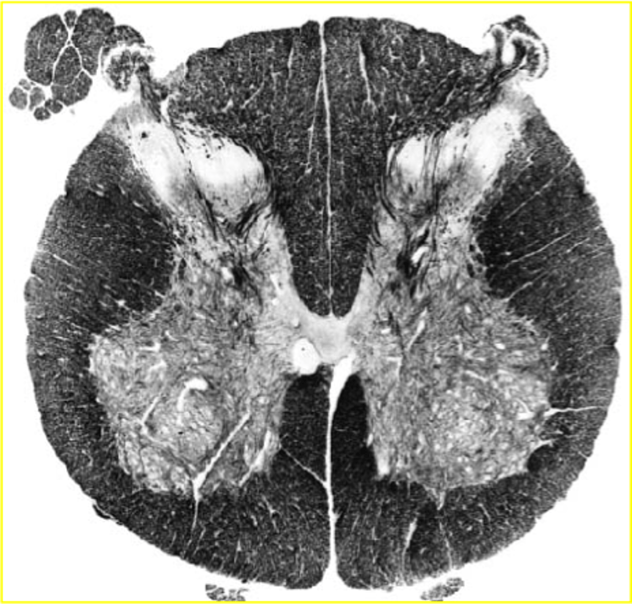
sacral spinal cord segments
small in diameter, but relatively large amounts of grey matter
Function of ascending tracts of spinal cord
carry sensory information from the body to the brain
Function of descending tracts in spinal cord
Deliver motor (output) instructions
Is spinal cord white matter sensory or motor?
both
How are white matter columns in spinal cord named?
according to where a tract begins(soma) and where it ends(axon terminals)
How does CSF flow?
CSF is formed by the choroid plexus in all main ventricles. From the right and left lateral ventricles, CSF flows into the third ventricle. From there, it flows through the narrow channel of the midbrain called the cerebral aqueduct and into the fourth ventricle. From there, CSF travels into the central canal of the spinal cord AND out of the one median and two lateral apertures of the 4th ventricle into the subarachnoid space surrounding both the brain and spinal cord. Inside the brain, CSF is moved into the superior sagittal sinus through arachnoid villi. These sinuses will join together to form the internal jugular veins.