Ankle Anatomy/X-rays/Fractures
1/13
There's no tags or description
Looks like no tags are added yet.
Name | Mastery | Learn | Test | Matching | Spaced | Call with Kai | Chat |
|---|
No analytics yet
Send a link to your students to track their progress
14 Terms
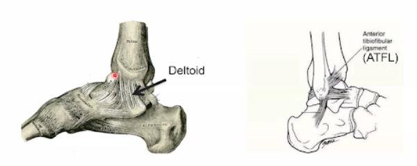
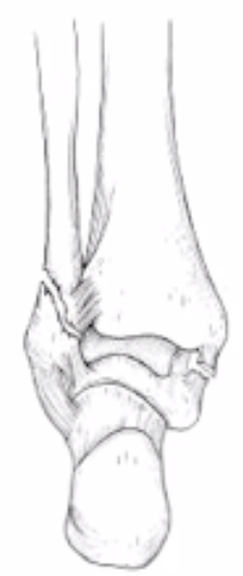
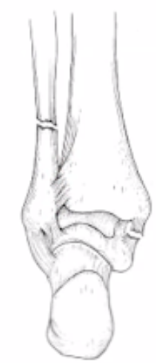
What view is the Deltoid
Medial

What view is Syndesmosis
Lateral
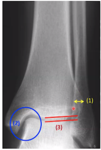
What is shown in this view?
AP view
1. Tibiofibular overlap
Fibula is posterior to the tibia
2. Medial clear space
3. Tibiotalar joint space
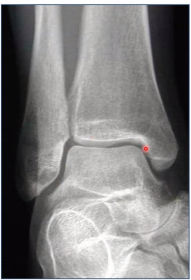
What is being shown in this x-ray
Mortise x-ray
Leg internally rotated 15-25 degrees
Visualization of the space between the tibia and the fibula
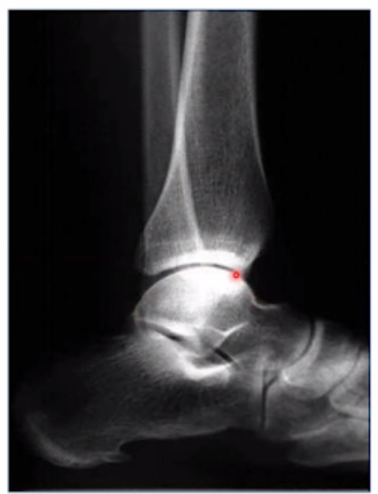
What is this view showing?
Lateral view
Tibiotalar joint space is uniform
What fracture is this?
Bi-malleolar
Commonly the medial and lateral malleoli
What fracture is this?
Tri-malleolar
Medial, lateral, posterior malleoli
What fracture is this?
Pilon fracture
Plafond is severely fractured
What is the plafond?
The articular surface at the bottom of the tibia
What fracture is this?
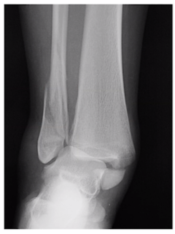
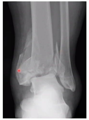
Weber A fracture
Usually transverse, avulsion fracture
What fracture is this?
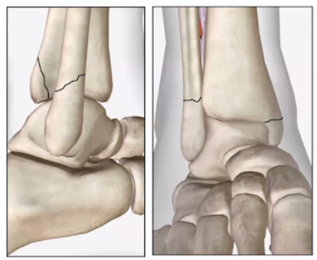
Weber B
Usually a spiral fracture
What fracture is this?
Weber c
Unstable fracture
Syndesmosis disruption
Above ankle joint
What is a syndesmotic injury?
Tibia/Fibula relationship disrupted
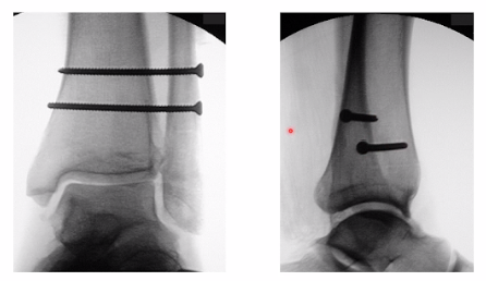
How to treat syndesmotic injuries
30 degree screw insertion from fibula into tibia (fibula is posterior to tibia)
screws
fibula plate
fibulink