Di Imaging II - Exam 2 (Extremity Trauma)
 Call Kai
 Learn
Learn Practice Test
 Spaced Repetition
 Match
Match Flashcards
 Knowt Play
1/76

There's no tags or description
Looks like no tags are added yet.
Name | Mastery | Learn | Test | Matching | Spaced | Call with Kai | Chat |
|---|
No analytics yet
Send a link to your students to track their progress
77 Terms
Types of fractures
transverse, oblique, spiral, comminuted, greenstick, avulsion
Incomplete fractures
greenstick and torus
Forces involved in production of fx
angulation (transverse fx)
compression/impaction (oblique fx)
rotation (spiral fx)
shear
tension (transverse fx)
Transverse fx typically result from
direct blows or tensile forces
pathologic bone conditions
Oblique fx typically result from
angulation and compression
Spiral fx typically result from
torque and axial compression
ends are pointed like a pen
Distal fragment compared to proximal fragment
ALWAYS
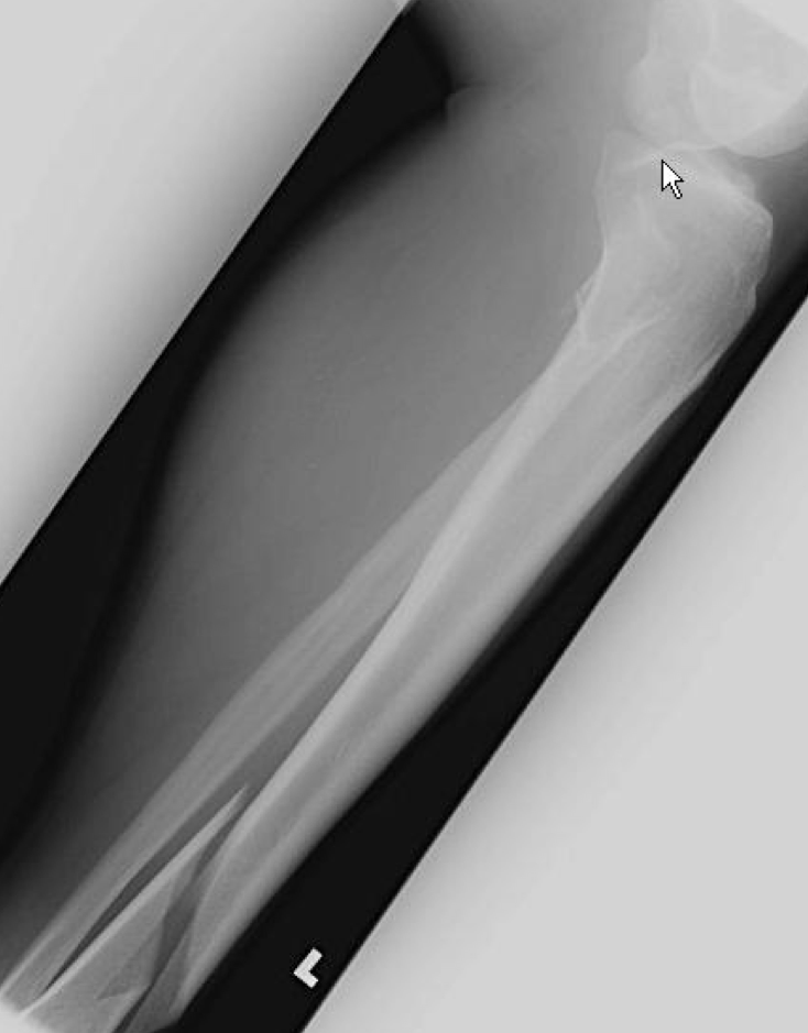
What type of fx is this
spiral fx of distal tibia w/posterior displacement

What type of fx is this
spiral fx with overriding deformity, posterior and lateral displacement and rotational misalignment
Alignment
describes the ANGULATION of the distal fragment at the fx site
Apposition
describes the CLOSENESS of the bony contact at fx site
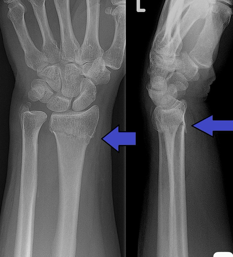
What type of fx is this
transverse fx of distal radius, good apposition (heal easier)
Epiphyseal (weakest part of bone) injuries are MC btw the ages of
10-16yo and elderly
Three phases of healing
inflammatory/circulatory phase
reparative/metabolic phase
remolding phase
The first radiographic visualization of a Callus formation is
14 days
Clinical union
callus is sufficiently developed to allow weight bearing
What is the MC complication of a fx
non-union
Salter-Harris Fx (involving active physis/growth plate) types
1 = complete shear injury of physis
2 = fx through physis and metaphysis
3 = fx through physis and epiphysis
4 = fx through physis, metaphysis, epiphysis
5 = compression fx of physis
What is the MC Salter fx
type 2
What is the LC Salter fx
type 5
What should you do to confirm if someone has a Salter fx
x-ray the other extremity to compare
Salter 1
wide growth plate and soft tissue swelling
Salter 2
metaphyseal fragment = Thurston Holland sign
Salter 3 and 4 are
intraarticular
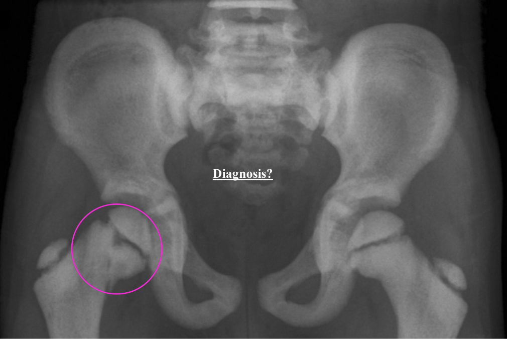
What is the dx
osteopetrosis and Salter type 2
Normal AC width
<5mm
Normal distance btw coracoid and clavicle
11-13mm
Grade 1 AC sprain
slight widening btw acromion and clavicle w/o elevation
acromioclavicular ligament stretched/partially torn
Grade 2 AC sprain
acromioclavicular ligament completely torn
coracoclavicular ligaments intact
Grade 3 AC sprain
acromioclavicular ligament and coracoclavicular ligament completely torn
>50% displacement
100% elevation
What is the MC fx to occur during birth
clavicle fx (MC middle 1/3)
Complication of clavicle fx
post traumatic osteolysis (resorption of clavicle)
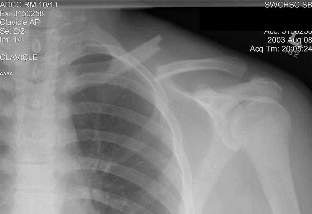
Transverse mid-clavicle fx can get an
overhanging/bayonet deformity
Three subcategories of anterior shoulder dislocations
subcoracoid, subglenoid, intrathoracic
What is the MC anterior shoulder dislocation
subcoracoid anterior shoulder dislocation
Bankart lesion
fx through ANTEROINFERIOR labrum involving inferior bony glenoid
Hill Sachs defect
impaction fx of humeral head and glenoid fossa
notch like defect of SUPEROLATERAL aspect of humeral head
What does a hill sachs suggest is in a pt’s history
previous anterior shoulder dislocation
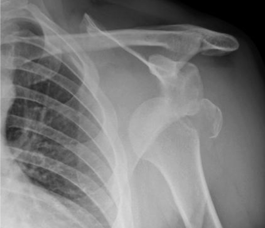
What is the dx
anterior subcoracoid shoulder dislocation w/flap fracture
Posterior shoulder dislocation
Rim sign = widening of joint space >6mm
Trough line sign = impaction fx of anterior medial humeral head on posterior glenoid rim
Tennis racqet appearance of humeral head or lightbulb sign
Rotator cuff injury
supraspinatus (MC)
infraspinatus
teres minor
subscapularis
What is the gold standard for dx of RC injury
MRI - tears are usually hyperintense in the tendon on T1 weighted
RC tears are typically located in the
critical zone (decreased vascular supply)
What is the MC fx of elbow in children
supracondylar fx
What is the MC fx of elbow in adults
radial head/neck
Hints to know if elbow has a fx - Fat pads
elevated supinator fat pad
Sail sign = elevated anterior fat pad
can see posterior fat pad
Hints to know if elbow has a fx - Radio-capitellar line (adults)
drawn down middle of radius and should intersect middle 1/3 capitulum
looks for radius dislocations
Hints to know if elbow has a fx - Anterior humeral line
drawn down anterior cortex of humerus and should intersect middle 1/3 capitulum
looks for supracondylar fx (via FOOSH/hyperextension)
Which of the two lines drawn to find a fx is very important in kids
anterior humeral line
If there is a positive fat pad sign in children, there is a 90% chance there will be an associated
intraarticular fx
Radial head/neck fractures (chisel fx) are MC in
adults
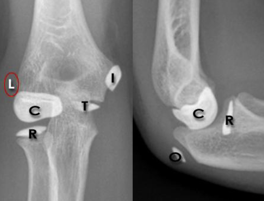
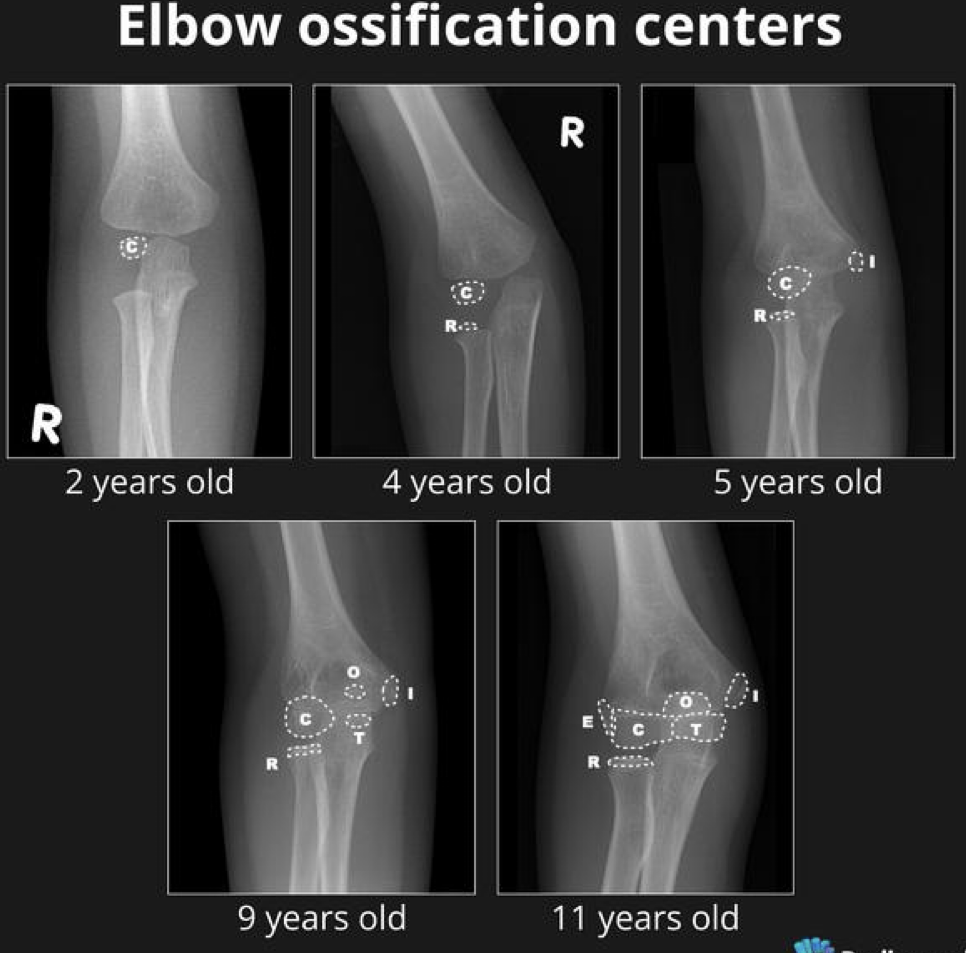
Ossification centers of the elbow (CRITOE + 1, 3, 5, 7, 9, 11)
capitellum
radial head
internal epicondyle
trochlea
olecranon
external epicondyle
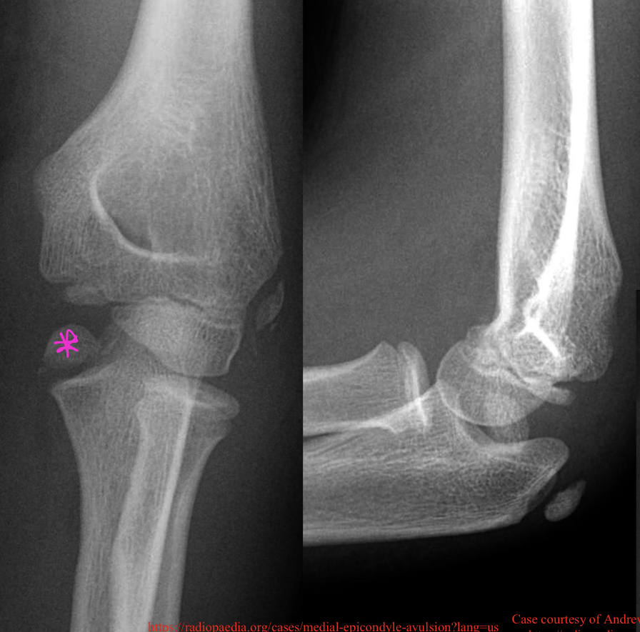
What is the dx
avulsion fx of internal epicondyle
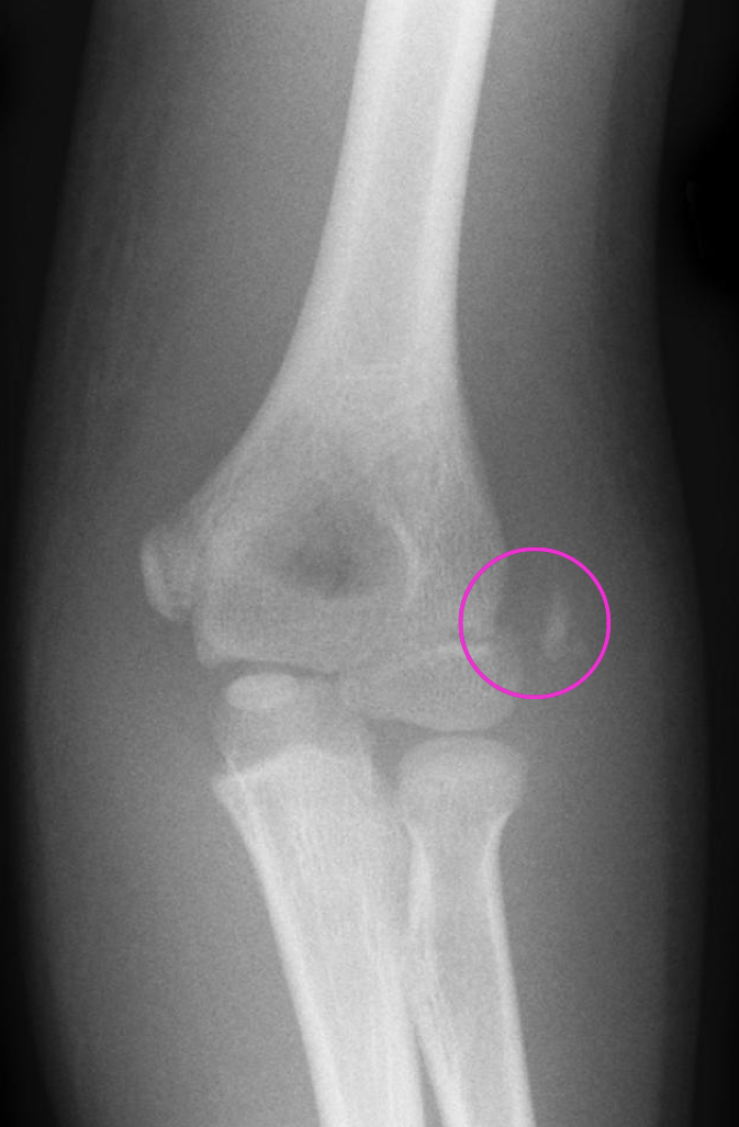
What is the dx
lateral epicondyle avulsion fx
What is the 3rd MC site of dislocation in adults
elbow
What is the MC site of dislocation in children
elbow
What is the MC dislocation of the elbow
posterior/posterior lateral
When an injury to the forearm occurs, the MC fx occurs in
both ulna and radius
A Nightstick fx is a fx of what bone
ulna
A Galeazzi fx is defined as
distal radius fx + slight dislocation of distal radio-ulnar articulation
A Monteggia fx is defined as
ulna fx + anterior dislocation of radius at proximal radio-ulnar articulation
Greenstick fx
incomplete fx
force is greater than torus fx
tendency for re-angulation following initial reduction
What is the MC forearm fx in children 6-10yo (typically in radius)
torus/buckle fx
Normal volar/palmer tilt of radius
10-15 deg anterior
Colles fx
radius fx w/posterior displacement of distal fragment
dinner fork deformity
Colles and Barton’s fx are caused by
FOOSH - typically elderly
Smith’s fx
radius fx w/anterior (volar/palmer) displacement of distal fragment
Smith’s fx are caused by
fall onto the back of the hand
Barton’s fx
posterior rim of radius fx w/carpal dislocation
Hutchinson’s fx/Cheufer’s fx
radial styloid fx
What is the MC carpal fractured
scaphoid (triquetrum 2nd, hamate 3rd)
Scaphoid fractures typically heal without periosteal callus, which means
non-union, slow healing, often gets AVN
Where is the MC site for an occult fx (can’t see it)
scaphoid
Complications with a scaphoid fx
AVN - especially in proximal pole
MUST SAY WHERE FX IS IN SCAPHOID
distal pole
waist (MC)
proximal pole
Steps if suspect a scaphoid fx
take ulnar flexion x-ray
immobilize wrist
repeat x-ray 7-10 days post injury
What carpal fx has a “pooping duck sign” on the lateral wrist x-ray
triquetral