Genes and Cancer Biology
 Call Kai
 Learn
Learn Practice Test
 Spaced Repetition
 Match
Match Flashcards
 Knowt Play
1/95

Earn XP
Description and Tags
9 / 11 lecture
Name | Mastery | Learn | Test | Matching | Spaced | Call with Kai | Chat |
|---|
No analytics yet
Send a link to your students to track their progress
96 Terms
chromosomes: somatic cells vs germline cells
within the nucleus of every cell is the genetic code responsible for the structure and function of the human body
cells are subdivided into
germline cells (gametes: sperm and egg cells)
somatic cells (all other cells)
chromosomes: numbers
somatic cells contain 23 pairs of chromosomes
22 pairs of autosomes
each pair is nearly identical in appearance and DNA sequence
they are homologous
1 pair of sex chromosomes
2 homologous X chromosomes in people AFAB
1 non-homologous pair of X and Y chromosomes in people AMAB
germline cells contain 23 single copies of chromosomes
autosomes
code for every protein and enzyme that your body produces
you get one copy of chromosomes from one parent and another copy from your other parent
genes and DNA: components
each chromosome is composed of deoxyribonucleic acid (DNA) and histone proteins
DNA provides the genetic code for the synthesis of proteins
histone proteins help DNA fold into chromosomes
each strand of DNA is made up of sequences of 4 nucleotides
nucleotide: nitrogenous base, deoxyribose, phosphate
four nitrogenous bases:
two purines: adenine and guanine
two pyrimidines: cytosine and thymine
purines
adenine
guanine
pyrimidines
cytosine
uracil
thymine
genes and DNA: hydrogen bonds
strands of DNA are held together by hydrogen bonds connecting the nitrogenous bases
adenine forms a bond with thymine
guanine forms a bond with cytosine
resulting structure is a double helix
cell division: the cell cycle
DNA needs to replicate to preserve genetic information when cells divide
the cell cycle represents the process of cell division
G0 → the resting and repairing phase
most cells are in this phase and carrying out the job they were specialized to do
G1 → cell is preparing for DNA synthesis
S phase → DNA synthesis
G2 → cell is preparing for mitosis
M → mitosis
prophase, metaphase, anaphase, telophase
every step that isn’t mitosis is considered interphase
Cell Division: S phase
DNA molecule unwinds and opens up to expose the nucleotide bases
DNA polymerase moves along the template strand of DNA and creates a complementary strand of DNA based on the nucleotides in the template
cytosine and guanine line up
adenine and thymine line up
DNA polymerase “proofreads” the complementary strand
if the base is not complementary it is excised and replaced
once the strand has been completely replicated, there are two identical chromosomes
cell division: Mitosis (M phase) - prophase
once DNA replication has completed (during interphase), the cell can divide
the DNA coils, shortens and thickens to produce the chromosomes
the chromosomes are visible as two identical halves (chromatids) attached at a single point (centromere)

cell division: Mitosis (M phase) - metaphase
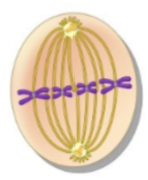
the chromosomes line up at the cellular equator in the middle of the cell
spindle fibers radiate from centrioles at each pole of the cell to the centromere
cell division: Mitosis (M phase) - anaphase
the centromere split and the sister chromatids are pulled apart toward opposite poles

cell division: Mitosis (M phase) - telophase
the new nuclear envelope is formed
spindle fibers disappear
cell divides
the end result is two daughter cells that contain the same genetic information (diploid)

cell division: meiosis I
the process by which gametes (sex cells) are formed
will end up with half of the genetic information
the DNA replicates during interphase
prophase I: replicated chromosomes coil up
metaphase I: homologous chromosomes line up on equator of cell opposite one another
spindle fibers radiate from the centrioles and attach to centromere
anaphase I: homologous chromosomes are pulled to opposite poles of the cell
telophase I: nuclear envelope forms and cell divides into two daughter cells
cell division: meiosis II
metaphase II: chromosomes line up along the equator again
spindle fibers radiate from the centrioles and attach to centromere
anaphase II: chromatids are pulled apart and move toward opposite poles
telophase II: cell divides into two germ cells
each germ cell now has half the genetic information (haploid)
mutations during cell division: polyploidy
cells that have a multiple of the normal number of chromosomes (23)
triploidy: three copies of each chromosome
tetraploidy: four copies of each chromosome
nearly all triploidy and tetraploidy pregnancies are spontaneously aborted
mutations during cell division: aneuploidy
cells that DO NOT have a multiple of 23 chromosomes
usually due to nondisjunction (when chromosomes fail to separate during meiosis I)
trisomy: three copies of one chromosome (trisomy 13, 18, or 21 can survive)
monosomy: one copy of one chromosome (all are lethal)
fertilized eggs containing no X chromosome cannot survive
trisomy 21: down syndrome
1 in 800 live births
intellectually disabled
clinical manifestations
low nasal bridge
epicanthal folds
protruding tongue
flat, low set ears
poor muscle tone
short stature
~50% have congenital heart defects **
increased risk of respiratory illness
54-90% have obstructive sleep apnea **
almost all develop symptoms of alzheimer’s disease by age 40 **
turner syndrome (45, X)
syndrome characterized by presence of one X chromosome without a homologous X or Y chromosome
absence of Y chromosome means these individuals are usually AFAB
usually sterile: gonadal streaks rather than ovaries
clinical manifestations
short stature **
webbing of the neck (~50%) **
sparse body hair **
normal intellectual development usually **
widely spaced nipples
coarctation (narrowing) of the aorta
Klinefelter syndrome (47, XXY)
syndrome characterized by presence of two or more X chromosomes in addition to a Y chromosome
presence of Y chromosome means these individuals are usually AMAB
usually sterile
clinical manifestations **
elevated stature
gynecomastia
small testes
sparse body hair
high pitched voice
some intellectual disability is common
abnormalities associated with chromosome structure
some disorders are the result of abnormalities within the chromosome
parts of chromosomes can be duplicated or lost during DNA replication and cell division
oftentimes less severe
missing information is usually more problematic than excess information
abnormal chromosome structure: deletion
loss of a portion of the genetic material
deleton
abnormal chromosome structure: duplication
redundant genetic material in the chromosome
duplicacation
abnormal chromosome structure: inversion
reversal of the order of the genetic code resulting from 2 breaks
insrevion
abnormal chromosome structure: translocation
interchanging of material from 2 non-homologous chromosomes

abnormal chromosome structure: fragile sites
microscopic breaks and gaps in DNA
cri du chat
syndrome associated with deletion of the short arm of chromosome 5
clinical manifestations
high pitched, cat-like cry **
wide-set eyes with prominent epicanthal fold **
intellectual disability **
low-set posterior rotated ears
low birth weight
smaller than normal head
heart defects
survival is good
fragile X syndrome
syndrome associated with multiple (> 200) repeats of a 3-nucleotide sequence in long arm of X chromosome (1 in 4000 males / 1 in 8000 females)
leads to silencing of the genes, many of which are responsible for neuronal synaptic connections
second most common cause of intellectual disability (after down syndrome) and autism
clinical manifestations **
elongated face
enlarged genitals
intellectual disabilities (language delay, poor attention, signs of autism)
long hands and feet
large ears
mood disturbances (tantrums, anxiety, aggression)
sensory and motor deficits
fragile X phenotype
F → face elongated
R → repeats (CGG, CGG, CGG…)
A → ADHD
G → giant genitals
I → intellectual impairment
L → large hands/feet
E → ears protruding
X → extensible joints
DNA → genes → polypeptides
sequences of nucleotides in DNA make up genes
the basic units of inheritance
genes code for all the body’s proteins
structural proteins
enzymes
proteins are made up of sequences of amino acids (there are 20 amino acids)
specific combination of amino acids creates a polypeptide
polypeptides are bound together and folded in a certain way (alpha helices or beta pleated sheets are most common) to form a protein
protein synthesis
cookbook = DNA
recipe = RNA
dinner = protein
protein synthesis: transcription
the cell needs to decipher the genetic code to know what protein to make
this creates a messenger ribonucleic acid (mRNA) template (the recipe) from the DNA (the cookbook) for protein synthesis
RNA polymerase binds to promoter site on DNA
specifies the beginning of a genre
RNA polymerase opens the DNA to expose the nitrogenous bases
RNA nitrogenous bases that are complementary to the DNA nitrogenous bases line up to form mRNA
cytosine and guanine line up
adenine pairs up with uracil (similar to thymine)
termination sequence in DNA marks the end of the gene and terminates transcription
now you have a strand of mRNA
protein synthesis: removal of introns
before moving into the cytoplasm, some RNA sequences are removed from the mRNA (introns)
the function of these is unknown
protein synthesis: translation
the mRNA molecule travels to a ribosome to build a polypeptide
each sequence of 3 nucleotides on the mRNA (codon) codes for a specific amino acid
molecules of transfer RNA (tRNA) in the cytoplasm have a sequence of 3 nucleotides (anticodon) that is complementary to the codon on the mRNA
each tRNA carries one amino acid
translation
mRNA binds to ribosome
the anticodon of a tRNA lines up with the codon on a mRNA
The ribosome will look at the first 3 nucleotides (codon) and it corresponds to a specific amino acid (the amino acid is attached to a specific tRNA)
subsequent tRNA molecules bind to subsequent codons on mRNA
amino acids on adjacent tRNA molecules bind together to form a polypeptide
As each tRNA attaches to the codon, the amino acid that is attached to each individual tRNA will string together and make a polypeptide chain
dominant and recessive alleles
each gene occupies a locus on a chromosome
different forms of a gene are called alleles
alleles may be dominant or recessive
dominant → characterized by capital letters, stronger expression
recessive → characterized by lowercase letters, weaker expression
a person receives one allele from each parent
genotype
refers to the genetic makeup of the alleles
used to describe a characteristic
if the alleles from each parent are identical (both dominant or both recessive), a person is homozygous (BB or bb)
need to specify
homozygous dominant (BB)
homozygous recessive (bb)
if the alleles are NOT identical (one dominant and one recessive), they are heterozygous
phenotype
refers to the observable trait in a person
a dominant trait will be observed when paired with another dominant allele (BB) or when paired with a recessive allele (Bb)
a recessive trail will only be observable when paired with another recessive allele (bb)
disease carriers
a person is a carrier of a disease if they have the allele for a disease but are phenotypically normal
often this is because the disease causing allele is recessive
a homozygous dominant person (BB) does not carry the trait
a heterozygous person (Bb) carries the trait but will not exhibit signs of the disease
to express disease, both recessive alleles have to be present (bb)
ex
tay sachs
cystic fibrosis
cystic fibrosis
AUTOSOMAL RECESSIVE
mutation results in abnormal an ion channel for Cl- and HCO3- (decreased number and abnormal function)
primarily affects:
lungs
pancreas
GI tract
sweat glands
vas deferens
cystic fibrosis clinical manifestations
mucous accumulation in the lungs
increased reabsorption of Na and water from lumen of airway
mucous in airway is thick and tenacious
persistent airway obstruction
recurrent infections
increased mucin production in GI tract
plugging of pancreatic ducts
GI disruption
increased Na+ and Cl- in sweat due to poor absorption
cystic fibrosis diagnosis
trypsinogen test for newborns: trypsinogen is elevated in CF
sweat test
administer a medication to induce sweating
measure Cl- in sweat
Huntington Disease
AUTOSOMAL DOMINANT
repeats of a 3-nucleotide sequence contributes to extra long protein that is prone to fragment
result is neuronal dysfunction and eventually death
Huntington Disease Clinical Manifestations
onset of symptoms around age 45
many have already had children (their children are likely to have it too)
motor signs (diagnostic)
involuntary movement (choreiform movements)
impaired voluntary movement (bradykinesia and lack of coordination)
cognitive impairment
depression
death usually occurs 10-30 years after onset of symptoms
background on cancer
second leading cause of death in the US (first leading cause is heart disease)
incidence rate: the number of NEW CASES in a given time
incidence rates have increased since the 1960s
most likely a result of more screening rather than more diseases
prevalence: the number of cases at a point in time
prevalence of many cancers has increased since the 1960s
aging has led to more deaths due to cancer compared to other causes
neoplasm = new growth
incidence / prevalence males vs females
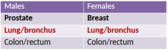
mortality males vs females
we don’t have good screenings for lung cancer
many of the screenings only find something if it is late stage

etiology of cancer
cancer is a complex, genetic disease
genetic changes occur with age
may occur by mutation or by epigenetic mechanisms
point mutations (one nucleotide)
translocations (large amounts of DNA)
repetition of certain gene sequences can increase expression of some genes (gene amplification)
epigenetics (methylation, etc.) may suppress expression of some genes
these mutations change a normal cell into a malignant cell (malignant transformation)
etiology of cancer: oncogenes
mutations of proto-oncogenes
normal genes that code for cellular growth and development (growth factors, growth factor receptors, transcription factors, etc.)
mutations convert proto-oncogenes into oncogenes
oncogenes increase cellular growth and development
ex
RAS: overexpression can lead to colorectal cancer and lung cancer
HER-2: overexpression can lead to breast cancer
etiology of cancer: tumor suppressor genes
underactivity or silencing of tumor suppressing genes
genes that normally block cell division when DNA is damaged and induce apoptosis
mutation prevents activation of these genes
cancerous cells are not eliminated and cancer is allowed to grow
ex
BRCA-1 and BRCA-2 defect: promotes breast and ovarian cancer
APC gene mutation: promotes familial adenomatous polypoposis
influence of genetic changes in cancer
will have an effect on:
proliferation → the process of cell division
differentiation → the process by which cells become specialized
maturity → differentiated cells that have reached full functional potential
differentiation
regulated by genes and external stimuli (environment exposure to substances, cytokines, growth factors, etc.)
all cells contain all genes
normally, only the genes for the specific, differentiated cell are active
as cells divide, they normally lose their ability to develop characteristics of other specialized cells
cancer doesn’t play by the rules
cancer’s effect on proliferation
cells also normally lose their ability to proliferate
instead, tissues rely on progenitor/stem cells to grow
however, cancer doesn’t play by the rules
stem cells: two properties
self-renewal
potency
cancer cell characteristics
abnormal, rapid proliferation
loss of cell differentiation
genetic instability
growth factor independence
failure of cell density-dependent inhibition
loss of cell cohesiveness and adhesion
loss of anchorage dependence
loss of cell-to-cell communication
antigen expression
production of enzymes, hormones, etc
cytoskeletal changes
“unlimited” life span
loss of cell differentiation (anaplasia) in cancer
cells do not exhibit same features of differentiated cells
more similar to embryonic cells rather than tissue or origin
greater numbers of cells in mitosis due to rapid proliferation
odd mitotic features (spindles)
nuclei are odd in shape, may have abnormal number of chromosomes
clumped chromatin
large nucleoli
classified from Grade I (well differentiated) to IV (poorly differentiated)
genetic instability in cancer
inherent instability that contributes to development and progression of cancer
high number of genetic mutations that would normally be corrected in normal tissue
lost/gained chromosomes, DNA insertions/deletions, etc
growth factor independence in cancer
ability to proliferate without growth factor
some produce their own growth factor
failure of cell density dependent inhibition in cancer
cessation of growth when cells reach a pre-determined density is not active in cancer cells
cancer cells will replicate in the absence of growth factor or just make their own growth factor
loss of cell cohesiveness and adhesion in cancer
loss of tendency for cells to stick together
loss of anchorage dependence in cancer
do not require anchorage to neighboring cells or extracellular matrix to grow and develop
When the cancer cell breaks free, it can still multiply even though it is away from other tissues
Especially prevalent in ovarian cancer
If one cell from the ovary were to make its way to the peritoneal lining, it will spread everywhere
antigen expression in cancer
produce antigens immunologically different than host
cancer cells can secrete cytokines that suppress the immune response
production of enzymes, hormones, etc. in cancer
enzymes, clotting mechanisms, hormones that aid in metastasis
production of hormones normally produced in other parts of the body (pituitary, etc.)
can lead to paraneoplastic syndromes
when you have a cancer that is producing a substance that is usually produced somewhere else
“unlimited” life span in cancer cells
shortening of the telomere normally limits life span of a cell
cancer cells produce telomerase → enzyme that prevents telomere shortening and prolongs cell life
cancer growth
normal adult cells reach equilibrium between cell production and death
cancer cells continue to divide until limits in blood supply and nutrients become a factor
benign tumors
named by adding -oma to the type of parenchymal tissue from which the growth originated
adenoma → benign neoplasm of glandular tissue
osteoma → benign neoplasm of bone
malignant neoplasm of epithelial tissue
end in suffix -carcinoma
adenocarcinoma → malignant neoplasm of glandular epithelial tissue
malignant neoplasm of mesenchymal origin
end in suffix -sarcoma
osteosarcoma → malignant neoplasm of bone
liposarcoma → malignant neoplasm of adipose tissue
(mesenchymal refers to things that create the structure of the body [like ligaments, bone, etc.])
benign cancer key characteristics
cells are
well differentiated
resemble tissue of origin
rate of growth
slow
may even stop
mode of growth
grows by expansion
usually encapsulated
no metastasis
potential for death is low
malignant cancer key characteristics
cells are
poorly differentiated
little resemblance to tissue of origin
rate of growth
variable
more undifferentiated, more rapid
mode of growth
grows by invasion
metastasis
will spread with access to blood and lymph channels
potential for death is high
categories of malignant neoplasms: solid tumors
confined to tissue or organ (ex. lymphoma)
cells detach from original tumor, invade surrounding tissue, and enter bloodstream/lymphatic system
categories of malignant neoplasms: hematologic cancers
involve cells already in circulation (ex. leukemia)
categories of malignant neoplasms: carcinoma in situ
localized, pre-invasion lesion
has not broken through the basement membrane of the tissue
more easily targeted
cancer growth: DNA repair defects
inability of body to repair non-lethal mutations
proto-oncogenes are repaired and preserved
cancer growth: defects in growth factor signaling pathway
normally cell proliferation requires signal from growth factor to nucleus
some cells bypass this
cancer growth: evasion of apoptosis
normally cells undergo apoptosis to destroy abnormal cells that could lead to malignancy
cancer cells can avoid apoptosis
many mechanisms
cancer growth: evasion of cellular senescence
cancer cells secrete telomerase
telomerase promotes synthesis of telomere
prevents the chromosomes from shortening and unraveling
cancer growth: development of sustained angiogenesis
cancer cells produce vascular endothelial growth factor (VEGF)
promotes production of new blood vessels
cancer growth: invasion and metastasis
MET proto-oncogene promotes metastasis
host factors that promote cancer growth: heredity
genetic predisposition
BRCA 1 and BRCA2 mutations
HER2 mutations
host factors that promote cancer growth: hormones
unclear mechanism
may be related to stimulation of proliferation of cells with malignant phenotype
host factors that promote cancer growth: immunology
immune system works to suppress tumors
white blood cells and antibodies have ability to attack tumor cells and destroy them (NK cells, cytotoxic T cells, etc.)
cancer survival may be related to impaired ability to suppress tumor growth
immunocompromised
elderly
environmental factors that promote cancer growth
chemical carcinogens
many due to lifestyle (smoking, diet, alcohol, etc.)
radiation
ionizing
ultraviolet (UVA penetrates deeper)
bacteria
helicobacter pylori
viruses
HPV, EBV, HBV
clinical manifestation of cancer
initial manifestations often associated with disruption of normal parenchymal (functional) tissue
cancer of the lung associated with impairment of respiratory function initially
tumors compress and erode blood vessels
may lead to frank bleeding or hemorrhage
tumors may produce enzymes and toxins that destroy surrounding tissue
development of effusions (fluid) in peritonea/pleural/pericardial space
anorexia cachexia syndrome and cancer
an energy imbalance disorder in which energy intake is decreased and energy usage is increased
loss of appetite and impaired taste associated with cancer (mostly solid tumors)
hypermetabolic state, protein breakdown and apoptosis of muscle cells
visualized as muscle wasting
fatigue and cancer
early sign of malignancy
multifactorial
poor sleep quality
anemia and cancer
multifactorial
nutritional deficiency
bone marrow failure
inflammatory cytokines
response to chemotherapy
blood loss or hemolysis
other clinical manifestations of cancer
GI disturbances
diarrhea and stomatitis - due to chemo therapy destroying highly proliferative cells
alopecia
due to chemotherapy destroying highly proliferative cells in hair follicles
lymphedema
accumulation of fluid in tissue 2o disruptions in lymphatic system due to surgery, radiation or metastasis
pain
intensifies with disease progression - 2o direct pressure, obstruction, stretching, inflammation
paraneoplastic syndromes and cancer
cancer can produce manifestations in sites not affected by the primary cancer
most commonly the result of hormones secreted by the tumor
most common in lung, breast, and hematologic cancers

screening and diagnosis of cancer
screening is secondary prevention
observation, palpation, and lab tests (pap smear, colonoscopy, mammography, PSA testing, etc.)
had lead to early detection of many cancers
identified at earlier stages
more easily treated
screening and diagnosis of cancer: tumor markers

types of screening for cancer
Papanicolaou test
microscopic examination for abnormal cells
used for cervical cancer and for evaluating body secretions (nipple drainage, pleural/peritoneal fluid) for cancer
tissue biopsy
removal of tissue sample for microscopic evaluation
immunohistochemistry
use of antibodies to detect cell products or surface markers on cancer cells
three factors in cancer staging
used to determine how advanced cancer is
size of tumor (T)
degree of local spread to lymph nodes (N)
extent of metastases (M)
this info is usually combined with other factors (tumor marker data, etc) to establish a staging in the four stage system
four stage system in cancer
stage 0: carcinoma in situ
stage 1: cancer confined to organ or origin
stage 2: cancer that is starting to show signs of local spread within local tissue
stage 3: cancer that has spread to regional nodes
stage 4: cancer that has spread to distal organs (liver, brain, bone, etc.)
methods of cancer spread
direct invasion
direct extension
seeding → one cell breaks free and implants itself somewhere else
metastasis
cancer cell breaks loose and gains access to blood or lymph channel
blood → venous system
lymph → sentinel node and then adjacent nodes
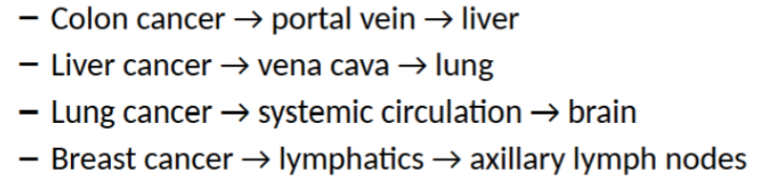
cancer spread: sites of metastasis
venous and lymphatic drainage of primary tumor often determines location of metastasis
not always true
modes of cancer treatment
surgery
often first treatment for solid tumors
radiation
destroy or damage cancer cells by creating free radicals
rapidly proliferating cancer cells more susceptible
chemotherapy
prevent cell growth, inhibit DNA, RNA, protein synthesis
hormone therapy
disrupt hormonal environment of cancer cells (antiestrogens, androgen blockers, etc.)
biotherapy
change person’s immune response to cancer