ACBS 400A Skeletal Structure: Cartilage and Joints
 Call Kai
 Learn
Learn Practice Test
 Spaced Repetition
 Match
Match Flashcards
 Knowt Play
1/51

There's no tags or description
Looks like no tags are added yet.
Name | Mastery | Learn | Test | Matching | Spaced | Call with Kai | Chat |
|---|
No analytics yet
Send a link to your students to track their progress
52 Terms
What are chondrocytes?
Cells responsible for cartilage formation that become trapped in lacunae within the cartilage matrix.
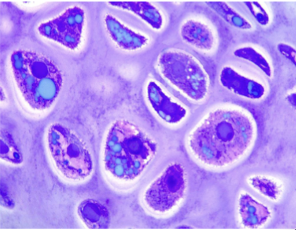
What is cartilage matrix made of?
An amorphous, gel-like matrix secreted by chondroblasts containing collagens, proteoglycans, and non-collagenous proteins
How are proteoglycans formed in cartilage?
Chondroitin sulfate polysaccharides combine with proteins in the ground substance to form proteoglycans.
What happens to chondrocytes after they secrete cartilage matrix?
They become trapped in spaces called lacunae within the matrix.
Can young chondrocytes divide after becoming isolated in lacunae?
Yes. Daughter cells secrete new matrix between them, causing them to move apart into separate lacunae.
Define what chondroblasts and chondrocytes are
Chondroblast = builds/secretes matrix
Chondrocyte = mature cartilage cell living in lacuna
What is the perichondrium?
A structure similar to the periosteum that surrounds cartilage. It is a dense fibrous layer lined by cells capable of secreting hyaline matrix.
What does the outer fibrous layer of the perichondrium do?
Provides protection, mechanical support, and attaches cartilage to other structures.
What does the inner cellular layer of the perichondrium do?
It is involved in the growth and maintenance of cartilage.
Does cartilage contain nerves or blood vessels?
No. Cartilage lacks nerves and blood vessels; oxygen is supplied through capillary beds at its edge.
What is cartilage generally resistant to?
Compression
What is hyaline cartilage and where is it found?
It’s most abundant type of cartilage in animals. It’s found at the ventral ends of ribs, tracheal rings, larynx, articulating surfaces of bones, and growth plates
What are the characteristics of hyaline cartilage?
Collagen fibers are randomly oriented and there is little elastin.
Where is elastic cartilage found?
In the external ear and epiglottis.
What is the main characteristic of elastic cartilage?
It contains many elastic fibers embedded in the cartilage.
Where is fibrocartilage found?
At the insertion of many large ligaments onto bone, in intervertebral disks, and at the pubic symphysis.
What are the structural characteristics of fibrocartilage?
It has dense collagen bundles oriented mostly parallel, with cells in lacunae arranged in rows between coarse collagen bundles.
Give a summarized version of what perichondrium and cartilage is
Perichondrium: protection + support + growth/maintenance
Cartilage: no nerves or blood vessels → resistant to compression
Give a summarized version of what hyaline and elastic is
Hyaline: most common → ribs, trachea, larynx, joints, growth plates
Elastic: ear + epiglottis → lots of elastic fibers
Give a summarized version of what fibrocartilage is
Fibrocartilage: ligaments, intervertebral disks, pubic symphysis → thick collagen bundles
Why is cartilage growth especially important during embryonic development?
It forms the cartilage models for future bones.
Where do chondrocytes come from and how do they contribute to cartilage growth?
Chondrocytes come from mesenchymal cells, undergo mitosis, and spread out, causing cartilage to expand from within.
What do chondrocytes produce as they grow?
They surround themselves with a specialized mixture of proteins that forms the interstitial matrix.
How are collagen fibrils formed in cartilage?
Cells secrete tropocollagen into the matrix, which aggregates and assembles into collagen fibrils.
What is interstitial growth?
Growth through the addition of new cells within existing tissue. It involves mitosis, increases tissue size by adding cells, and occurs throughout life.
What is appositional growth?
Growth through the addition of new cells to the surface of tissue. It involves stem cell differentiation, increases tissue size by adding new layers, and occurs during growth.
What is the main difference between interstitial and appositional growth?
Interstitial = growth from within → mitosis → throughout life
Appositional = growth on the surface → stem cell differentiation → during growth
What makes elastic cartilage flexible?
Elastin fibers in the ground substance of the matrix.
What makes fibrocartilage strong?
Increased collagen content, with thick, tough collagen bundles aligned in one direction to improve tensile strength.
What makes up the interstitial matrix of hyaline cartilage?
Chondroitin 4-sulfate and chondroitin 6-sulfate
What is the function of proteoglycans and glycosaminoglycans in cartilage?
They attract water electrostatically, maintaining hydration and providing resilience and compressive strength.
What is the main type of collagen found in cartilage?
Type II collagen
How much of the dry weight of most cartilaginous tissues is Type II collagen?
More than 50%.
Where is Type II collagen found?
It’s found only in cartilage
What does Type IX collagen do?
It’s located on the surface of Type II collagen and regulates the spatial arrangement of Type II collagen.
Does Type IX collagen form fibrils?
No
Where is Type X collagen found?
In chondrocytes within the growth plates of long bones.
Summarize a short way to differentiate the Types of collagens
Type II = main cartilage collagen → >50% dry weight → only cartilage
Type IX = on Type II → regulates arrangement → no fibrils
Type X = chondrocytes in growth plates
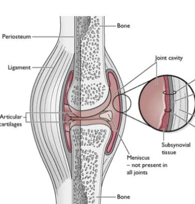
What surrounds a synovial joint?
A capsule with an outer fibrous layer made of collagen fibers that run from the periosteum of one bone to the other.
What does the outer fibrous layer of a synovial joint provide?
side to side stability
What are ligaments?
Special extensions of the fibrous capsule that can be located inside or outside the joint capsule.
Synovial Membrane: What is the inner surface of the joint capsule made of?
Synovial membranes that produce synovial fluid.
What is synovial fluid made of?
An ultrafiltrate of blood plasma with hyaluronic acid and lubricin added by synovial cells
What is the function of synovial fluid?
It lubricates the joint and reduces wear between opposing surfaces.
What are ligaments and what do they do?
Tough, elastic connective tissue that connects bones, supports joints, and limits movement.
What are tendons and what do they do?
Tough connective tissue that connects muscles to bones and helps control joint movement.
What are bursas and what is their function?
Fluid-filled sacs between bones, ligaments, or nearby structures that cushion friction in a joint.
What are menisci and what do they do?
Areas of dense fibrocartilage between articulating surfaces, commonly in weight-bearing areas, that cushion compressive forces.
What are the five types of synovial joints?
Pivot, hinge, ball-and-socket, condyloid, and plane.
What movement does a pivot joint allow, and what are examples?
Rotation around a single axis. Examples: atlas-axis joint and radius-ulna.
What movement does a hinge joint primarily allow, and what are examples?
Movement mainly in one plane, such as up/down or side-to-side. Examples: toes, elbows, stifles, and tarsi.
What are the characteristics and examples of ball-and-socket, condyloid, and plane joints?
Ball-and-socket: rounded bone surface moves within a depression; allows a wide range of movement. Examples: shoulders and hips.
Condyloid: ovoid convex surface fits into an ellipsoidal cavity. Example: jaw.
Plane: flat or nearly flat surfaces slide over each other. Example: metacarpals and cuneiform bones of the foot.