Organisation of Eukaryotic Genome

0.0(0)
Studied by 3 people
call kaiCall Kai
Locked
learnLearn
examPractice Test
spaced repetitionSpaced Repetition
heart puzzleMatch
flashcardsFlashcards
GameKnowt Play
Card Sorting

1/29

encourage image

There's no tags or description

Looks like no tags are added yet.

Last updated 8:59 AM on 8/10/26
Name
Mastery
Learn
Test
Matching
Spaced
Call with Kai
Chat

No analytics yet

Send a link to your students to track their progress

30 Terms

1
New cards

Genome

  • genome: genetic material of an organism or virus; the complete complement of an organism’s or virus’s genes along with its non-coding nuclei acid sequences (coding + non-coding)

  • all organisms have a genome comprised of genetic material (DNA or RNA) that contains all genetic information needed to direct the development and maintenance of that organism

    • the entire complete set (or complement) of genetic information for all the proteins and RNA that the organism will ever synthesise

    • most genomes including the human genome and those of all other cellular life forms are made of DNA

    • viruses may contain DNA or RNA genomes

  • prokaryotes - genome residues usually in a single DNA molecule; eukaryotic genome is much larger (human genome contains more than 5x no. of genes of a typical prokaryote)

  • eukaryotic cells have a nuclear genome and a mitochondrial genome

    • for plants and algae - chloroplast genome

2
New cards

Eukaryotic genome

  • biological information contained in a genome is encoded in its DNA and is functionally divided into discrete units called genes

  • gene: a gene is a section of the DNA that contains the information in the form of a specific sequence of nucleotides/base to direct the synthesis of one polypeptide chain or RNA

    • unit of inheritance located in a fixed position (locus) on the chromosome which specifies a particular character of an organism

  1. Genes are carried on chromosomes

  • each gene resides in a specific location along the chromosome called the gene locus

  1. Most eukaryotic genes are distributed among a species-specific number of linear chromosomes

  • recall: DNA structure and replication

    • each chromosome is composed of a single DNA molecule (double helix) packaged with various histone and nonhistone proteins (e.g. scaffold proteins) found in the nucleus

    • a small proportion of eukaryotic DNA is found in the mitochondria

    • in the case of photosynthetic organisms, DNA is also found in the chloroplasts

  1. Every eukaryotic cell has a complete copy of the nuclear genome

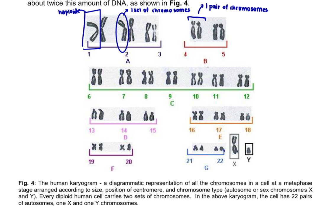
  • each cell nucleus (except gametes) contains 2 sets of chromosomes, one from each parent - diploid cells

  • either set of chromosomes is known as haploid set of chromosomes

  • a compete eukaryotic genome comprises

    • one complete copy of genetic information carried by a haploid set of linear chromosomes in the nucleus (nuclear genome)

    • mitochondrial genome (consists of a single small circular DNA molecule)

    • chloroplast genome (in photosynthetic organisms only) which is also composed of one small circular DNA molecule

  1. The human nuclear genome

  • in humans, a complete copy of the nuclear genome comprises of ~3 × 10^9 DNA nucleotide base pairs distributed over 22 different autonomies and one of the two sex chromosomes (X or Y chromosomes)

  • human genome sequence refers to the complete nucleotide sequence of DNA in these 23 chromosomes

    • diploid - a human somatic cell (i.e. cell that is not a germ cell or gamete) contains about twice this amount of DNA
<ul><li><p>biological information contained in a genome is encoded in its DNA and is functionally divided into discrete units called genes </p></li><li><p>gene: a gene is a section of the <strong>DNA</strong> that contains the information in the form of a <strong><u>specific</u> sequence of nucleotides/bas</strong>e to<strong> direct the synthesis</strong> of one polypeptide chain or RNA </p><ul><li><p>unit of inheritance located in a fixed position (locus) on the chromosome which specifies a particular character of an organism </p></li></ul></li></ul><ol><li><p><mark data-color="yellow" style="background-color: yellow; color: inherit;">Genes are carried on chromosomes</mark> </p></li></ol><ul><li><p>each gene resides in a specific location along the chromosome called the <strong>gene locus</strong> </p></li></ul><p></p><ol start="2"><li><p><mark data-color="blue" style="background-color: blue; color: inherit;">Most eukaryotic genes are distributed among a species-specific number of </mark><strong><mark data-color="blue" style="background-color: blue; color: inherit;">linear chromosomes</mark> </strong></p></li></ol><ul><li><p>recall: DNA structure and replication </p><ul><li><p>each chromosome is composed of a single DNA molecule (double helix) packaged with various histone and nonhistone proteins (e.g. scaffold proteins) found in the nucleus </p></li><li><p>a small proportion of eukaryotic DNA is found in the mitochondria </p></li><li><p>in the case of photosynthetic organisms, DNA is also found in the chloroplasts </p></li></ul></li></ul><p></p><ol start="3"><li><p><mark data-color="red" style="background-color: red; color: inherit;">Every eukaryotic cell has a complete copy of the nuclear genome</mark> </p></li></ol><ul><li><p>each cell nucleus (except gametes) contains 2 sets of chromosomes, one from each parent - diploid cells </p></li><li><p>either set of chromosomes is known as <strong>haploid</strong> set of chromosomes </p></li><li><p>a <u>compete</u> eukaryotic genome comprises </p><ul><li><p>one complete copy of genetic information carried by a <strong>haploid</strong> set of linear chromosomes in the nucleus (nuclear genome) </p></li><li><p><strong>mitochondrial</strong> <strong>genome</strong> (consists of a single small circular DNA molecule) </p></li><li><p><strong>chloroplast</strong> <strong>genome</strong> (in photosynthetic organisms only) which is also composed of one small circular DNA molecule </p></li></ul></li></ul><p></p><ol start="4"><li><p><mark data-color="purple" style="background-color: purple; color: inherit;">The human nuclear genome</mark> </p></li></ol><ul><li><p>in humans, a complete copy of the nuclear genome comprises of ~3 × 10^9 DNA nucleotide base pairs distributed over 22 different autonomies and one of the two sex chromosomes (X or Y chromosomes) </p></li><li><p>human genome sequence refers to the complete nucleotide sequence of DNA in these 23 chromosomes</p><ul><li><p>diploid - a human somatic cell (i.e. cell that is not a germ cell or gamete) contains about twice this amount of DNA </p></li></ul></li></ul><p></p>
3
New cards

human karyogram

<p></p><p></p>
4
New cards

The Complexity of the Eukaryotic genome

a. More complex organisms tend to have larger genome sizes

  1. Genome size is usually expressed as the total number of base pairs (by) per haploid genome

  • usually expressed in kilobases (kb) for base pairs in thousands or megabases (Mb) base pairs in millions

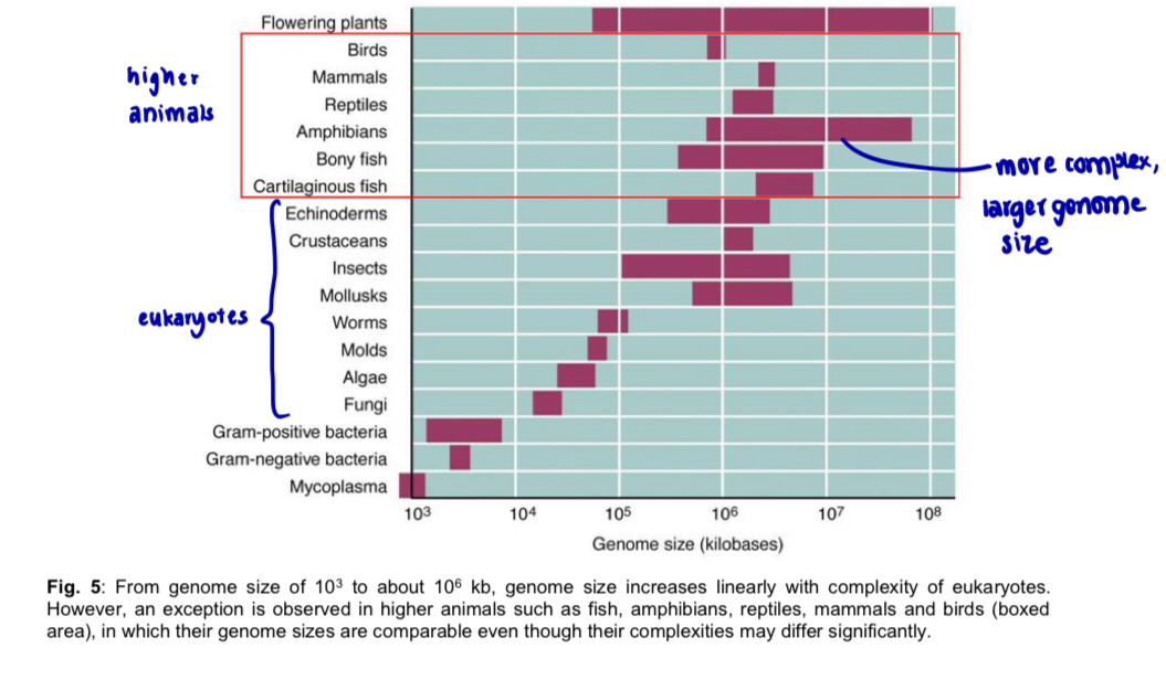
  1. More complex organisms tend to have larger genome sizes compared to simpler organisms (e.g. prokaryotes)

  • there is a correlation between an organism’s genome size and its apparent biological complexity because more genes and gene products are required to direct the development and maintenance of more complex organisms

  1. Gene size is also larger in more complex organisms due to the increase in proportion of regulatory sequences needed for more complex control of gene expression (e.g. alternative splicing)

note: prokaryotes tend to have only one chromosome and a significantly smaller genome; also lack many regulator sequences present in eukaryotes

  • e.g. in fig 5, genome size increases linearly with complexity of eukaryotes

  • but this correlation is not observed between higher eukaryotes
<p>a. <u><mark data-color="yellow" style="background-color: yellow; color: inherit;">More complex</mark></u><mark data-color="yellow" style="background-color: yellow; color: inherit;"> organisms tend to have </mark><u><mark data-color="yellow" style="background-color: yellow; color: inherit;">larger genome sizes</mark> </u></p><ol><li><p><strong>Genome size</strong> is usually expressed as the <strong>total number of base pairs (by) per haploid genome </strong></p></li></ol><ul><li><p>usually expressed in kilobases (kb) for base pairs in thousands or megabases (Mb) base pairs in millions </p></li></ul><ol start="2"><li><p>More complex organisms tend to have larger genome sizes compared to simpler organisms (e.g. prokaryotes) </p></li></ol><ul><li><p>there is a correlation between an organism’s genome size and its apparent biological complexity because <u>more genes and gene products </u>are required to <u>direct the development and maintenance of more complex organisms </u></p></li></ul><ol start="3"><li><p>Gene size is also larger in more complex organisms due to the <strong>increase in proportion of regulatory sequences</strong> needed for <strong>more complex <u>control</u> </strong>of gene expression (e.g. alternative splicing) </p></li></ol><p>note: prokaryotes tend to have only one chromosome and a significantly smaller genome; also lack many regulator sequences present in eukaryotes </p><ul><li><p>e.g. in fig 5, genome size increases linearly with complexity of eukaryotes</p></li><li><p>but this correlation is not observed between higher eukaryotes </p></li></ul><p></p>
5
New cards

Complexity of eukaryotic genome

b. no correlation between biological complexity of an organism and number of genes in its genome

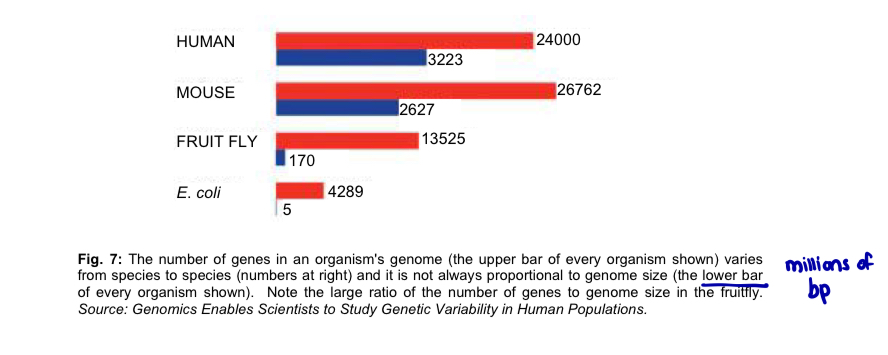
  1. Genome size is not necessarily proportional to number of genes in the genome

  • e.g. humans have greater genome size but fewer genes than Pufferfish

  1. There are other mechanisms at play to generate high biological complexity from a limited pool of genes

  • action of different regulatory proteins interacting with specific regulatory elements to alter gene expression

6
New cards

Complexity of eukaryotic genome

c. Prokaryotic genomes have much higher gene densities than that of eukaryotes

  1. the measure of the number of genes per million base pairs (Mb) in the genome —> gene density

  2. human genome has an estimated 100-fold lower gene density than that of a typical prokaryote, despite its approximately 1000 times larger genome size
<p>c. <mark data-color="red" style="background-color: red; color: inherit;">Prokaryotic genomes have much higher gene densities than that of eukaryotes</mark></p><ol><li><p>the measure of the <strong>number of genes per million base pairs (Mb)</strong> in the genome —&gt; <strong>gene density</strong></p></li><li><p><strong>human</strong> genome has an estimated 100-fold <strong>lower</strong> gene density than that of a typical prokaryote, despite its approximately 1000 times larger genome size</p></li></ol><p></p><p></p>
7
New cards

Complexity of eukaryotic genomes

d. The more complex eukaryotes generally have lower gene density than lower eukaryotes (higher eukaryotes = lower gene density)

  1. decreased gene density especially in higher eukaryotes attributed to the large proportion of non-coding intergenic DNA relative to genes present in their genomes

  • sequences are less compact and don’t belong in genes

  • don’t encode any expressed protein or RNA product

note: prokaryotes typically have 85-90% of their genomes containing structural genes as compared to only 1-5% for the eukaryotic genome

8
New cards

Overview of DNA in eukaryotic genome

knowt flashcard image
9
New cards

Packing of DNA in eukaryotic chromosome

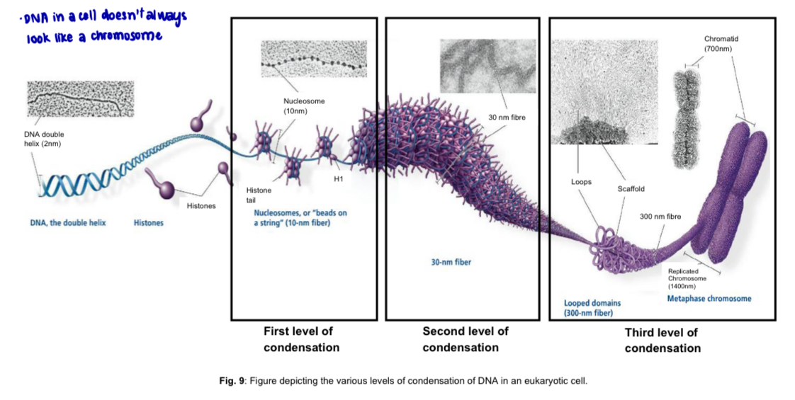
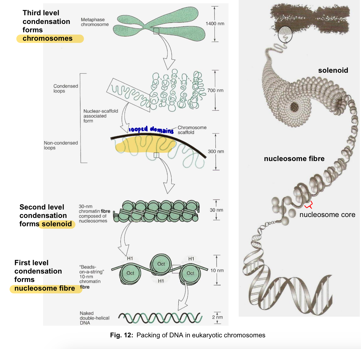
First level of condensation (nucleosome fibre) —> Second level of condensation (solenoid) —> Third level of condensation (chromosomes)
<p>First level of condensation (nucleosome fibre) —&gt; Second level of condensation (solenoid)  —&gt; Third level of condensation (chromosomes) </p><p></p>
10
New cards

First level of condensation

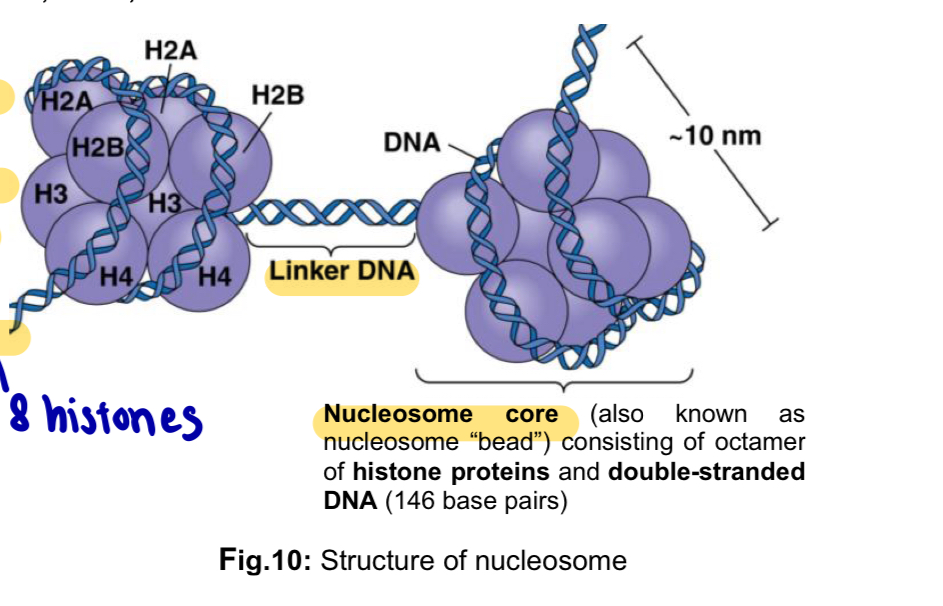
  1. Nucleosomes packing process involves a molecule of DNA coiled around an octamer of histone proteins, two each of histones H2A, H2B, H3 and H4

  • histones are small proteins with a high concentration of positively-charged residues e.g. lysine and arginine, which form ionic bonds with the negatively-charged sugar-phosphate backbone of DNA

  • histones assemble into an octomer (8 histones) to form a core upon which 146 base pairs of DNA is bound

  • double-stranded DNA is coiled around the histone core, forming a nucleosome core (gives chromatin a ‘beads-on-a-string’ look

  • completed chromatin subunit consists of the nucleosome core, the linker DNA and the associated non-histone chromosomal proteins

  1. Multiple nucleosomes are packed together to produce the 10-nm chromatin fibre also known as nucleosome fibre
<ol><li><p>Nucleosomes packing process involves <mark data-color="yellow" style="background-color: yellow; color: inherit;">a molecule of DNA coiled around an octamer of histone proteins</mark>, two each of histones H2A, H2B, H3 and H4</p></li></ol><ul><li><p><strong>histones</strong> are small proteins with a high concentration of <strong>positively-charged residues</strong> e.g. <u>lysine</u> and <u>arginine</u>, which form <strong>ionic bonds </strong>with the <strong>negatively-charged sugar-phosphate backbone </strong>of<strong> </strong>DNA</p></li><li><p><strong>histones</strong> assemble into an <strong>octomer</strong> (8 histones) to form a core upon which 146 base pairs of DNA is bound</p></li><li><p>double-stranded <strong>DNA</strong> is coiled around the <strong>histone</strong> core, forming a <strong>nucleosome core </strong>(gives chromatin a ‘beads-on-a-string’ look</p></li><li><p>completed chromatin subunit consists of the <strong>nucleosome core</strong>, the <strong>linker DNA </strong>and the associated non-histone chromosomal proteins</p></li></ul><ol start="2"><li><p><mark data-color="blue" style="background-color: blue; color: inherit;">Multiple nucleosomes </mark>are packed together to <mark data-color="blue" style="background-color: blue; color: inherit;">produce the 10-nm chromatin fibre also known as nucleosome fibre</mark></p></li></ol><p></p><p></p>
11
New cards

Second level of condensation

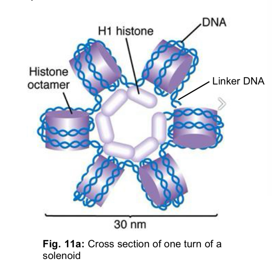
  1. DNA is further folded or coiled to produce the 30-nm chromatin fibre, known as solenoid

  2. Histone H1 and linker DNA are involved in this coiling of the 10-nm nucleosome fibre to produce the 30-nm chromatin fibre
<ol><li><p>DNA is further<strong> folded or coiled</strong> to produce the 30-nm <strong>chromatin fibre</strong>, known as <strong>solenoid</strong></p></li><li><p><strong>Histone H1 </strong>and <strong>linker DNA </strong>are involved in this coiling of the 10-nm nucleosome fibre to produce the <strong>30-nm chromatin fibre</strong></p></li></ol><p></p>
12
New cards

Third level of condensation

  1. Non-histone proteins known as scaffold proteins are involved in condensing the 30-nm chromatin fibre to form looped domains

  2. In mitotic and meiotic chromosomes, the looped domains themselves coil and fold, further compacting all the chromatin to produce the characteristic metaphase chromosome

  • width of one chromatid is 700nm

note: particular genes always end up located at the same places in mitotic and meiotic chromosomes, indicating that the packing steps are highly specific and precise

Role of condensation

  • to organise and pack the giant DNA molecules of eukaryotic chromosomes into structures that will facilitate their segregation onto daughter nuclei

  • DNA molecules of different chromosomes will not be entangled and as a consequence, break during separation at anaphase (prevent breakage)
<ol><li><p>Non-histone proteins known as <strong>s<mark data-color="yellow" style="background-color: yellow; color: inherit;">caffold proteins</mark></strong><mark data-color="yellow" style="background-color: yellow; color: inherit;"> are involved in </mark><strong><mark data-color="yellow" style="background-color: yellow; color: inherit;">condensing</mark></strong><mark data-color="yellow" style="background-color: yellow; color: inherit;"> the 30-nm chromatin fibre to form </mark><strong><mark data-color="yellow" style="background-color: yellow; color: inherit;">looped domains</mark></strong></p></li><li><p>In mitotic and meiotic <strong>chromosomes</strong>, the <mark data-color="blue" style="background-color: blue; color: inherit;">looped domains themselves coil and fold, further compacting all the chromatin to produce the characteristic metaphase chromosome</mark></p></li></ol><ul><li><p>width of one chromatid is 700nm</p></li></ul><p>note: particular genes always end up located at the same places in mitotic and meiotic chromosomes, indicating that the packing steps are <u>highly specific</u> and <u>precise</u></p><p>Role of condensation</p><ul><li><p>to <strong>organise and pack</strong> the giant DNA molecules of eukaryotic chromosomes into structures that will <strong>facilitate their segregation onto daughter nuclei</strong></p></li><li><p>DNA molecules of different chromosomes will <strong>not be entangled</strong> and as a consequence, <strong>break</strong> during separation at anaphase (prevent breakage)</p></li></ul><p></p>
13
New cards

Organisation of eukaryotic genome

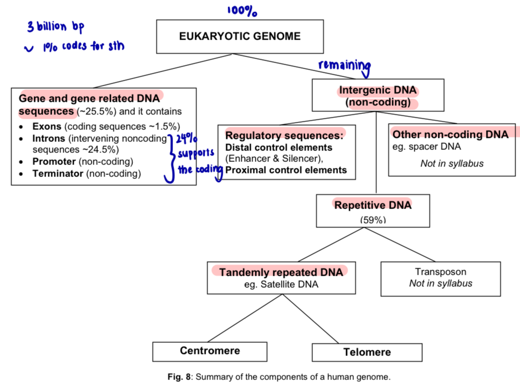
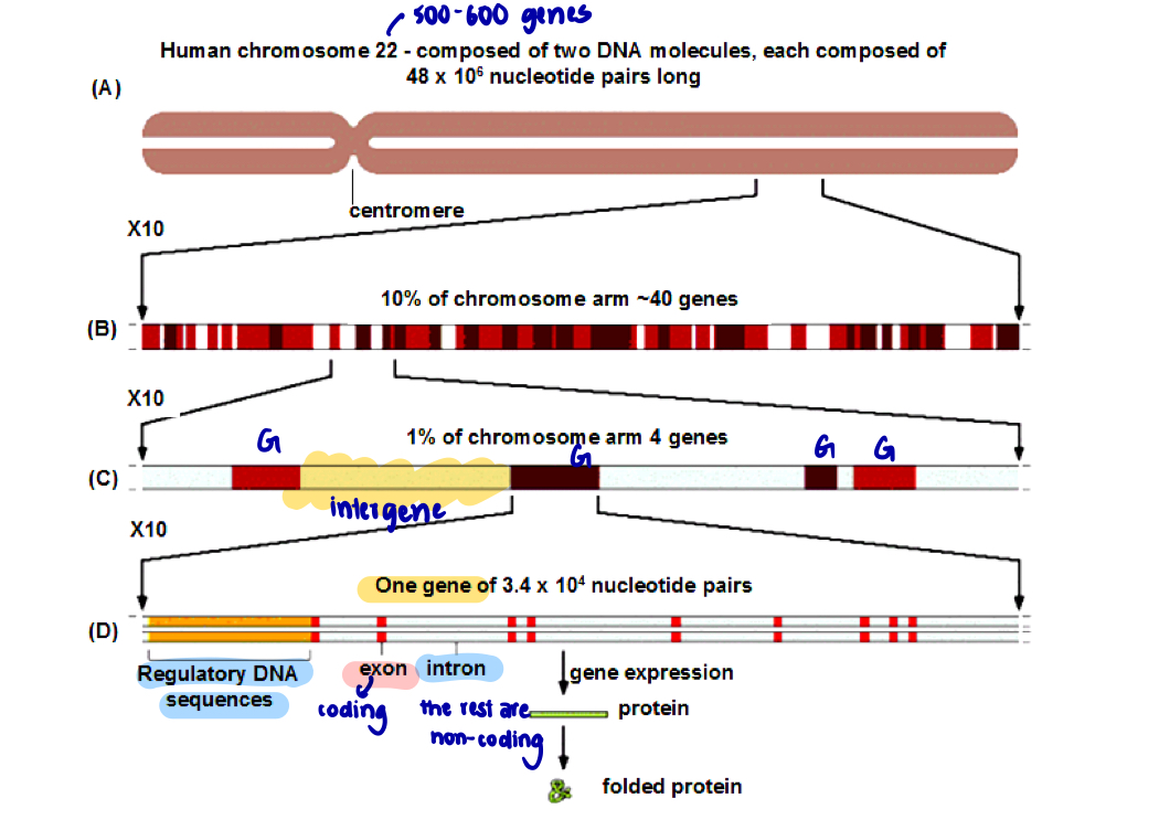
  1. Eukaryotic genomes comprise of coding (i.e. genes) and non-coding DNA sequences

  • a eukaryotic gene includes not only the coding sequences that encode a functional gene product, but also non-coding regulatory nucleotide sequences required for proper expression of the gene

  1. Total number of genes represented in the human genome is estimated to be approximately 1%; remaining DNA is non-coding

ref. to fig

(A) human chromosome 22, shown in its replicated form consisting of 2 chromatids joined at the centromere

(B) a ten-fold expansion of a portion of chromosome 22, with about 40 genes (dark regions) interspersed with intergenic DNA white regions)

(C) an expanded portion of (B) shows the entire length of 4 genes (dark regions), separated by intergenic DNA (gray regions)

(D) close-up of a gene in (C) showing the arrangement of non-coding introns (gray regions), coding exons (dark regions) and regulatory DNA sequences characteristic of a eukaryotic gene
<ol><li><p><mark data-color="purple" style="background-color: purple; color: inherit;">Eukaryotic genomes comprise of </mark><strong><mark data-color="purple" style="background-color: purple; color: inherit;">coding</mark></strong><mark data-color="purple" style="background-color: purple; color: inherit;"> (i.e. genes) and </mark><strong><mark data-color="purple" style="background-color: purple; color: inherit;">non-coding</mark></strong><mark data-color="purple" style="background-color: purple; color: inherit;"> DNA sequences </mark></p></li></ol><ul><li><p>a eukaryotic gene includes not only the <u>coding</u> sequences that encode a functional gene product, but also<u> non-coding regulatory</u> nucleotide sequences required for proper expression of the gene </p></li></ul><ol start="2"><li><p><mark data-color="green" style="background-color: green; color: inherit;">Total number of </mark><strong><mark data-color="green" style="background-color: green; color: inherit;">genes</mark></strong><mark data-color="green" style="background-color: green; color: inherit;"> represented in the human genome is estimated to be approximately </mark><strong><mark data-color="green" style="background-color: green; color: inherit;">1%</mark></strong><mark data-color="green" style="background-color: green; color: inherit;">; remaining DNA is non-coding </mark></p></li></ol><p>ref. to fig </p><p>(A) human chromosome 22, shown in its replicated form consisting of 2 chromatids joined at the centromere </p><p>(B) a ten-fold expansion of a portion of chromosome 22, with about 40 genes (dark regions) interspersed with <strong>intergenic DNA </strong>white regions) </p><p>(C) an expanded portion of (B) shows the entire length of 4 genes (dark regions), separated by intergenic DNA (gray regions) </p><p>(D) close-up of a gene in (C) showing the arrangement of non-coding introns (gray regions), coding exons (dark regions) and regulatory DNA sequences characteristic of a eukaryotic gene </p>
14
New cards

organisation of eukaryotic gene at DNA level flowchart
knowt flashcard image
15
New cards

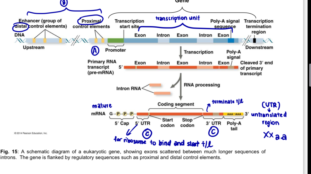
Organisation of eukaryotic gene at DNA level

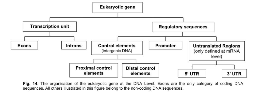
  1. Eukaryotic protein-coding gene

  • eukaryotic protein-coding gene requires the following DNA sequences for the proper expression of the gene

a. coding exons and non-coding introns —> transcription unit

b. non-coding DNA regulatory sequences

(see image)

  1. Transcription unit

  • as exons are interrupted by introns, exons are described as discontinuous coding DNA sequences of eukaryotic gene

  • each exon codes for a particular portion (amino acid sequence) of the polypeptide while introns are not represented in the amino acid sequence of the protein gene product (i.e. introns are non-coding DNA sequences)

  • number and sizes of introns per gene varies

  • the amount of DNA in the intron sequences is often greater than the exons

note: prokaryote genomes are arranged in operons where multiple genes are clustered together under the control of a single promoter and regulatory region; no introns present and post-transcriptional modifications are not necessary prior to translation

  1. Non-coding DNA regulatory sequences

  • Regulatory sequences: regions of DNA sequence where gene regulatory proteins bind to control the rate of assembly of protein complexes required for gene expression

  • regulatory sequences include

a. Promoter

  • a series of DNA sequences located upstream of the transcriptional start site

  • RNA polymerase and transcription factors bind to the promoter to initiate transcription

b. Control elements

  • segments of DNA involved in regulating the initiation and rate of transcription by binding particular proteins

  • include proximal and distal control elements that are located near to and far from the promoter respectively

i. Proximal control elements: sequences where gene regulatory proteins called general (or basal) transcription factors bind to initiate transcription

ii. Distal control elements consists of enhancers, DNA sequences that bind specific regulatory proteins known as activators to increase transcription rate, and silencers which interact with other specific regulatory proteins known as repressors to decrease transcription rate

enhancer (element) + activator (protein)

silencer (element) + repressor (protein)

note: elements —> DNA

factors —> proteins

c. Untranslated regions (UTRs)

  • found in the exons of the mRNA but are not translated into polypeptide sequence

i. 5’ UTR

  • starts at the +1 position on DNA template strand where transcription begins and ends one nucleotide before the start codon

  • contains DNA sequence which is transcribed into a ribosome binding site on mRNA - ribosome binds to the mRNA and initiate translation

  • contains DNA sequence which is transcribed into binding sites on mRNA for proteins which regulate the mRNA’s stability for translation

ii. 3’ UTR

  • starts after the stop codon

  • contains DNA sequence which is transcribed into a polyadenylation signal on mRNA, which is needed for termination of transcription
<ol><li><p><mark data-color="yellow" style="background-color: yellow; color: inherit;">Eukaryotic protein-coding gene</mark></p></li></ol><ul><li><p>eukaryotic protein-coding gene requires the following DNA sequences for the proper expression of the gene</p></li></ul><p>a. coding <strong>exons</strong> and non-coding <strong>introns</strong> —&gt; <strong>transcription unit</strong></p><p>b. non-coding DNA<strong> regulatory sequences</strong></p><p>(see image)</p><ol start="2"><li><p><mark data-color="blue" style="background-color: blue; color: inherit;">Transcription unit</mark></p></li></ol><ul><li><p>as exons are <strong>interrupted by introns</strong>, exons are described as<strong> discontinuous coding DNA sequences </strong>of eukaryotic gene</p></li><li><p>each<strong> exon codes</strong> for a particular portion (amino acid sequence) of the polypeptide while <strong>introns are not represented</strong> in the amino acid sequence of the protein gene product (i.e. introns are non-coding DNA sequences)</p></li><li><p>number and sizes of introns per gene varies</p></li><li><p>the amount of DNA in the intron sequences is often greater than the exons</p></li></ul><p>note: <strong>prokaryote</strong> genomes are arranged in operons where multiple genes are clustered together under the control of a single promoter and regulatory region; <strong>no introns present</strong> and <strong>post-transcriptional modifications are not necessary </strong>prior to translation</p><ol start="3"><li><p><mark data-color="red" style="background-color: red; color: inherit;">Non-coding DNA regulatory sequences</mark></p></li></ol><ul><li><p><strong>Regulatory sequences</strong>: regions of DNA sequence where gene regulatory proteins bind to <strong>control the rate of assembly of protein complexes</strong> required for gene expression</p></li><li><p>regulatory sequences include</p></li></ul><p>a. <strong>Promoter</strong></p><ul><li><p>a series of DNA sequences located upstream of the transcriptional start site</p></li><li><p>RNA polymerase and transcription factors bind to the promoter to initiate transcription</p></li></ul><p>b. <strong>Control elements</strong></p><ul><li><p>segments of DNA involved in regulating the initiation and rate of transcription by binding particular proteins</p></li><li><p>include <strong>proximal and distal control elements </strong>that are located near to and far from the promoter respectively</p></li></ul><p>i. <strong>Proximal control elements</strong>: sequences where gene regulatory proteins called <u>general (or basal) transcription factors bind </u>to <u>initiate</u> <u>transcription</u></p><p>ii. <strong>Distal control elements</strong> consists of <strong>enhancers</strong>, DNA sequences that bind specific regulatory proteins known as <strong>activators</strong> to<u> increase transcription rate</u>, and <strong>silencers</strong> which interact with other specific regulatory proteins known as <strong>repressors</strong> to <u>decrease transcription rate</u></p><p>enhancer (element) + activator (protein)</p><p>silencer (element) + repressor (protein)</p><p>note: elements —&gt; DNA</p><p>factors —&gt; proteins</p><p></p><p>c. <strong>Untranslated regions (UTRs)</strong></p><ul><li><p>found in the exons of the mRNA but are not translated into polypeptide sequence</p></li></ul><p>i. <strong>5’ UTR</strong></p><ul><li><p><strong>starts at the +1 position</strong> on DNA template strand where transcription begins and <strong>ends one nucleotide before the start codon</strong></p></li><li><p>contains DNA sequence which is transcribed into a <strong>ribosome</strong> binding site on mRNA - ribosome binds to the mRNA and initiate translation</p></li><li><p>contains DNA sequence which is transcribed into binding sites on mRNA for <strong>proteins</strong> which <strong>regulate the mRNA’s stability for translation</strong></p></li></ul><p>ii. <strong>3’ UTR</strong></p><ul><li><p>starts after the stop codon</p></li><li><p>contains DNA sequence which is transcribed into a <strong>polyadenylation signal on mRNA</strong>, which is needed for termination of transcription</p></li></ul><p></p>
16
New cards

Organisation of the eukaryotic intergenic DNA

  • these DNA sequences which are located between genes are termed intergenic DNA sequences consisting mainly of repetitive DNA

Repetitive DNA

  • Repetitive DNA refers to sequences present in multiple copies in the genome

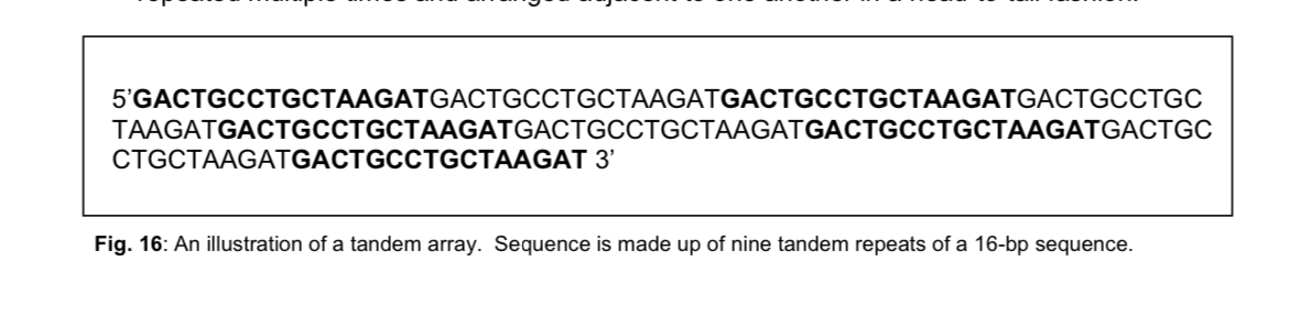
  • Tandemly repeated DNA (or tandem arrays) consist of DNA sequences repeated multiple times and arranged adjacent to one another in a head-to-tail fashion

    • Tandemly repeated DNA: Satellite DNA - the most highly repeated tandem sequences found so far in mammals, simple sequence DNA, also referred to as satellite DNA, makes up about 3-6% of the human genome

  1. Satellite DNA mostly consists of relatively short sequences (of between 1 to 500 bp) repeated many times in tandem to form a long array or cluster in a localised area of the genome

  2. preferentially located in regions of heterochromatin including centromeres, telomeres, and specific locations within the arms of particular chromosomes, satellite DNA is divided, based on length of the repeating unit, into 3 types

  • regular satellite DNA - centromeres

  • minisatellites - telomeres

  • (FYI) microsatellites - variable number tandem repeats (VNTR)
<ul><li><p>these DNA sequences which are located between genes are termed <strong>intergenic</strong> DNA sequences consisting mainly of repetitive DNA</p></li></ul><p></p><p><u>Repetitive DNA</u></p><ul><li><p>Repetitive DNA refers to sequences present in <strong>multiple copies</strong> in the genome</p></li><li><p><strong>Tandemly repeated DNA</strong> (or tandem arrays) consist of DNA sequences repeated multiple times and arranged adjacent to one another in a head-to-tail fashion</p><ul><li><p><strong>Tandemly repeated DNA: Satellite DNA </strong>- the most highly repeated tandem sequences found so far in mammals, <strong>simple sequence DNA</strong>, also referred to as satellite DNA, makes up about 3-6% of the human genome</p></li></ul></li></ul><ol><li><p>Satellite DNA mostly consists of <u>relatively short sequences</u> (of between 1 to 500 bp) <u>repeated many times in tandem</u> to form a <u>long array or cluster</u> in a localised area of the genome</p></li><li><p>preferentially located in regions of <strong>heterochromatin</strong> including <strong>centromeres, telomeres</strong>, and specific locations within the arms of particular chromosomes, satellite DNA is divided, based on <u>length of the repeating unit</u>, into 3 types</p></li></ol><ul><li><p><strong>regular satellite DNA </strong>- <mark data-color="yellow" style="background-color: yellow; color: inherit;">centromeres</mark></p></li><li><p><strong>minisatellites</strong> - <mark data-color="blue" style="background-color: blue; color: inherit;">telomeres</mark></p></li><li><p>(FYI) microsatellites - variable number tandem repeats (VNTR)</p></li></ul><p></p><p></p><p></p>
17
New cards

Examples of satellite DNA: Centromere and Telomere

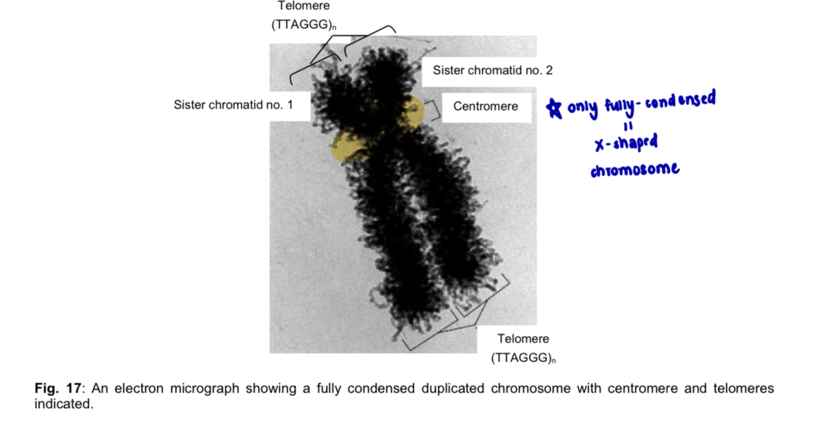
Comparisons btw centromere and telomere

  1. Region found on chromosome

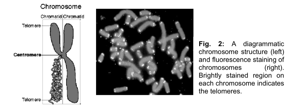
  • Centromere: centromere is the region where two sister chromatids are joined in a replicated chromosome during cell division

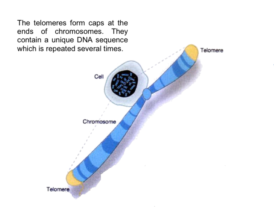
  • Telomere: telomere is located at the two physical ends or tips of a linear eukaryotic chromosome

  1. Form of DNA packaging

  • Centromere & telomere: heterochromatin (unexpressed DNA)

  1. Nature of DNA sequence

  • Centromere & telomere: consists of tandem repetitive, non-coding satellite DNA

  1. Category of satellite DNA

  • Centromere: regular satellite

  • telomere: minisatellite

note: prokaryote genomes are circular in nature

—> they will not contain telomeres at the ends of their chromosomes nor centromeres within their chromosome
<p>Comparisons btw centromere and telomere </p><ol><li><p><mark data-color="yellow" style="background-color: yellow; color: inherit;">Region found on chromosome </mark></p></li></ol><ul><li><p>Centromere: centromere is the region where <strong>two sister chromatids </strong>are <strong>joined</strong> in a <strong>replicated</strong> <strong>chromosome</strong> during cell division </p></li><li><p>Telomere: telomere is located at the two <strong>physical ends</strong> or tips of a linear eukaryotic chromosome </p></li></ul><p></p><ol start="2"><li><p><mark data-color="blue" style="background-color: blue; color: inherit;">Form of DNA packaging </mark></p></li></ol><ul><li><p>Centromere &amp; telomere: <strong>heterochromatin</strong> (unexpressed DNA) </p></li></ul><p></p><ol start="3"><li><p><mark data-color="red" style="background-color: red; color: inherit;">Nature of DNA sequence </mark></p></li></ol><ul><li><p>Centromere &amp; telomere: consists of <strong>tandem repetitive, non-coding satellite DNA </strong></p></li></ul><p></p><ol start="4"><li><p><mark data-color="purple" style="background-color: purple; color: inherit;">Category of satellite DNA </mark></p></li></ol><ul><li><p>Centromere: <strong>regular satellite </strong></p></li><li><p>telomere: <strong>minisatellite</strong> </p></li></ul><p>note: prokaryote genomes are circular in nature </p><p>—&gt; they will not contain telomeres at the ends of their chromosomes nor centromeres within their chromosome </p><p></p>
18
New cards

Structure and functions of the telomere

Structure of telomere

  • consists of specialised nucleoprotein which are complexes composed of telomeric DNA bound by specific proteins

  1. telomeric repeat sequence varies between organisms

  2. in humans and other vertebrates, telomeric DNA consists of long stretches of hundreds to thousands of tandem repeats of a short nucleotide sequence with a high G content

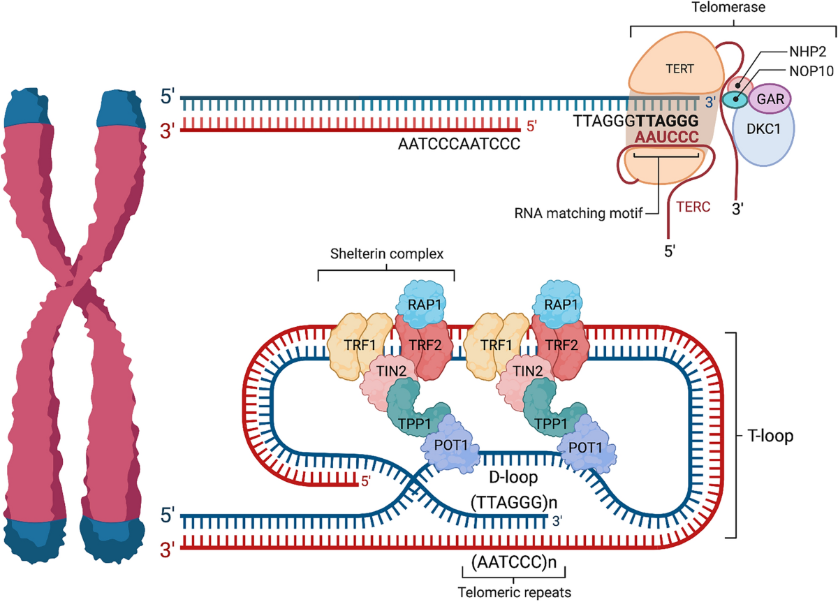
  3. human telomeres contain hundreds to as many as 2000 tandem repeats of the sequence 5’-TTAGGG-3’

  4. the 3’ end of the G-rich strand extends 12-16 nucleotides beyond the 5’ end of the complementary C-rich strand, forming a 3’ single-stranded overhang

(single-stranded - unstable; recall: end replication problem - no free 3’ OH = shorter daughter strand)

  • this overhang folds back on itself to form a hairpin loop called a telomere loop (t-loop)

  • single-stranded 3’ overhang invades an upstream telomeric repeat to displace the same sequence in an upstream region of the telomere and base pair with the complementary strand, forming a t-loop

    • formation and stabilisation of the t-loop involves various proteins

(more detailed explanation:

3’ overhang loops around and invades the preceding double-stranded telomeric DNA —> when it inserts itself into the DNA, it displaces one of the original strands, creating a small bubble known as D-loop)
<p>Structure of telomere</p><ul><li><p>consists of specialised <strong>nucleoprotein</strong> which are complexes composed of<strong> telomeric DNA bound by specific proteins</strong></p></li></ul><ol><li><p><mark data-color="yellow" style="background-color: yellow; color: inherit;">telomeric repeat sequence varies between organisms</mark></p></li><li><p><mark data-color="blue" style="background-color: blue; color: inherit;">in humans and other vertebrates, telomeric DNA consists of long stretches of hundreds to thousands of tandem repeats of a short nucleotide sequence with a </mark><strong><mark data-color="blue" style="background-color: blue; color: inherit;">high G content</mark></strong></p></li><li><p><mark data-color="red" style="background-color: red; color: inherit;">human telomeres contain hundreds to as many as 2000 tandem repeats of the sequence </mark><em><mark data-color="red" style="background-color: red; color: inherit;">5’-TTAGGG-3’</mark></em></p></li><li><p><mark data-color="purple" style="background-color: purple; color: inherit;">the 3’ end of the G-rich strand extends 12-16 nucleotides beyond the 5’ end of the complementary C-rich strand, forming a</mark><strong><mark data-color="purple" style="background-color: purple; color: inherit;"> 3’ single-stranded overhang</mark></strong></p></li></ol><p>(single-stranded - unstable; recall: end replication problem - no free 3’ OH = shorter daughter strand)</p><ul><li><p>this overhang folds back on itself to form a <strong>hairpin loop</strong> called a <strong>telomere loop (t-loop)</strong></p></li><li><p>single-stranded 3’ overhang invades an upstream telomeric repeat to displace the same sequence in an upstream region of the telomere and base pair with the complementary strand, forming a t-loop</p><ul><li><p>formation and stabilisation of the t-loop involves various proteins</p></li></ul></li></ul><p>(more detailed explanation: </p><p>3’ overhang loops around and invades the preceding double-stranded telomeric DNA —&gt; when it inserts itself into the DNA, it displaces one of the original strands, creating a small bubble known as D-loop) </p><p></p>
19
New cards

telomeres forming caps

knowt flashcard image
20
New cards

Structure and functions of telomere

Functions of telomere

  1. Protective function

  • Telomeric DNA forms t-loops with telomere-specific proteins forming a cap that

    • protect the 5’ ends and 3’ single stranded overhangs of linear chromosomes from degradation by cellular exonucleases (enzymes that cleave off nucleotides, one at a time, from the end of the polynucleotide chain)

    • prevents it from being recognised as a damaged DNA molecule by the cell’s repair machinery

  1. Maintaining stability

  • Telomeres confer stability to linear chromosomes as the t-loops prevent the chromosome tips from fusing to the ends of other chromosomes, thus ensuring that the ends of homologous chromosomes do not spontaneously fuse

  1. Preventing loss of genes

  • Telomeres protect the organisms’ genes, located within the interior of the linear chromosomes, from being eroded as the linear chromosome ends shorten with each successive round of DNA replication due to the end-replication problem

  • ensures that DNA replication can occur without the loss of important coding sequences

  1. Regulating replicative cell senescence

  • Each telomere shrinks with every successive cell division/DNA replication

  • when the telomeres have shortened to a critical length, the cell reaches the Hayflick limit and enters a period of replicative cell senescence - it withdraws permanently from the cell cycle and stops dividing

21
New cards

End replication problem

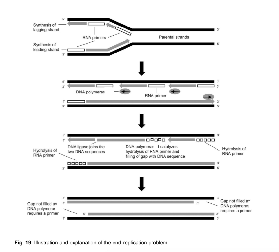
  1. chromosomes of eukaryotes are linear and will lead to the end-replication problem as DNA polymerase is incapable of completely replicating all the way to the ends of a linear chromosome, leading to shortening of telomeres with each successive cell division

  • each time a cell with linear chromosomes divides, a small section at the extreme 3’ end of the parental strand does not undergo DNA replication

  • due to inability of DNA polymerase to replicate the ends of the chromosome

  1. without telomeres, vital genetic information that is needed to sustain a cell’s activities will be lost
<ol><li><p>chromosomes of <strong>eukaryotes</strong> are <strong>linear</strong> and will lead to the end-replication problem as DNA polymerase is incapable of completely replicating all the way to the ends of a linear chromosome, leading to <strong>shortening of telomeres</strong> with each successive cell division </p></li></ol><ul><li><p>each time a cell with linear chromosomes divides, a small section at the extreme 3’ end of the parental strand does not undergo DNA replication </p></li><li><p>due to inability of DNA polymerase to replicate the ends of the chromosome </p></li></ul><ol start="2"><li><p>without telomeres, vital genetic information that is needed to sustain a cell’s activities will be lost </p></li></ol><p></p>
22
New cards

Replicative cell senescence

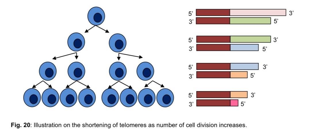
  • Replicative cell senescence: the period in which a cell withdraws permanently from the cell cycle and hence stops dividing after reaching Hayflick limit when it has divided for 25 to 50 cell divisions

  1. Hayflick limit: point at which a cell ceases to divide and grow, after 25 to 50 cell divisions

  • cell no longer divides and eventually dies (to prevent loss of genes)

  • complete loss of telomere repeated eventually triggers apoptosis (programmed cell death)

  1. if the cell does not obey the Hayflick limit, uncontrolled cell division will occur (KIV: Molecular basis of cancer)

  2. cells with longer telomeres tend to go through a higher number of cell divisions and survive longer when cultured in vitro (in an artificial environment) than cells with shorter telomeres

  3. telomere length thus appears to limit the number of times cells can divide and thereby regulate a cell’s life span

  • it acts as a reference to count cell division

  • research has shown that telomere shortening may be linked to the aging process of tissues/organisms

  1. germ cells, stem cells and cancer cells are able to counteract the gradual shortening of their linear chromosomes by adding telomeric repeat sequences to the ends of each chromosome with the aid of the enzyme telomerase

*end replication problem cannot be prevented and occurs with every round of DNA replication

*note: counteract NOT equals prevent
<ul><li><p><strong>Replicative cell senescence</strong>: the period in which a <u>cell withdraws permanently from the cell cycle </u>and hence stops dividing after reaching<strong> Hayflick limit</strong> when it has divided for 25 to 50 cell divisions</p></li></ul><ol><li><p><strong>Hayflick limit</strong>: point at which a cell ceases to divide and grow, after 25 to 50 cell divisions</p></li></ol><ul><li><p>cell no longer divides and eventually dies (to prevent loss of genes)</p></li><li><p>complete loss of telomere repeated eventually triggers apoptosis (programmed cell death)</p></li></ul><ol start="2"><li><p>if the cell does not obey the Hayflick limit, <mark data-color="green" style="background-color: green; color: inherit;">uncontrolled cell division will occur (</mark>KIV: Molecular basis of cancer)</p></li><li><p><mark data-color="purple" style="background-color: purple; color: inherit;">cells with longer telomeres tend to go through a higher number of cell divisions and survive longer </mark>when cultured in vitro (in an artificial environment) than cells with shorter telomeres</p></li><li><p><mark data-color="red" style="background-color: red; color: inherit;">telomere length thus appears to limit the number of times cells can divide and thereby regulate a cell’s life span</mark></p></li></ol><ul><li><p>it acts as a reference to count cell division</p></li><li><p>research has shown that telomere shortening may be linked to the aging process of tissues/organisms</p></li></ul><ol start="5"><li><p><mark data-color="blue" style="background-color: blue; color: inherit;">germ cells, stem cells and cancer cells are able to </mark><u><mark data-color="blue" style="background-color: blue; color: inherit;">counteract</mark></u><mark data-color="blue" style="background-color: blue; color: inherit;"> the gradual shortening of their linear chromosomes by adding telomeric repeat sequences</mark> to the ends of each chromosome with the aid of the <mark data-color="blue" style="background-color: blue; color: inherit;">enzyme telomerase</mark></p></li></ol><p>*end replication problem cannot be prevented and occurs with every round of DNA replication</p><p>*note: counteract NOT equals prevent </p><p></p>
23
New cards

The action of telomerase

  1. Telomere length cannot be maintained by normal DNA replication

  • it can be maintained by the enzyme telomerase

2. Telomerase does NOT prevent the end replication problem; it does not stop the shortening of chromosomal ends during replication

  • it only lengthens and thereby maintains, the chromosomal ends after the end replication problem has occurred

Characteristics of telomerase

  1. Telomerase generally found only in stem cells, germline cells and cancer cells

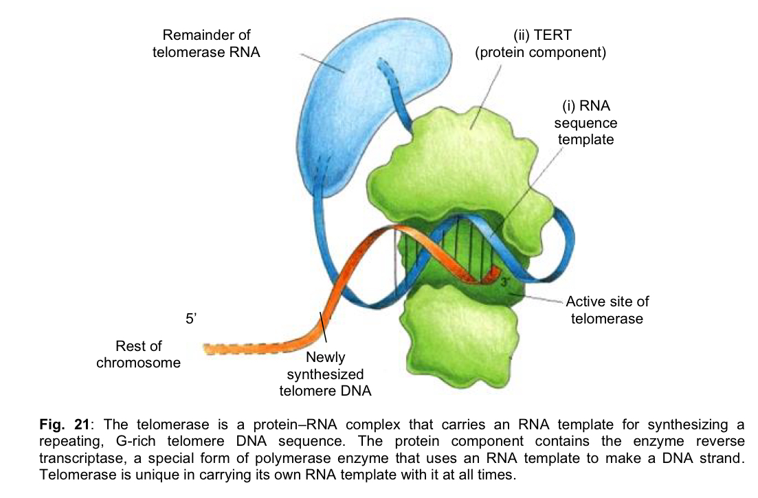
  2. Telomerase is a ribonucleoprotein (protein-RNA) complex that is made up of 2 components - RNA sequence template & protein component

  3. In mammals, the RNA sequence template has the sequence 3’ AAUCCC 5’, which is complementary to the telomere repeat sequence 5’ TTAGGG 3’

  • RNA template sequence will act as the template for the insertion of the deoxyribonucleotide sequence, 5’ TTAGGG 3’, onto the existing 3’ overhang strand end of the telomeres, effectively lengthening the overhang

  1. Protein component of telomerase is known as TERT (telomere reverse transcriptase)

  • TERT is a reverse transcriptase enzyme that provides the catalytic action to synthesise DNA from an RNA template
<ol><li><p><mark data-color="yellow" style="background-color: yellow; color: inherit;">Telomere length cannot be maintained by normal DNA replication</mark> </p></li></ol><ul><li><p>it can be maintained by the enzyme <strong>telomerase</strong> </p></li></ul><p>2. <mark data-color="blue" style="background-color: blue; color: inherit;">Telomerase does NOT prevent the end replication problem</mark>; it does not stop the shortening of chromosomal ends during replication </p><ul><li><p>it only <strong>lengthens</strong> and thereby <strong>maintains</strong>, the chromosomal ends <strong>after</strong> the end replication problem has occurred </p></li></ul><p></p><p>Characteristics of telomerase </p><ol><li><p>Telomerase generally found only in <strong>stem cells, germline cells and cancer cells </strong></p></li><li><p>Telomerase is a <strong>ribonucleoprotein</strong> (protein-RNA) complex that is made up of 2 components - <strong>RNA sequence template &amp; protein component </strong></p></li><li><p>In mammals, the <strong>RNA</strong> sequence template has the sequence 3’ AAUCCC 5’, which is <strong>complementary</strong> to the<strong> telomere repeat sequence </strong>5’ TTAGGG 3’ </p></li></ol><ul><li><p>RNA template sequence will act as the <u>template for the insertion of the deoxyribonucleotide sequence, 5’ TTAGGG 3</u>’, onto the existing 3’ overhang strand end of the telomeres, effectively <u>lengthening the overhang </u></p></li></ul><ol start="4"><li><p><strong>Protein</strong> component of telomerase is known as <strong>TERT</strong> (telomere reverse transcriptase) </p></li></ol><ul><li><p>TERT is a<u> reverse transcriptase enzyme </u>that provides the <u>catalytic action to synthesise DNA from an RNA template</u> </p></li></ul><p></p>
24
New cards

How telomerase maintains telomere length

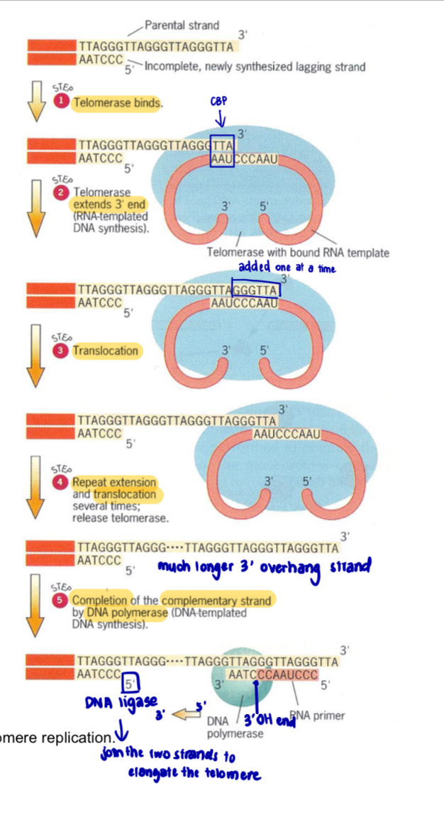
  1. Telomerase’s RNA template binds complementarily to the 3’ overhang of the parental DNA strand

  2. Telomerase extends the 3’ overhang of the parental DNA strand in the 5’ to 3’ direction by adding sequence repeats of 5’TTAGGG 3’ via complementary base-pairing

  3. By lengthening the 3’ overhang, the synthesis of the shorter daughter strand can be extended during the next round of DNA replication, resulting in a longer telomere (but still with a 3’ overhang)

  4. The action of telomerase helps to maintain the number of repeats at the telomeres, delaying the senescence of cells and enabling them to proliferate indefinitely

Step 1

  • Telomerase enzyme recognises and binds to the G-rich telomere sequence at the 3’ overhang on the parental strand

  • 3’ nucleotides are base-paired to the 5’ UAA 3’ sequence in the RNA template of the telomerase

Step 2

  • Through its reverse transcriptase activity, telomerase adds nucleotides to the 3’ end of the overhang using the bound RNA as a template (RNA-templated DNA synthesis), thereby extending the 3’ end of the parental strand

  • sequence 5’ GGGTTA 3’ is added one nucleotide at a time

Steps 3 & 4

  • Telomerase is translocated to the end of the extended overhang

  • Result is that the telomere is extended in a 5’ to 3’ direction, over repeated cycles of elongation and translocation

Step 5

  • Replication of the incomplete lagging daughter strand is completed by using these extensions as a template for synthesis of the complementary strand by DNA polymerase, leaving a 3’ overhang
<ol><li><p>Telomerase’s RNA template binds <strong>complementarily</strong> to the 3’ overhang of the parental DNA strand </p></li><li><p>Telomerase <strong>extends the 3’ overhang</strong> of the parental DNA strand in the <strong>5’ to 3’ direction</strong> by adding sequence repeats of 5’TTAGGG 3’ via <strong>complementary base-pairing </strong></p></li><li><p>By lengthening the 3’ overhang, the synthesis of the shorter daughter strand can be extended during the next round of DNA replication, resulting in a longer telomere (but still with a 3’ overhang) </p></li><li><p>The action of telomerase helps to <strong>maintain the number of repeats at the telomeres, delaying the senescence </strong>of cells and enabling them to <strong>proliferate indefinitely </strong></p></li></ol><p>Step 1</p><ul><li><p>Telomerase enzyme recognises and binds to the G-rich telomere sequence at the <strong>3’ overhang on the parental strand </strong></p></li><li><p>3’ nucleotides are base-paired to the 5’ UAA 3’ sequence in the RNA template of the telomerase </p></li></ul><p></p><p>Step 2 </p><ul><li><p>Through its reverse transcriptase activity, telomerase <strong>adds nucleotides to the 3’ end of the overhang </strong>using t<strong>he bound RNA as a template</strong> (RNA-templated DNA synthesis), thereby <strong>extending the 3’ end of the parental strand </strong></p></li><li><p>sequence 5’ GGGTTA 3’ is added one nucleotide at a time </p></li></ul><p></p><p>Steps 3 &amp; 4</p><ul><li><p>Telomerase is <strong>translocated to the end of the extended overhang</strong> </p></li><li><p>Result is that the telomere is <strong>extended in a 5’ to 3’ direction</strong>, over repeated cycles of elongation and translocation </p></li></ul><p></p><p>Step 5 </p><ul><li><p>Replication of the incomplete lagging daughter strand is completed by using these extensions as a template for <strong>synthesis of the complementary strand by DNA polymerase</strong>, leaving a <strong>3’ overhang </strong></p></li></ul><p></p>
25
New cards

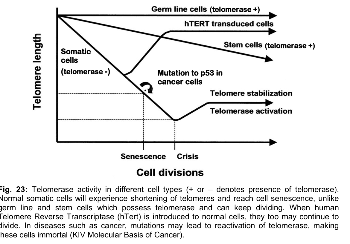
Telomerase activity in somatic and cancer cells

Somatic cells

  • In most human somatic cells that divide only a limited number of times, expression of the genes coding for the catalytic subunit of telomerase and the telomerase-associated RNA are switched off, or at least not fully activated

    • telomeres of these cells become shorter with every cell division

    • eventually, DNA damage occurs at the chromosome ends, triggering cell cycle arrest

    • cells then enter a non-dividing state from which they never recover

Germ cells and stem cells

  • genes encoding telomerase protein and telomerase-associated RNA template are active in germ cells and stem cells

  • same genes are reactivated in most human cancer/tumour cells, where telomerase is active and maintains telomere lengths so that the cancer cells divide indefinitely or are immortalised

    • this feature of immortality is seen in all human cancers, where multiple uncontrolled cell divisions give rise to tumours

    • telomerase appears to allow cancer cells to evade apoptosis i.e. programmed cell death, and it is thought that the abnormal retention of telomeres is involved in the development of some types of cancer

    • telomerase is likely to be an important factor in cancer implies its promising potential as a useful target for cancer diagnosis and treatment

      • stimulated search for inhibitors of human telomerase as potential agents for treating cancer
<p>Somatic cells</p><ul><li><p>In most human <strong>somatic cells</strong> that divide only a limited number of times, expression of the genes coding for the catalytic subunit of telomerase and the telomerase-associated RNA are <strong>switched off</strong>, or at least<strong> not fully activated</strong></p><ul><li><p>telomeres of these cells become <u>shorter</u> with every cell division</p></li><li><p>eventually, <u>DNA damage</u> occurs at the chromosome ends, triggering cell cycle arrest</p></li><li><p>cells then enter a <u>non-dividing state</u> from which they never recover</p></li></ul></li></ul><p>Germ cells and stem cells</p><ul><li><p>genes encoding <u>telomerase protein and telomerase-associated RNA template</u> are <strong>active</strong> in germ cells and stem cells</p></li><li><p>same genes are <strong>reactivated</strong> in most human <strong>cancer/tumour cells</strong>, where telomerase is active and <u>maintains telomere lengths </u>so that the cancer cells <u>divide indefinitely or are immortalised</u></p><ul><li><p>this feature of immortality is seen in all human cancers, where multiple uncontrolled cell divisions give rise to tumours</p></li><li><p>telomerase appears to allow cancer cells to <strong>evade</strong> <strong>apoptosis</strong> i.e. programmed cell death, and it is thought that the abnormal retention of telomeres is involved in the development of some types of cancer</p></li><li><p>telomerase is likely to be an important factor in cancer implies its promising potential as a useful target for cancer diagnosis and treatment</p><ul><li><p>stimulated search for <strong>inhibitors of human telomerase</strong> as potential agents for treating cancer</p></li></ul></li></ul></li></ul><p></p>
26
New cards

Centromere

Structure of centromere

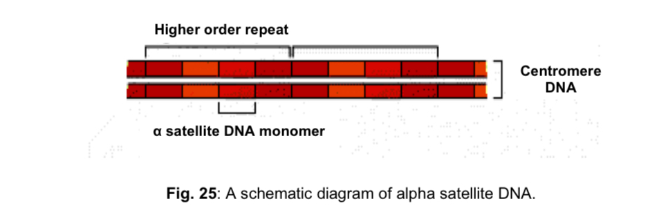
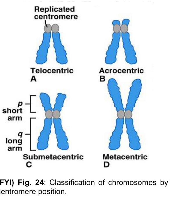
  1. Unlike telomeres, the single centromere found in each chromosome is not in a defined position

  • position of the centromere is unique for each chromosome

  1. Centromeres consist of satellite DNA

  • in humans, centromere satellite DNA is known as alpha satellite DNA

  • alpha satellite DNA consists of short, AT-rich sequences that are repeated thousands of times in tandem

  • numerous repeats vary slightly from one another in sequence; there is apparently no centromere-specific DNA sequence

  1. Centromeric DNA sequences vary greatly in length in different eukaryotic species

  • centromeres are more than 40 kb long in majority of eukaryotes
<p>Structure of centromere </p><ol><li><p>Unlike telomeres, the <strong>single</strong> centromere found in each chromosome is <strong>not in a defined position </strong></p></li></ol><ul><li><p><strong>position</strong> of the centromere is <strong>unique</strong> for each chromosome </p></li></ul><p></p><ol start="2"><li><p>Centromeres consist of<strong> satellite DNA </strong></p></li></ol><ul><li><p>in <u>humans</u>, centromere satellite DNA is known as <strong>alpha satellite DNA </strong></p></li><li><p>alpha satellite DNA consists of short,<strong> AT-rich </strong>sequences that are <strong>repeated thousands of times in tandem </strong></p></li><li><p>numerous repeats vary slightly from one another in sequence; there is apparently no centromere-specific DNA sequence </p></li></ul><p></p><ol start="3"><li><p>Centromeric DNA sequences <strong>vary greatly in length </strong>in different eukaryotic species </p></li></ol><ul><li><p>centromeres are more than 40 kb long in majority of eukaryotes </p></li></ul><p></p>
27
New cards

(FYI) classification of chromosomes by centromere position
knowt flashcard image
28
New cards

Structure of centromere

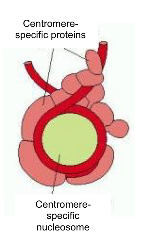
  1. Centromeres are embedded in a very large stretch of heterochromatin

  • centromeric DNA is bound by centromere-specific histones to form specialised nucleosomes, which are hetrochromatic in nature

  1. Folding of DNA into these specialised nucleosomes facilitates the assembly of other centromere-binding proteins to form the kinetochore that associates the centromere to the mitotic spindle
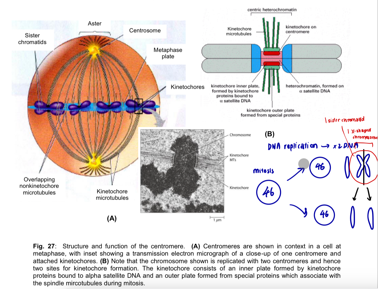
<ol start="4"><li><p>Centromeres are embedded in a very large stretch of <strong>heterochromatin</strong> </p></li></ol><ul><li><p>centromeric DNA is bound by <strong>centromere-specific histones</strong> to form<strong> specialised nucleosomes</strong>, which are hetrochromatic in nature </p></li></ul><p></p><ol start="5"><li><p>Folding of DNA into these specialised nucleosomes facilitates the assembly of other centromere-binding proteins to form the <strong>kinetochore</strong> that associates the centromere to the mitotic spindle </p></li></ol><p></p><p></p>
29
New cards
knowt flashcard image
30
New cards

Functions of centromere

  1. Sister chromatid adhesion

  • centromere is the region of a linear chromosome where the two sister chromatids join

  1. Kinetochore formation

  • centromere is the site of assembly of the kinetochore, a protein complex that attaches to the microtubules of the mitotic/meiotic spindle

  • sister chromatids are in turn joined via the centromere to the kinetochore microtubules

  1. Proper chromosome segregation

  • centromeres are essential for the correct segregation of the daughter chromosomes after DNA replication, so that one copy goes to each of the two daughter cells during cell division

  • the presence of only one centromere on each chromosome is critical

    • each kinetochore at the single centromere of each sister chromatid of a duplicated chromosome binds to kinetochore microtubules and is pulled towards one pole of the cell during anaphase

  • in the absence of a centromere, the daughter chromosomes segregate randomly, leading to loss or duplication of chromosomes in the daughter cells
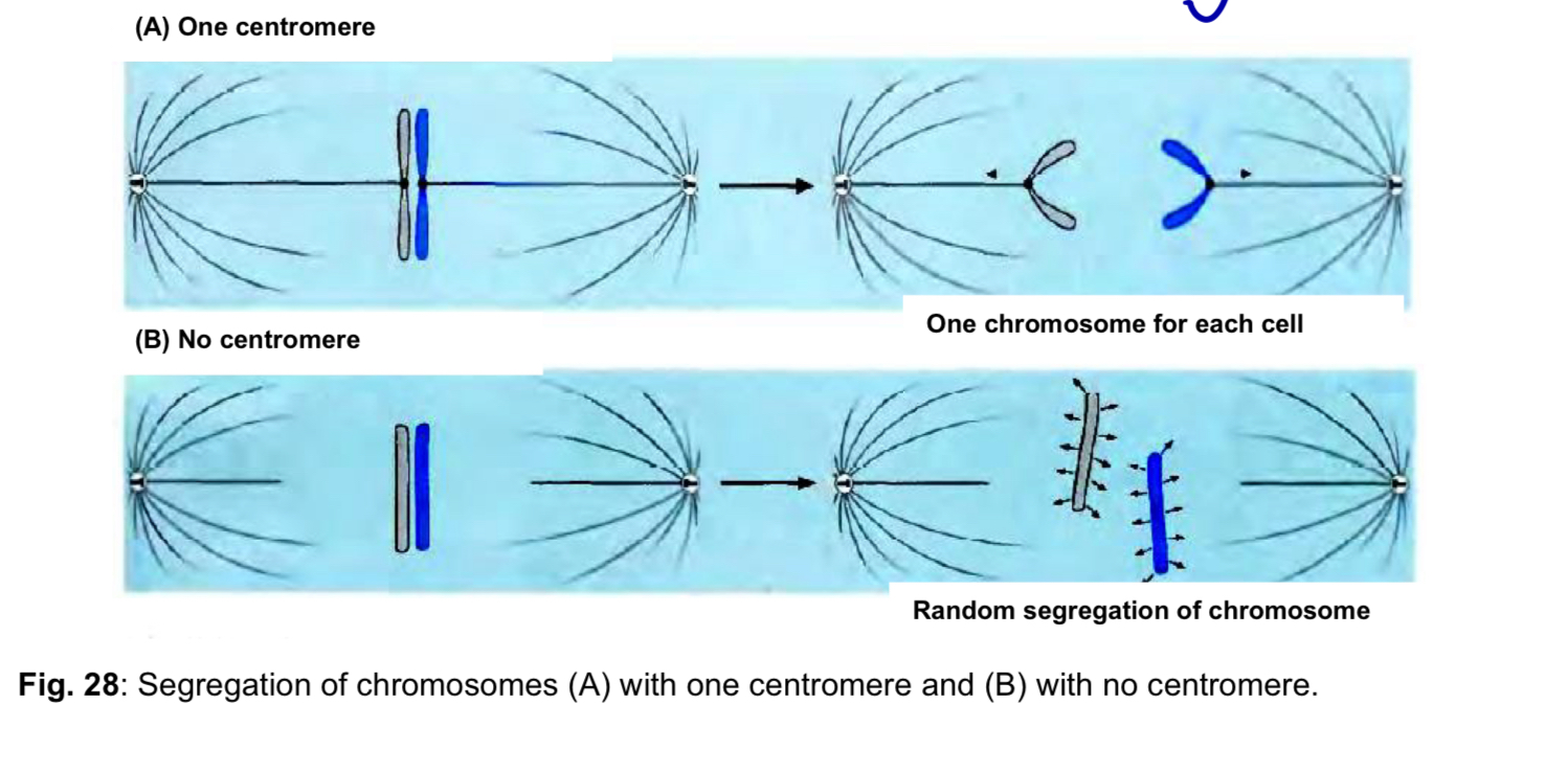
<ol><li><p><mark data-color="yellow" style="background-color: yellow; color: inherit;">Sister chromatid adhesion</mark> </p></li></ol><ul><li><p>centromere is the region of a linear chromosome where the two sister chromatids join </p></li></ul><ol start="2"><li><p><mark data-color="blue" style="background-color: blue; color: inherit;">Kinetochore formation</mark> </p></li></ol><ul><li><p>centromere is the site of assembly of the <strong>kinetochore</strong>, a protein complex that attaches to the <u>microtubules of the mitotic/meiotic spindle </u></p></li><li><p><strong>sister chromatids</strong> are in turn <u>joined via the centromere</u> to the <u>kinetochore microtubules </u></p></li></ul><ol start="3"><li><p><mark data-color="red" style="background-color: red; color: inherit;">Proper chromosome segregation</mark> </p></li></ol><ul><li><p>centromeres are essential for the correct segregation of the daughter chromosomes after DNA replication, so that one copy goes to each of the two daughter cells during cell division </p></li><li><p>the presence of only one centromere on each chromosome is critical </p><ul><li><p>each kinetochore at the single centromere of each sister chromatid of a duplicated chromosome binds to kinetochore microtubules and is pulled towards one pole of the cell during anaphase </p></li></ul></li><li><p>in the absence of a centromere, the daughter chromosomes segregate randomly, leading to <u>loss or duplication of chromosomes in the daughter cells </u></p></li></ul><p></p>