CSB063 MRI Upper and Lower Limb
1/72
There's no tags or description
Looks like no tags are added yet.
Name | Mastery | Learn | Test | Matching | Spaced |
|---|
No study sessions yet.
73 Terms
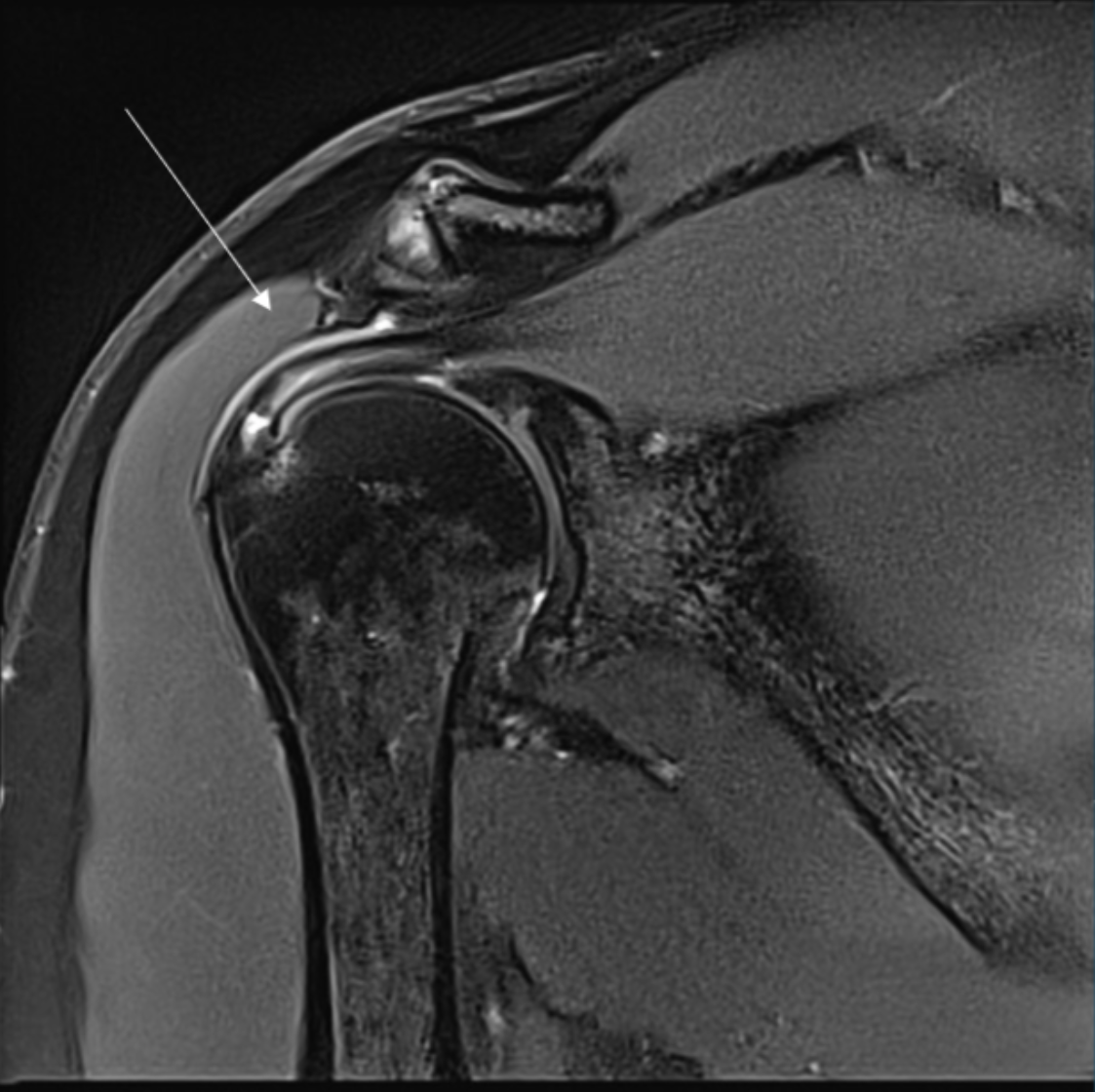
Describe the pathology indicated by the arrow (Cor T2 FS).
Supraspinatus tendon tear
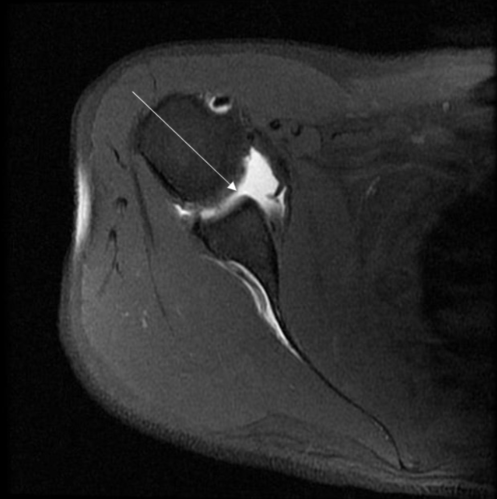
Describe the pathology indicated by the arrow (Ax T1 FS - arthrogram).
SLAP tear
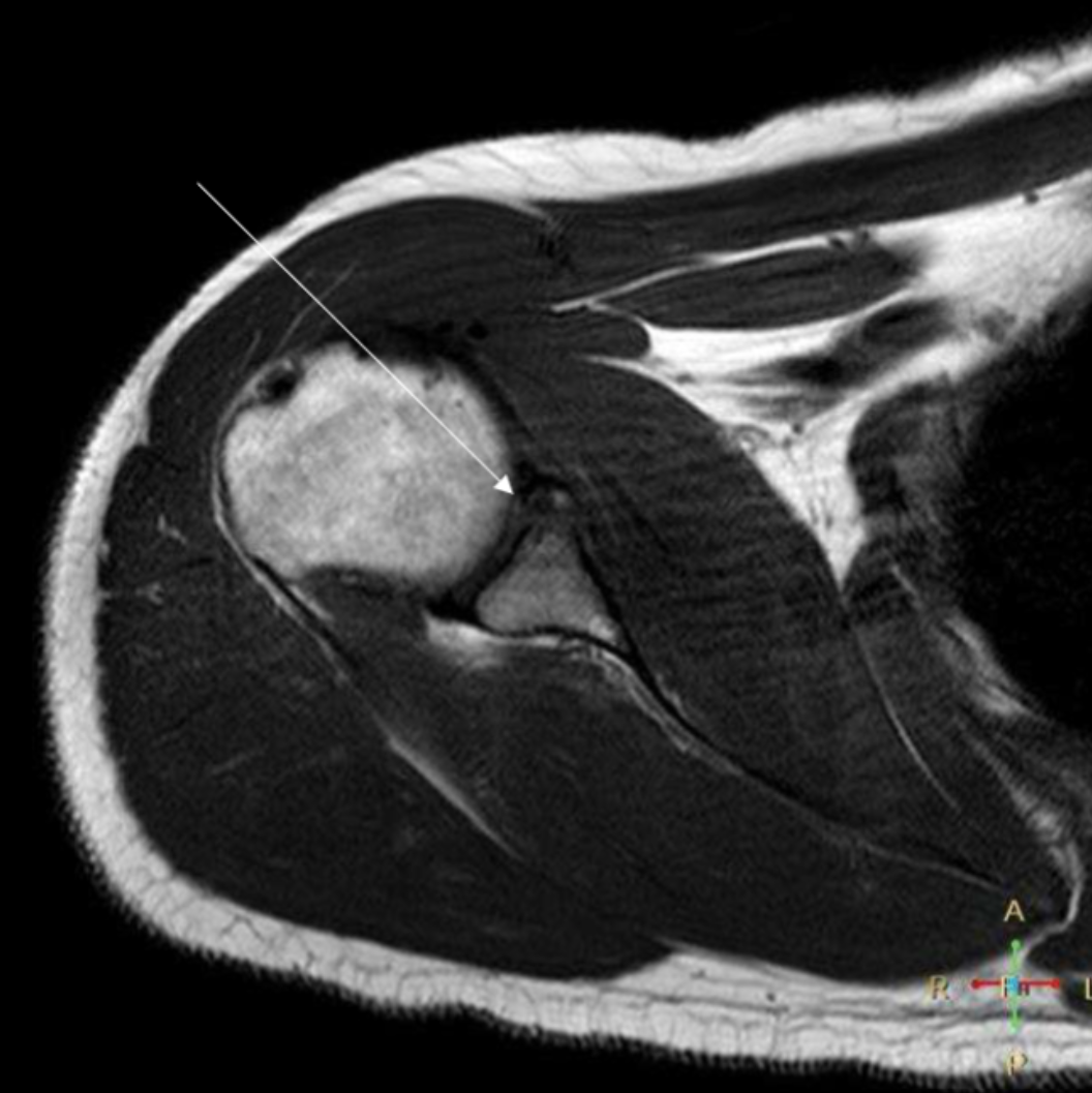
Describe the pathology indicated by the arrow (Ax T1).
Bankart lesion
Describe the pathology indicated by the arrow (Cor T1 FS +C).
Osteosarcoma
In an axial shoulder, how should the slices be angled?
Perpendicular to the glenoid surface
In an axial shoulder, what is the scan range?
Above ACJ to below GHJ capsule
In a coronal shoulder, how should the slices be angled?
Parallel to the supraspintus tendon
In a coronal shoulder, what is the scan range?
Coracoid process (anteriorly) to acromion (posteriorly)
In a sagittal shoulder, what is the scan range?
Greater tuberosity (laterally) to past glenoid (medially)
Coracoid process (anteriorly) to acromion (posteriorly)
In a sagittal shoulder, how should the slices be angled?
Parallel to the glenoid surface
In an axial elbow, what is the scan range?
Humeral metaphysis to radial tuberosity
In an axial elbow, how should the slices be angled?
Perpendicular to humeral shaft
In a coronal elbow, how should the slices be angled?
Parallel to the medial and lateral epicondyles
In a sagittal elbow, what is the scan range?
Humeral metaphysis to radial tuberosity
In a sagittal elbow, how should the slices be angled?
Perpendicular to the coronal plane (which is parallel to the medial and lateral epicondyles)
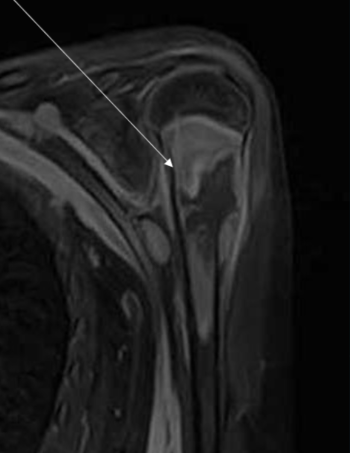
Describe the pathology indicated by the arrow (Sag T2 FS).
Biceps tendonitis
Describe the pathology indicated by the arrow (Cor T2 FS).
Lateral epicondylitis
Describe the pathology indicated by the arrow (Sag PD).
Biceps tendon rupture

Describe the pathology indicated by the arrow (Ax PD FS).
Ulnar nerve inflammation (from cubital tunnel syndrome)
In an axial wrist, what is the scan range?
CMCJ to below DRUJ
In an axial wrist, how should the slices be angled?
Parallel to the DRUJ
In a coronal wrist, what is the scan range?
CMCJ to below DRUJ
In a coronal wrist, how should the slices be angled?
Parallel to ulnar and radial styloids
In a sagittal wrist, what is the scan range?
Skin edge to skin edge
In a sagittal wrist, how should the slices be angled?
Perpendicular to wrist joint
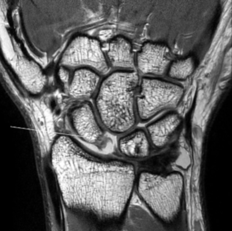
Describe the pathology indicated by the arrow (Cor PD).
TFCC tear
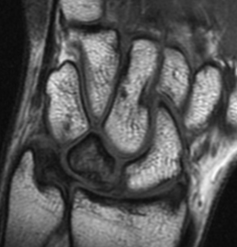
Describe the pathology indicated by the arrow (Cor PD).
Scapholunate ligament tear
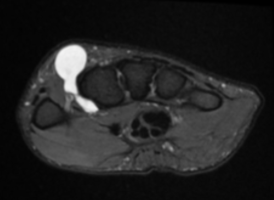
Describe the pathology indicated by the arrow (Ax T2 FS).
Dorsal ganglion cyst
Describe the pathology indicated by the arrow (Cor T1).
Lunate AVN
In an axial finger, what is the scan range?
Distal phalanx to MCMJ
In an axial finger, how should the slices be angled?
Parallel to the line of best fit for each IPJ space
In a coronal finger, what is the scan range?
Distal phalanx to MCPJ
In a coronal finger, how should the slices be angled?
Perpendicular to the line of best fit for each IPJ space
In a sagittal finger, what is the scan range?
Distal phalanx to MCPJ
In a sagital finger, how should the slices be angled?
Perpendicular to the line of best fit for each IPJ space
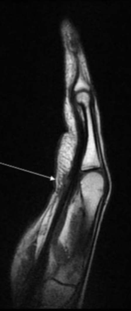
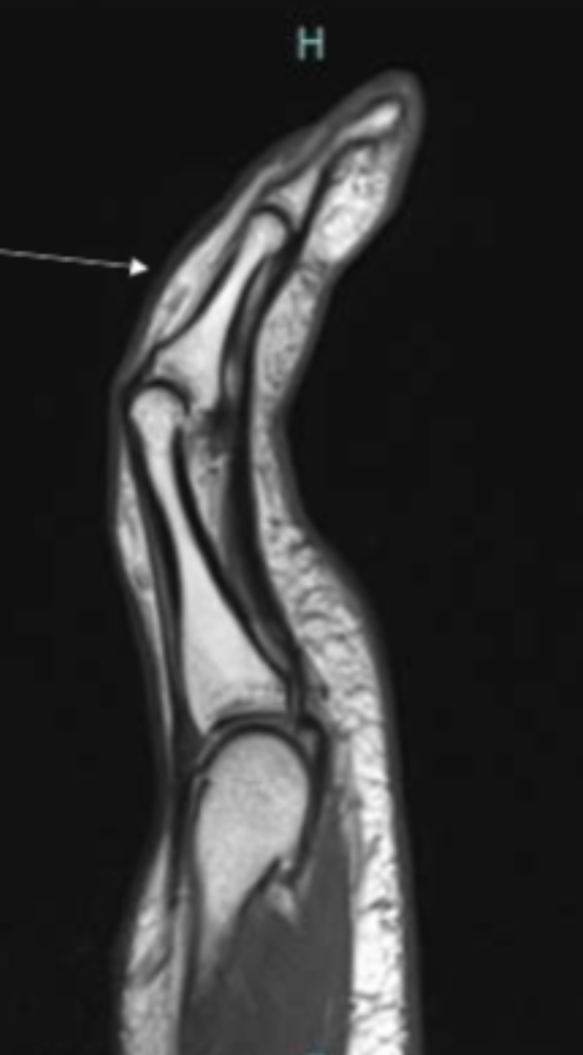
Describe the pathology indicated by the arrow (Sag PD).
Flexor tendon tear
Describe the pathology indicated by the arrow (Sag PD).
Extensor tendon tear
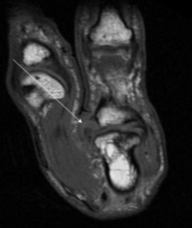
Describe the pathology indicated by the arrow (Sag PD).
UCL tear (Skier’s thumb)
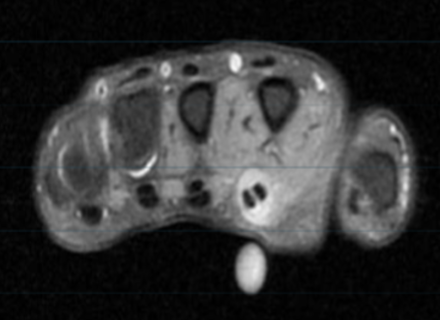
Describe the pathology (Ax T2 FS).
Tenosynovitis
In an axial oblique hip, what is the scan range?
Include entire acetabulum, femoral head and trochanters
In an axial oblique hip, how should the slices be angled?
Parallel to NOF
In a coronal hip, what is the scan range?
ASIS down to below lesser trochanter
In a coronal hip, how are the slices angled?
Parallel to NOF
In a sagittal hip, what is the scan range?
ASIS down to below lesser trochanter
In a sagittal hip, how are the slices angled?
Perpendicular to NOF
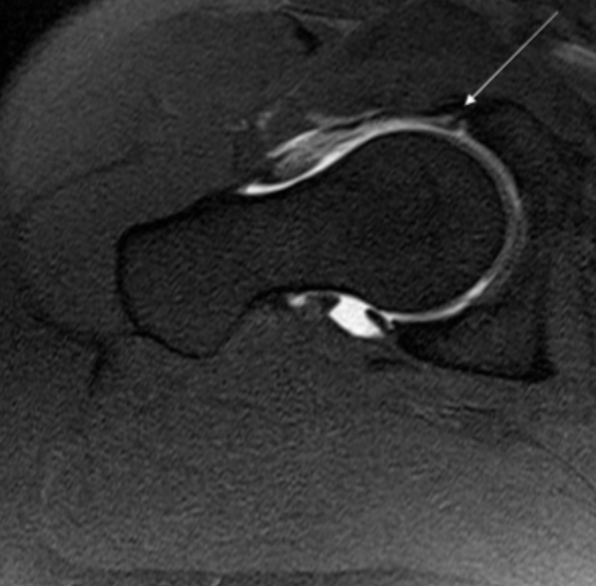
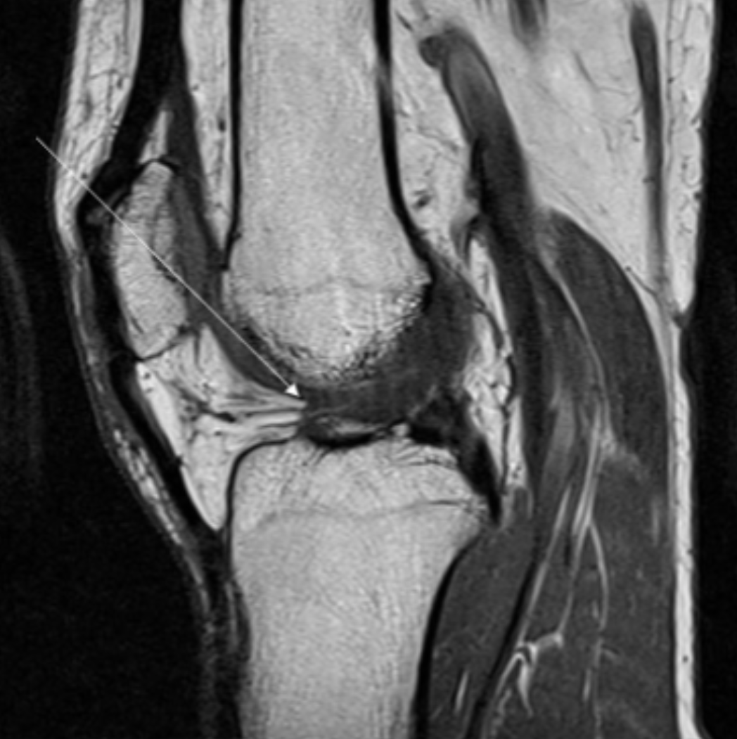
Describe the pathology indicated by the arrow (Ax T1 FS).
Labral tear
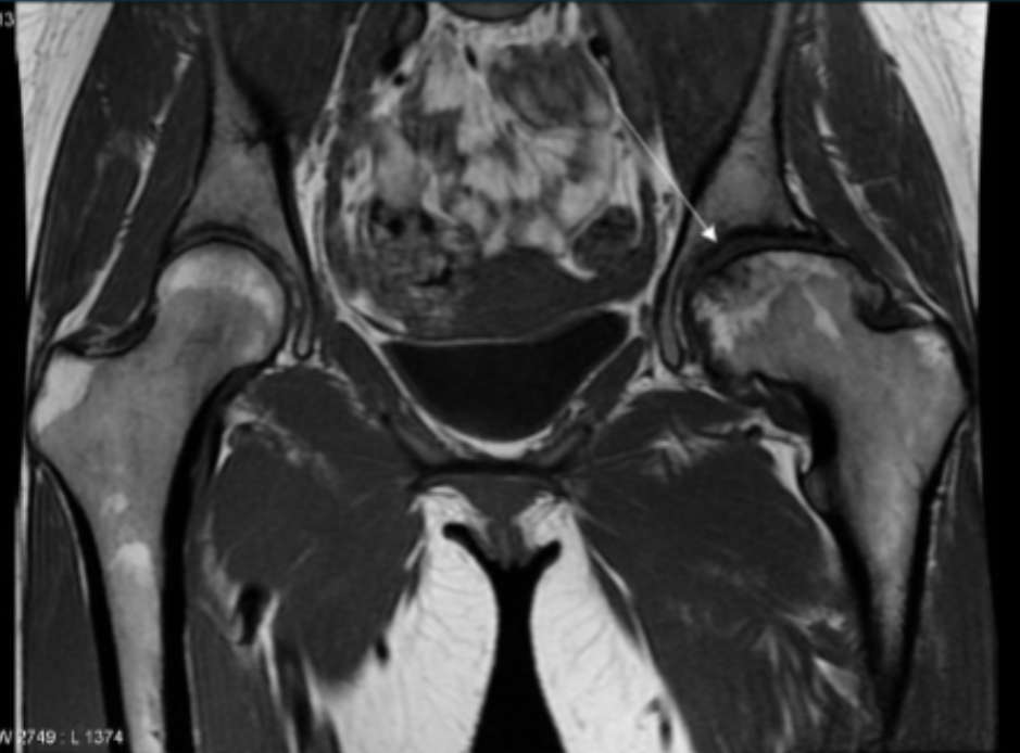
Describe the pathology indicated by the arrow (Cor T1).
AVN
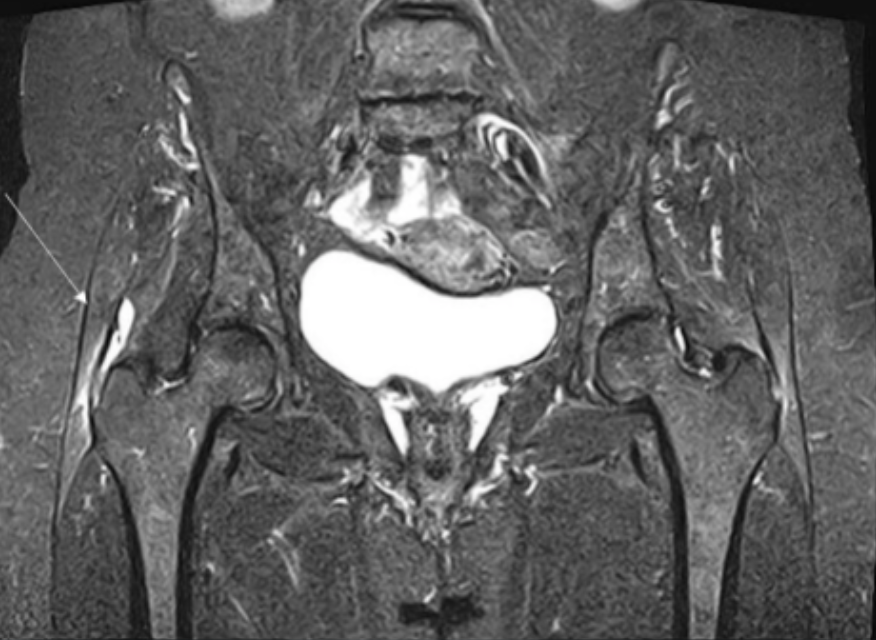
Describe the pathology (hint: it’s bone) (Ax T1 FS +C).
Osteochondroma
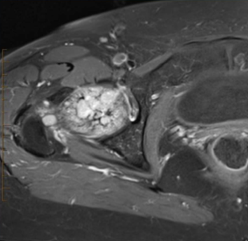
Describe the pathology indicated by the arrow (Cor T2 STIR pelvis).
Trochanteric bursitis
In an axial knee, what is the scan range?
Suprapatellar fat pad to tibial tuberosity
In an axial knee, how should the slices be angled?
Parallel to tibial plateau
In a coronal knee, what is the scan range?
Suprapatellar fat pad to tibial tuberosity
In a coronal knee, how should the slices be angled?
Perpendicular to the tibial plateau and parallel to the femoral condyles I
In a sagittal knee, what is the scan range?
Suprapatellar fat pad to tibial tuberosity
In a sagittal knee, how should the slices be angled?
Perpendicular to tibial plateau and parallel to ACL
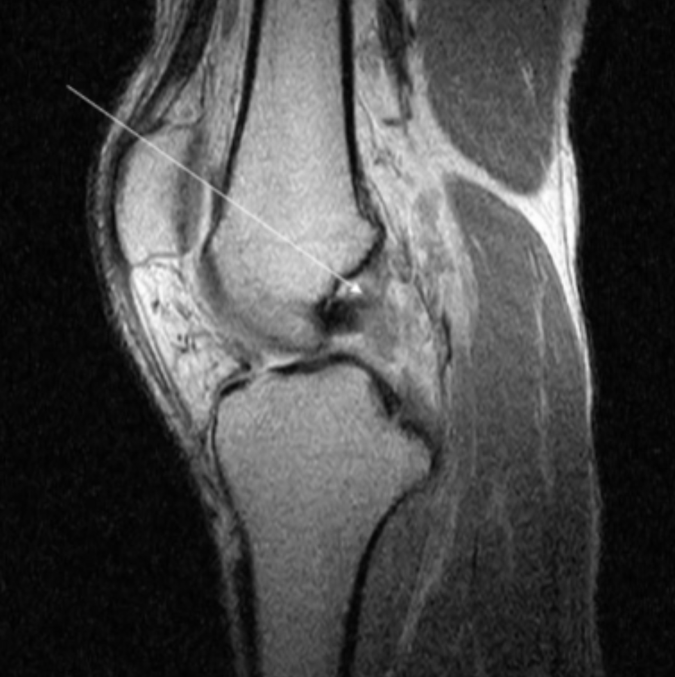
Describe the pathology indicated by the arrow (Sag PD).
ACL tear
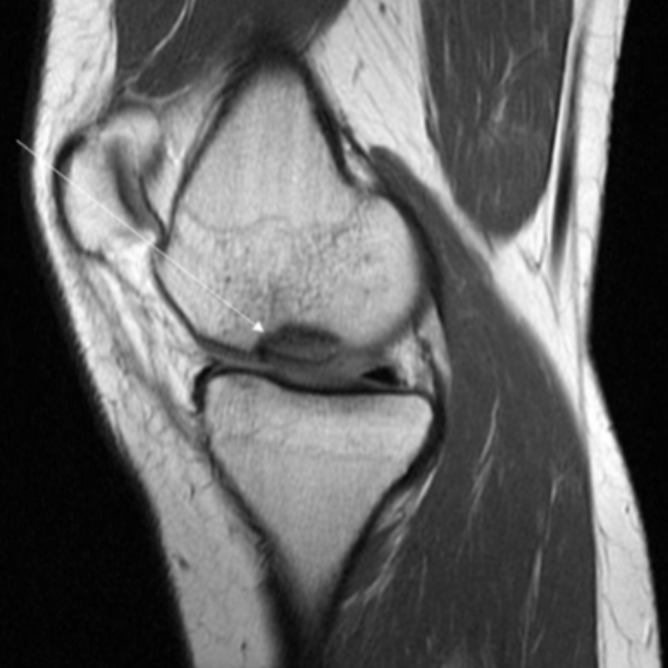
Describe the pathology indicated by the arrow (Sag PD).
PCL rupture
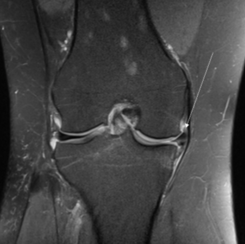
Describe the pathology indicated by the arrow (Cor PD FS).
MCL tear
Describe the pathology indicated by the arrow (Cor PD FS).
Medial meniscal tear
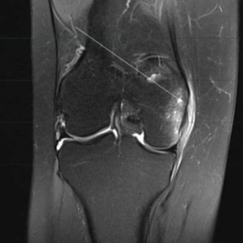
Describe the pathology indicated by the arrow (Sag PD).
Osteochondritis dissecans
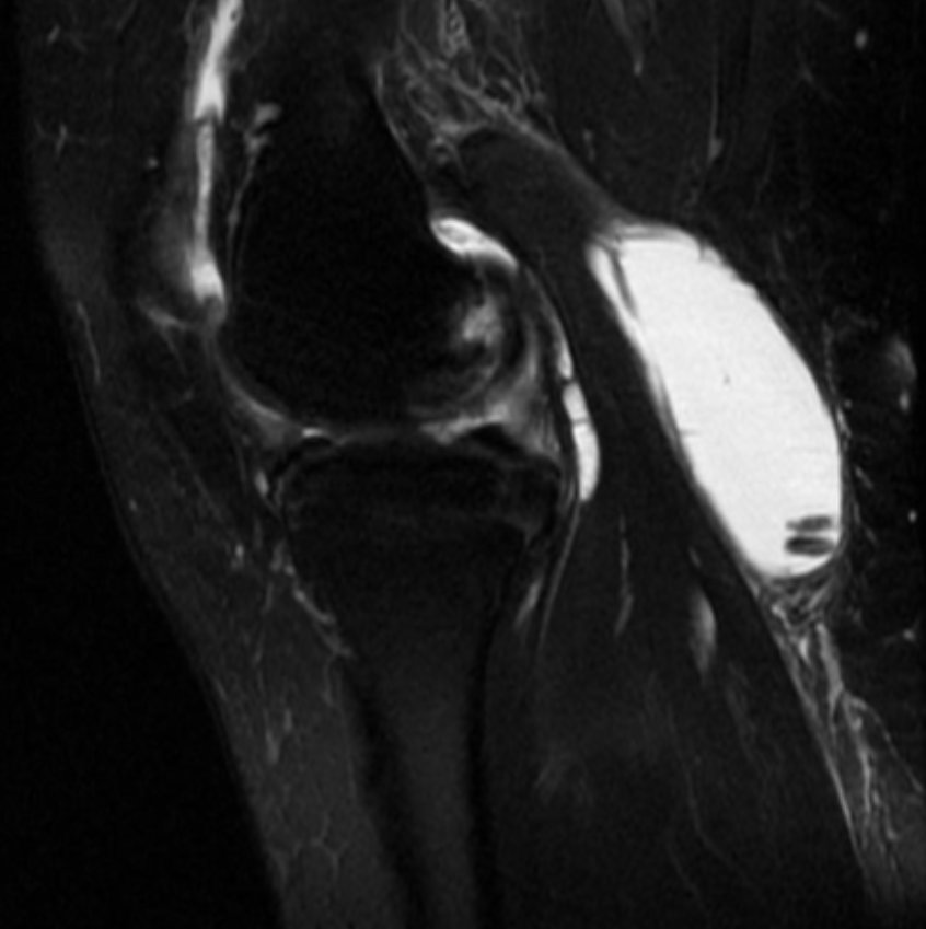
Describe the pathology (Sag T2 FS).
Baker’s cyst
In an axial ankle, what is the scan range?
Distal tib/fib syndesmosis to below ankle soft tissue
In an axial ankle, how should the slices be angled?
Parallel to mortise joint
In a coronal ankle, what is the scan range?
Distal tib/fib syndesmosis to below ankle soft tissue
In a coronal ankle, how should the slices be angled?
Perpendicular to medial and lateral malleoli
In a sagittal ankle, what is the scan range?
Distal tib/fib syndesmosis to below ankle soft tissue
In a sagittal ankle, how should the slices be angled?
Parallel to medial and lateral malleoli
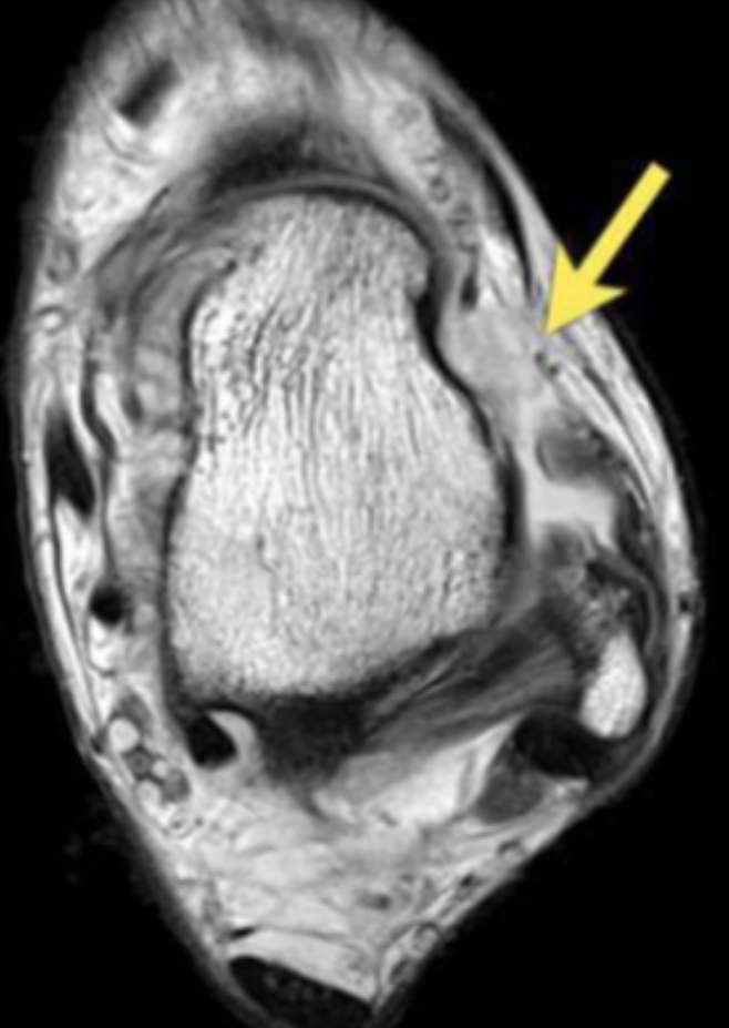
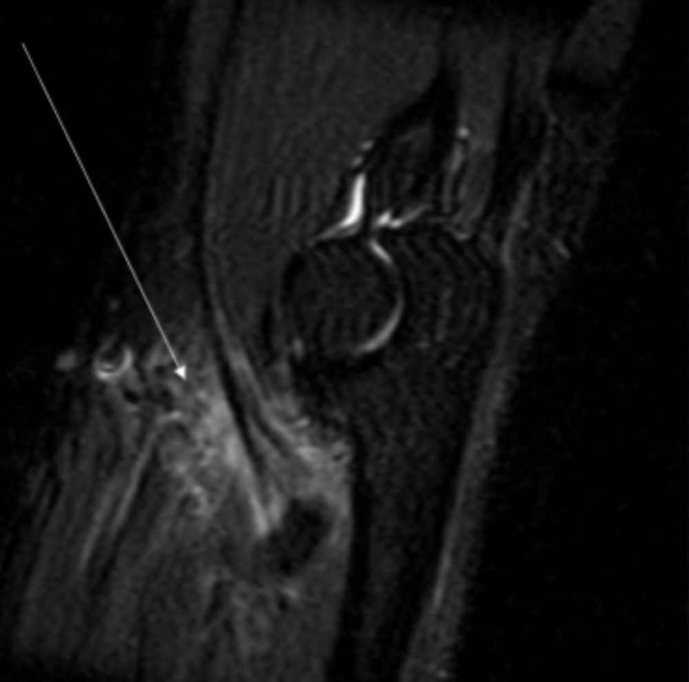
Describe the pathology indicated by the arrow (Ax PD).
Anterior talofibular ligament (ATFL) tear
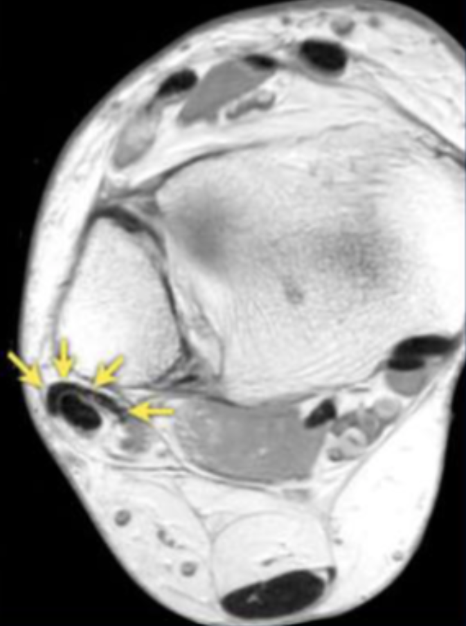
Describe the pathology indicated by the arrow (Ax PD).
Peroneal brevis tendon tear
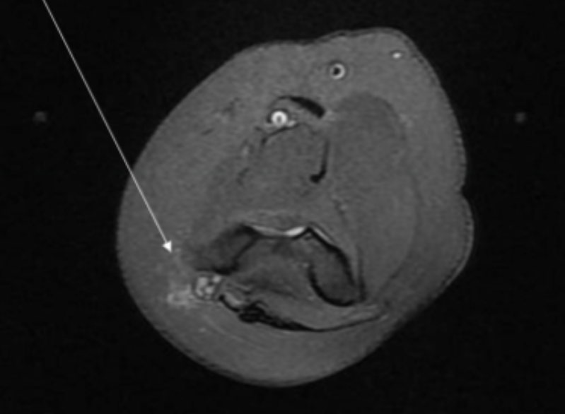
Describe the pathology indicated by the arrow (Sag PD).
Achilles rupture
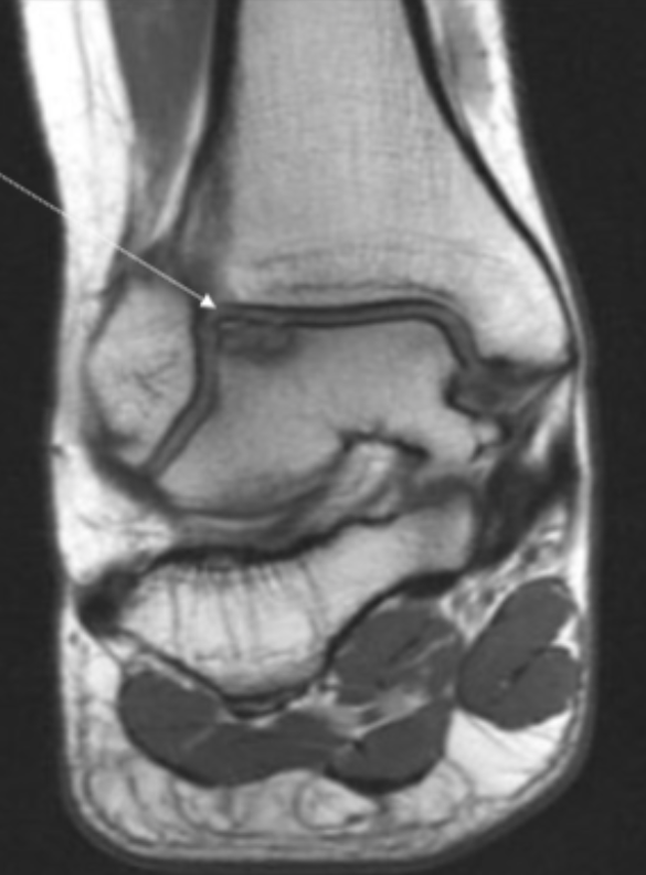
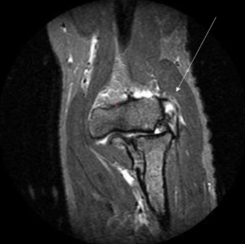
Describe the pathology indicated by the arrow (Cor PD).
Osteochondral defect
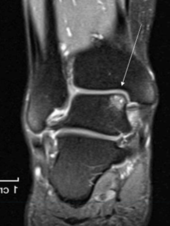
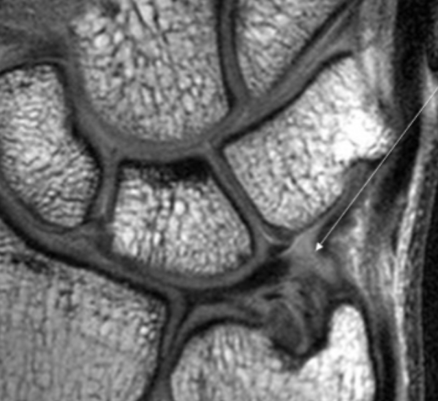
Describe the pathology indicated by the arrow (Cor T2 FS).
Osteochondral defect
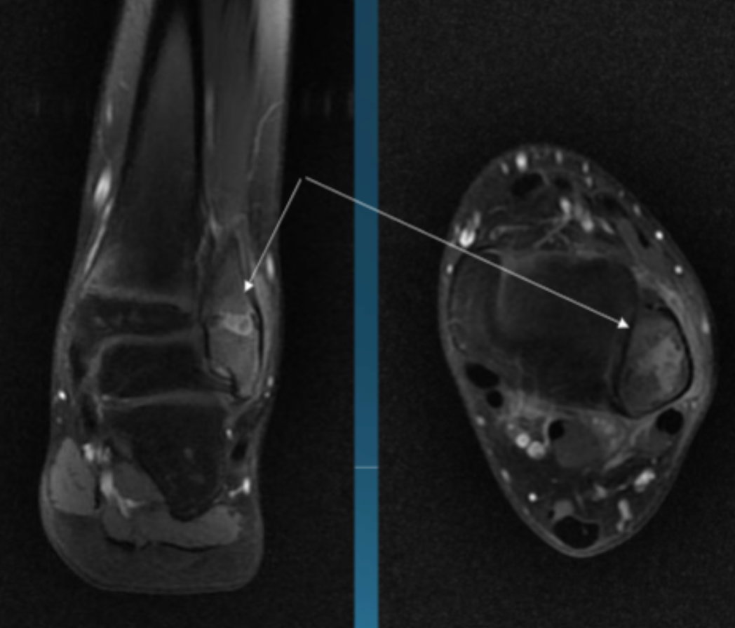
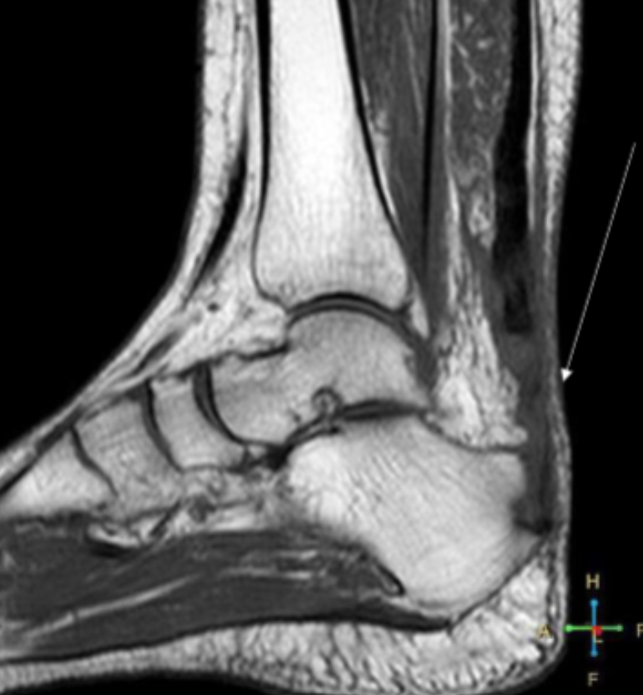
Describe the pathology indicated by the arrow (Cor T1 FS +C).
Osteomyelitis (focal)