OSU Anatomy - Unit 2
1/338
Earn XP
Description and Tags
Bones, muscles, veins, arteries, and nerves of the lower limbs
Name | Mastery | Learn | Test | Matching | Spaced |
|---|
No study sessions yet.
339 Terms
The most forward pieces of the pelvic girdle are the ______ _______ _______ _______ and the _______ _______
anterior superior iliac spine; pubic tubercles
The false pelvis runs between the _______ _______ and the _______ _______
pubic symphysis; sacral promontory
The true pelvis sits _______ the false pelvis
below
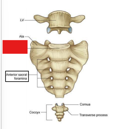
fill in the red box:
Promontory
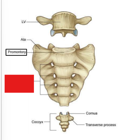
fill in the red box:
Anterior sacral foramina
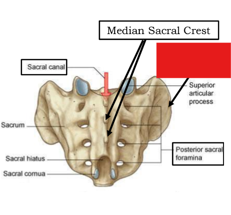
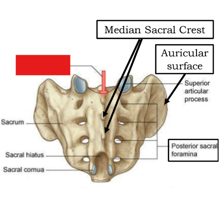
fill in the red box:
Median sacral crest
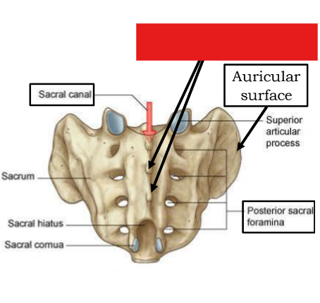
fill in the red box:
Auricular surface
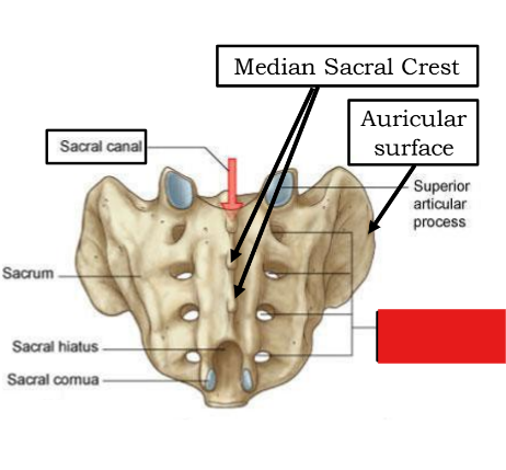
fill in the red box:
Sacral canal
fill in the red box:
Posterior sacral foramina
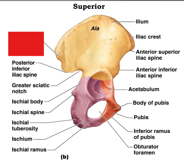
fill in the red box:
Posterior superior iliac spine
fill in the red box:
Posterior inferior iliac spine
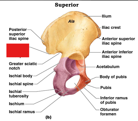
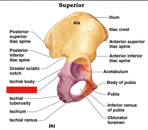
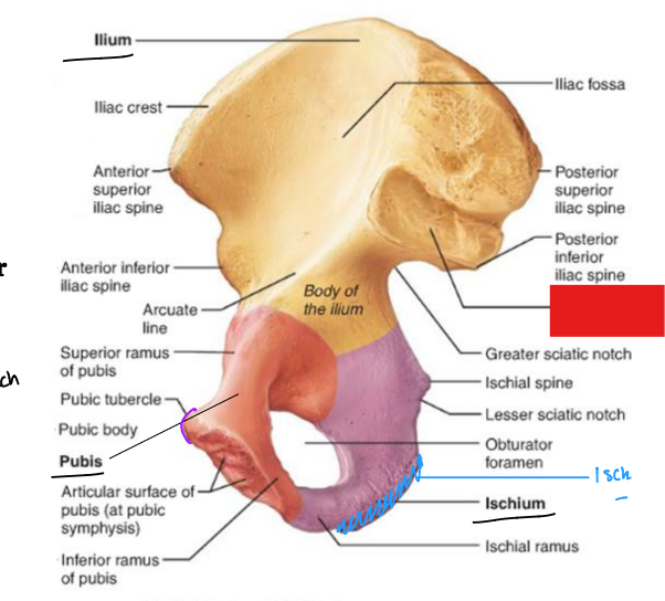
fill in the red box:
Greater sciatic notch
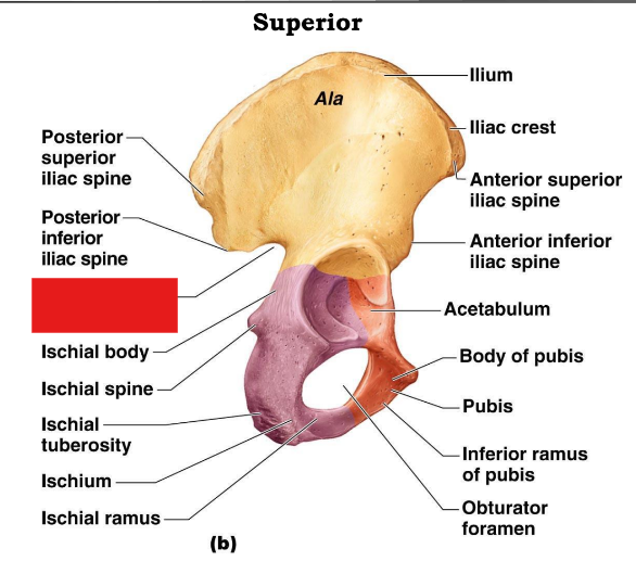
fill in the red box:
Ischial body
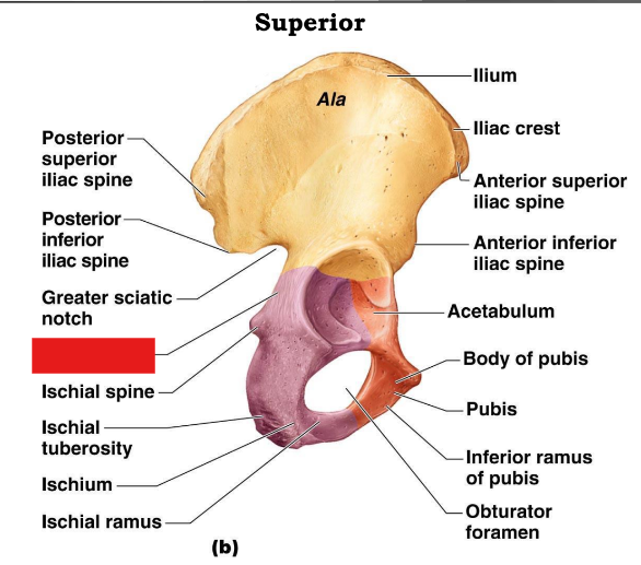
fill in the red box:
Ischial spine
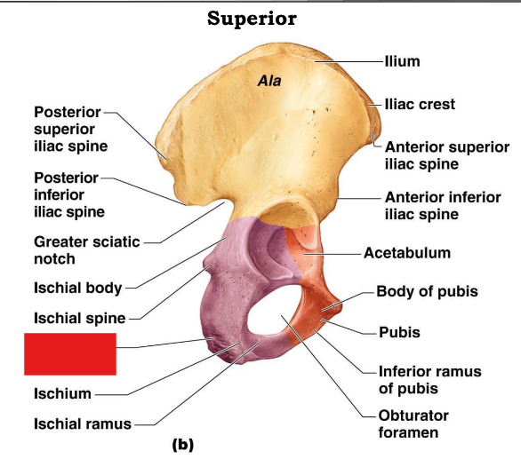
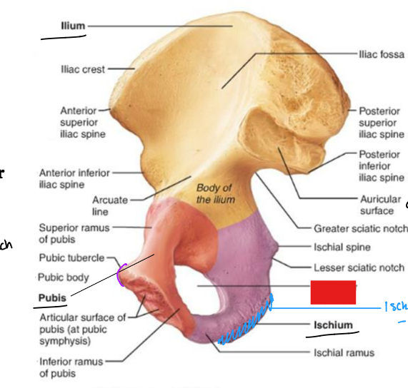
fill in the red box:
ischial tuberosity
fill in the red box:
Ischial ramus
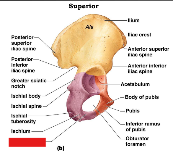
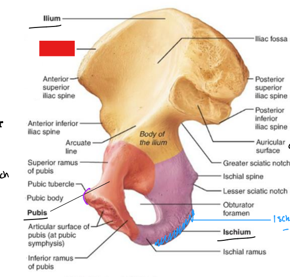
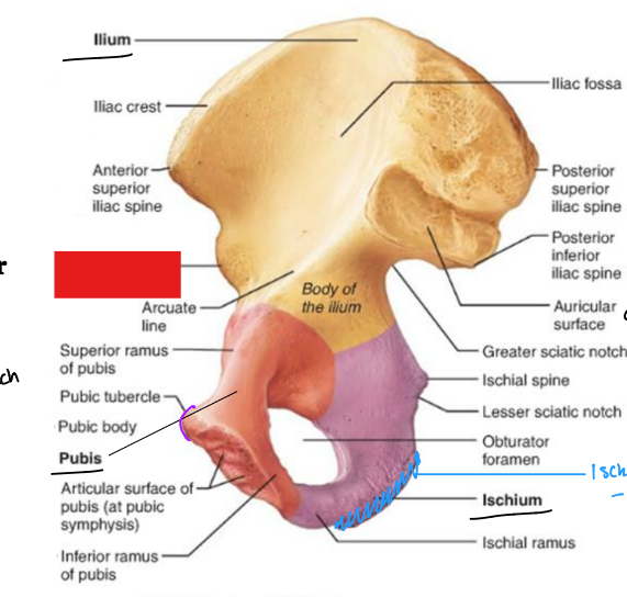
fill in the red box:
Iliac crest
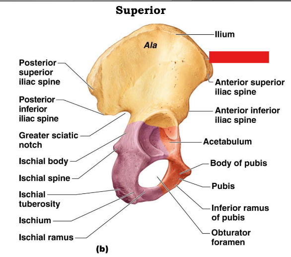
fill in the red box:
Anterior superior iliac spine
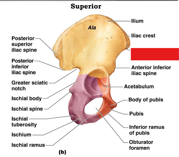
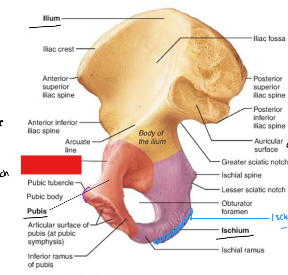
fill in the red box:
Anterior inferior iliac spine

fill in the red box:
Acetabulum
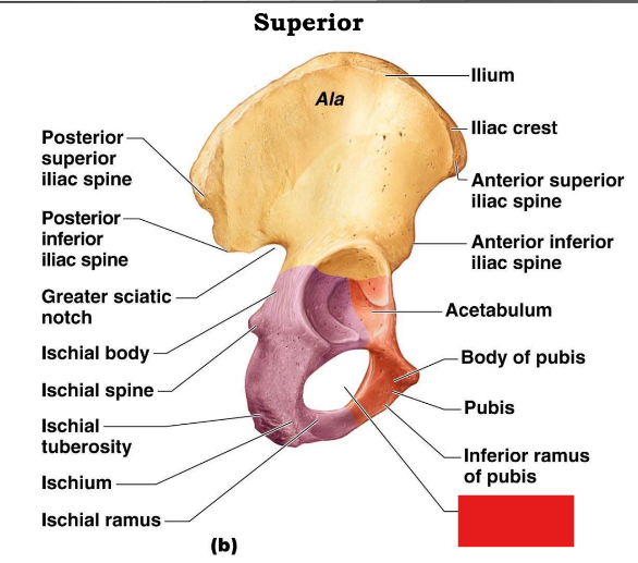
fill in the red box:
Inferior ramus of pubis
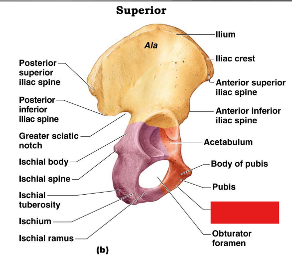
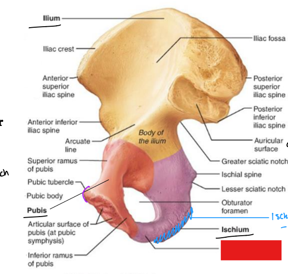
fill in the red box:
Obturator foramen
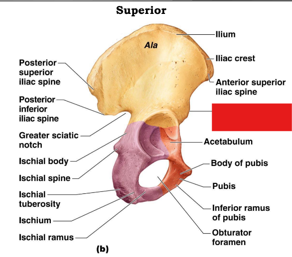
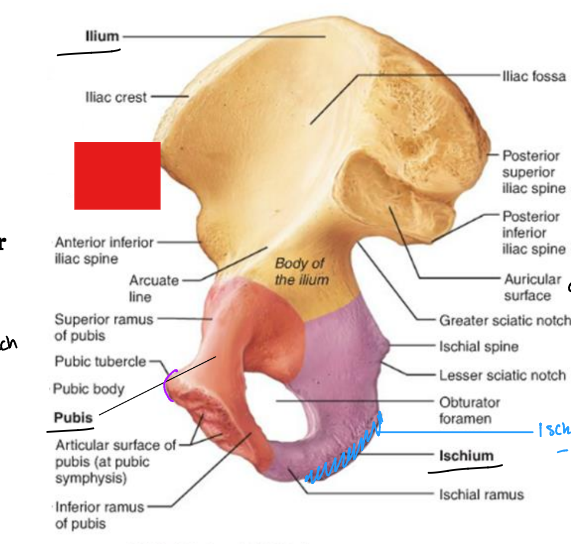
fill in the red box:
Iliac crest
fill in the red box:
Anterior superior iliac spine
fill in the red box:
Anterior inferior iliac spine
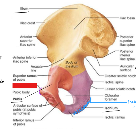
fill in the red box:
superior ramus of pubis
fill in the red box:
Pubic tubercle
fill in the red box:
Articular surface of pubis
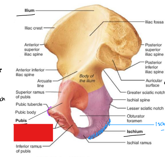
fill in the red box:
Inferior ramus of pubis
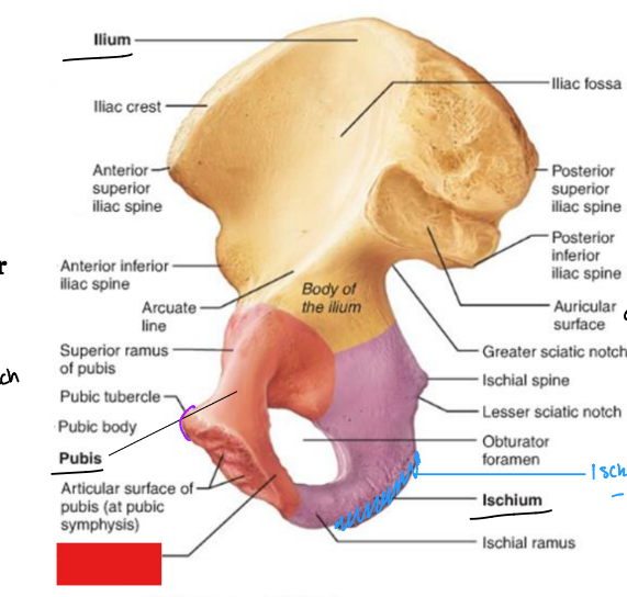
fill in the red box:
Iliac fossa
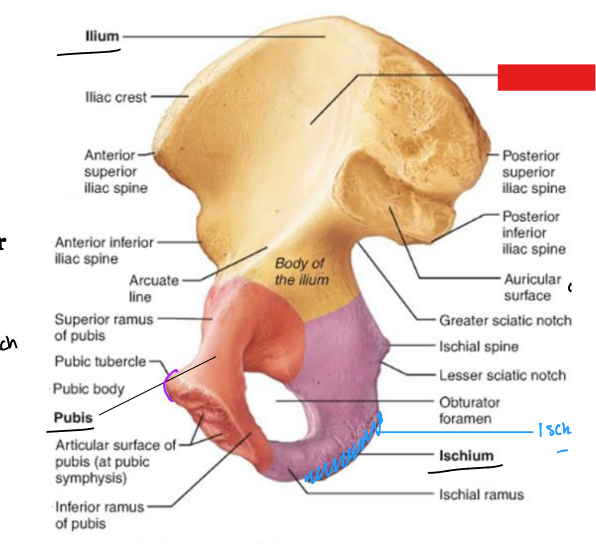
fill in the red box:
Posterior superior iliac spine
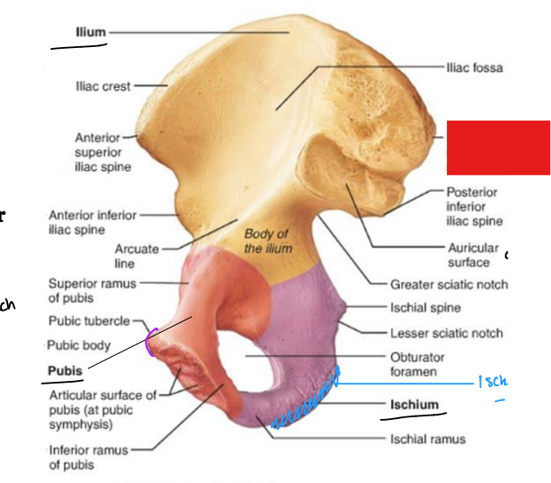
fill in the red box:
Posterior inferior iliac spine
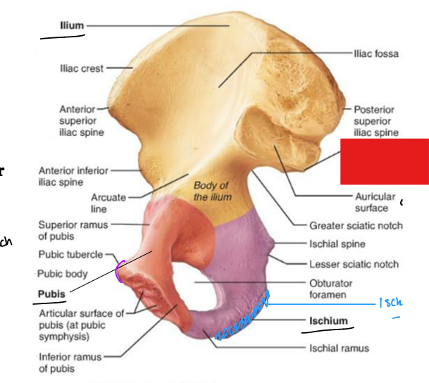
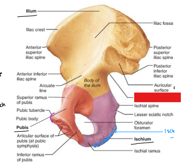
fill in the red box:
Auricular surface
fill in the red box:
Greater sciatic notch
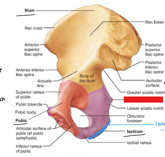
fill in the red box:
Ischial spine
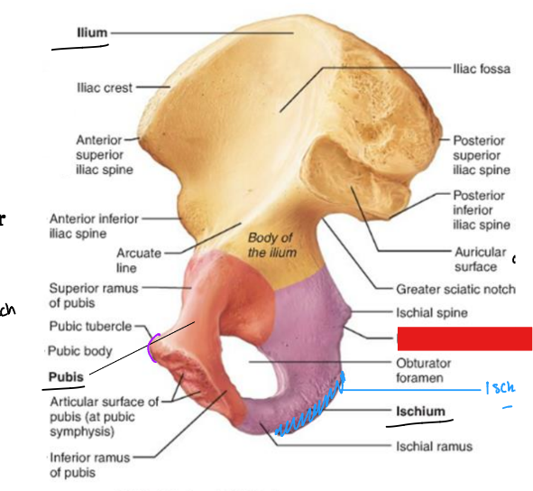
fill in the red box:
Lesser sciatic notch
fill in the red box:
Obturator foramen
fill in the red box:
Ischial ramus
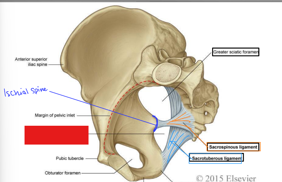
fill in the red box:
Lesser sciatic foramen
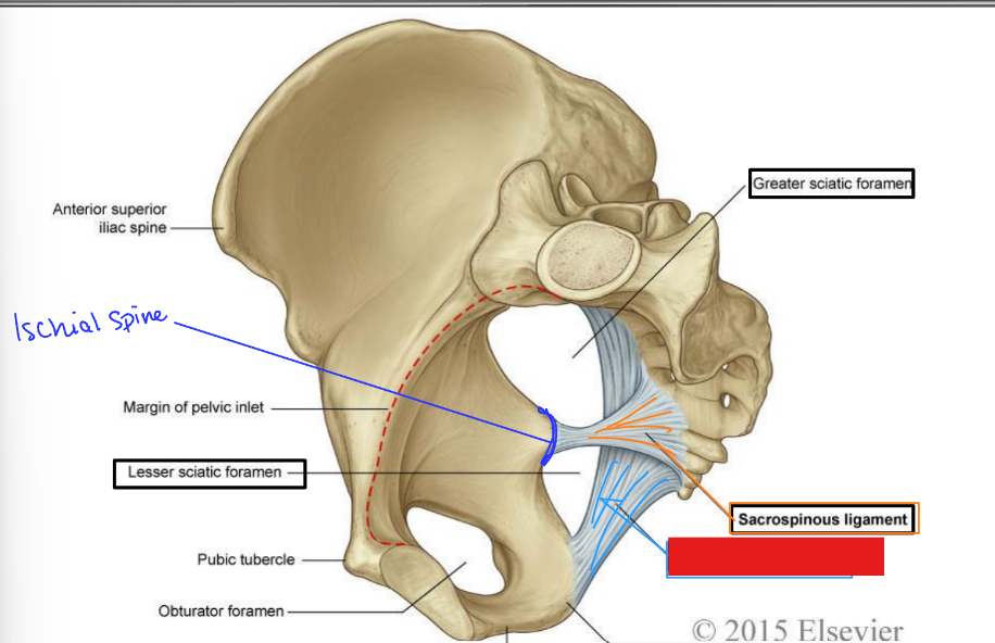
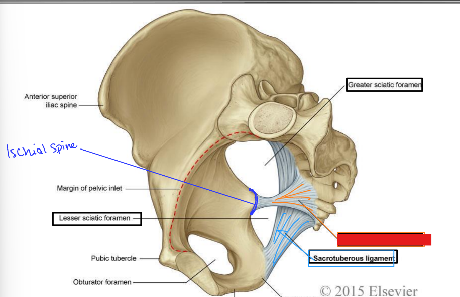
fill in the red box:
Sacrotuberous ligament
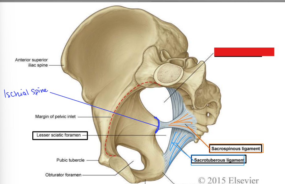
fill in the red box:
Sacrospinous ligament
fill in the red box:
Greater sciatic foramen
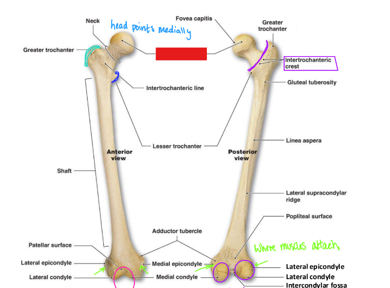
fill in the red box:
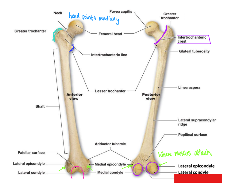
Femoral head
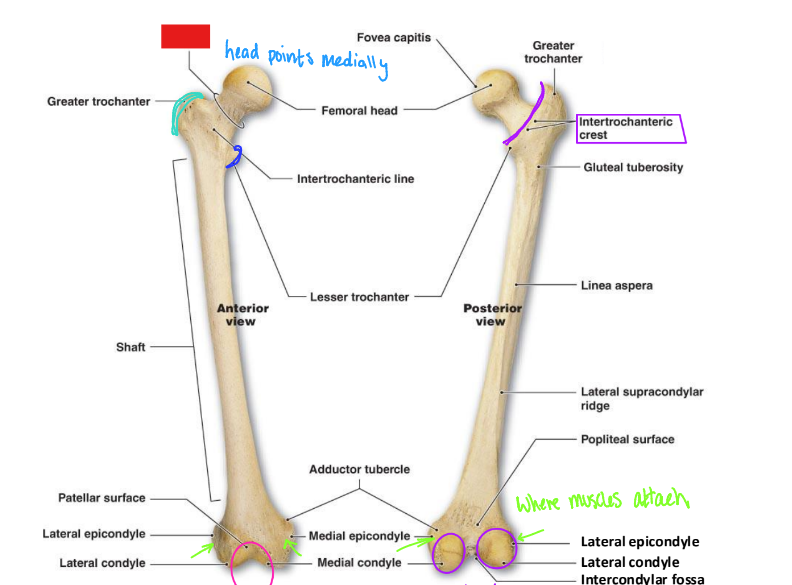
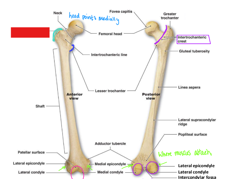
fill in the red box:
Neck
fill in the red box:
Greater trochanter
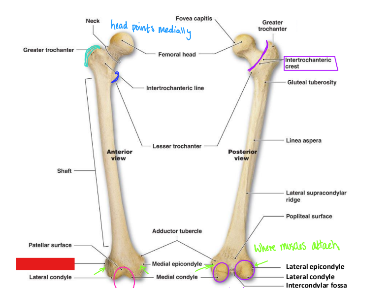
fill in the red box:
Patellar surface
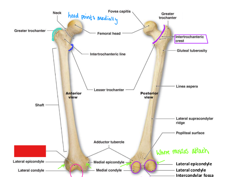
fill in the red box:
Lateral epicondyle
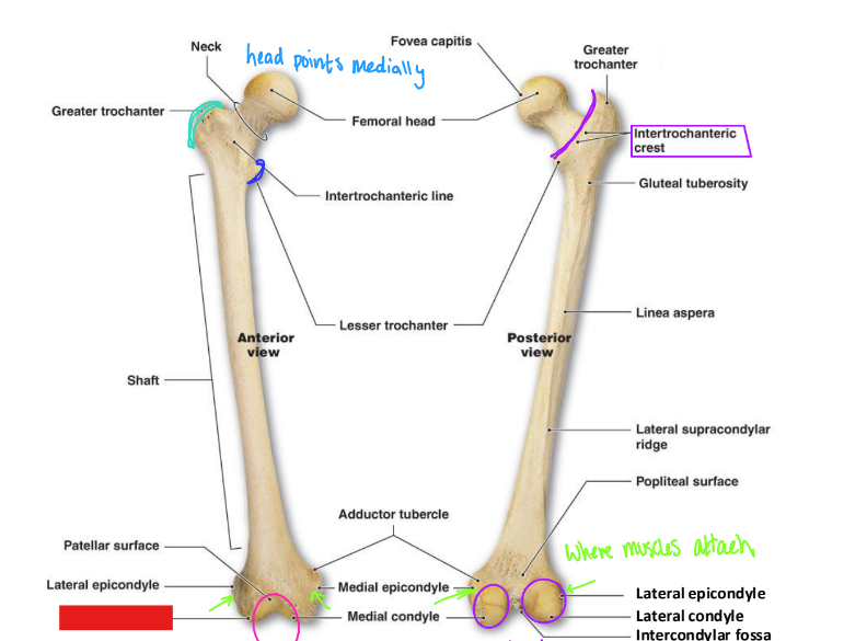
fill in the red box:
lateral condyle
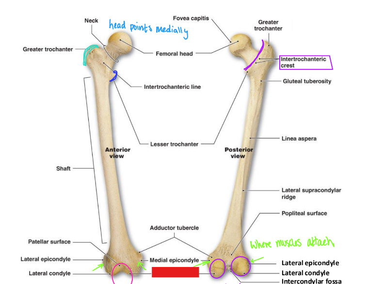
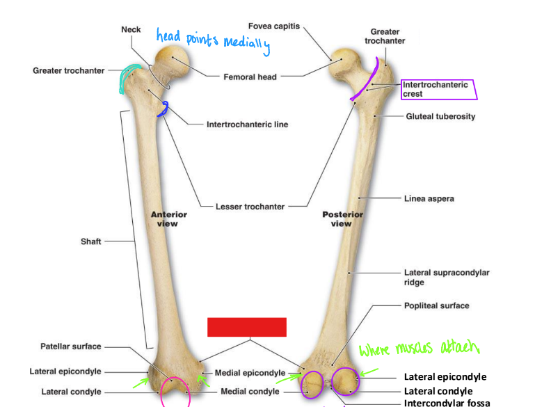
fill in the red box:
Medial condyle
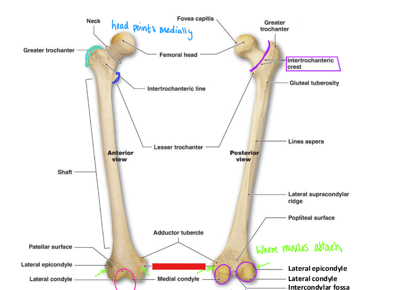
fill in the red box:
Medial epicondyle
fill in the red box:
Adductor tubercle
fill in the red box:
Lesser trochanter
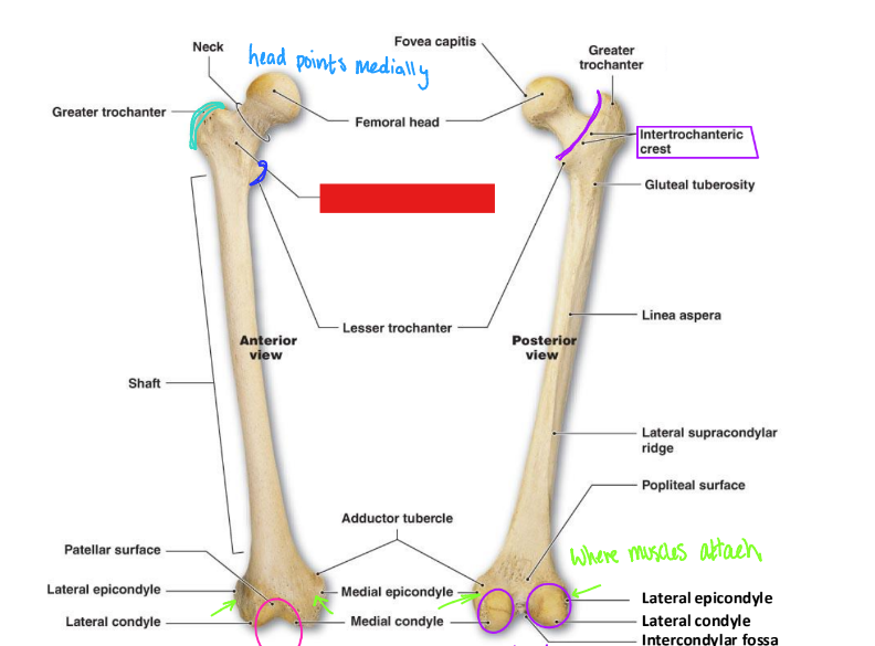
fill in the red box:
Intertrochanteric line
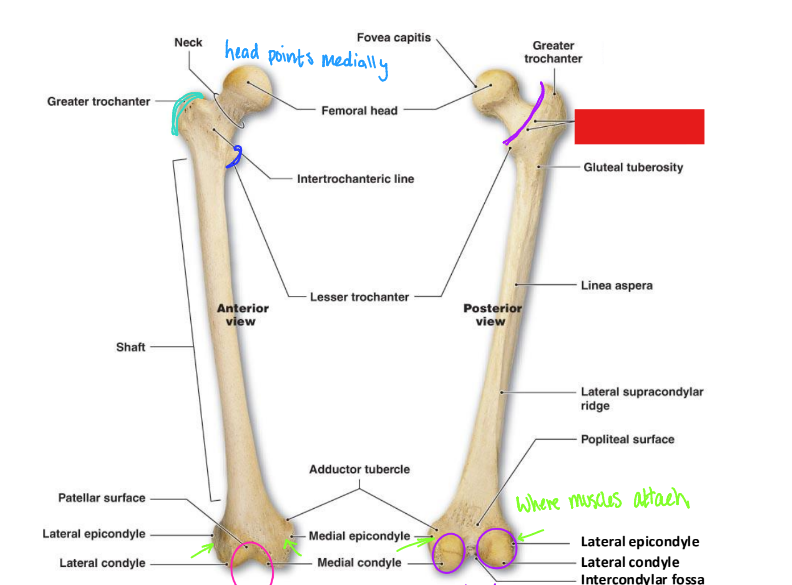
fill in the red box:
Intertrochanteric crest
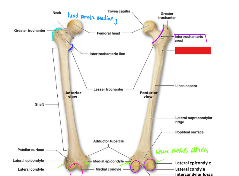
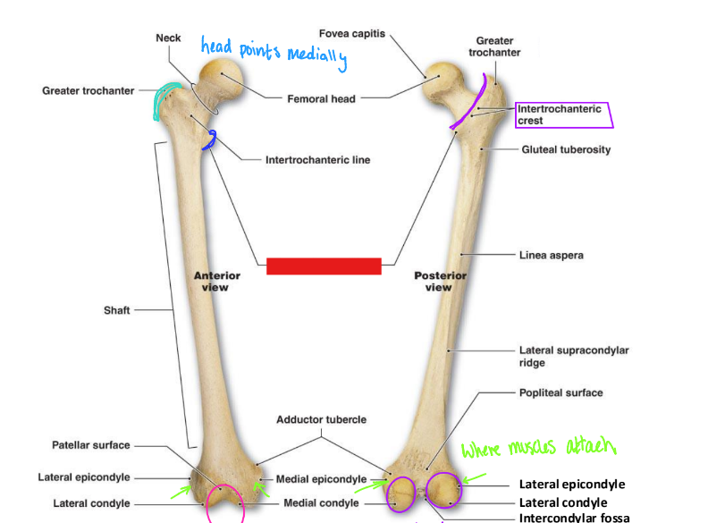
fill in the red box:
Gluteal tuberosity
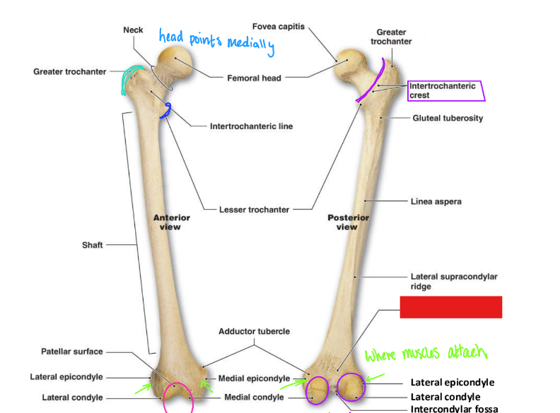
fill in the red box:
Popliteal surface
fill in the red box:
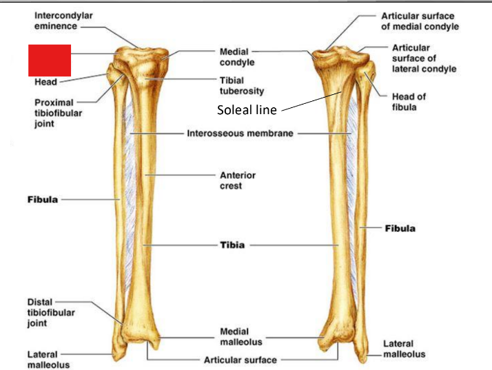
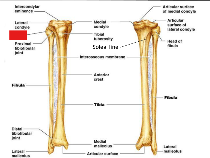
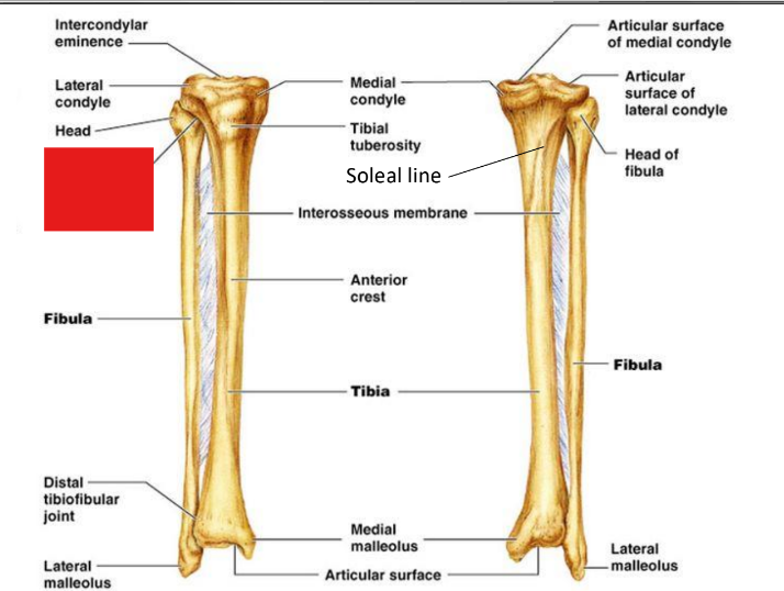
intercondylar fossa
fill in the red box:
Lateral condyle
fill in the red box:
head of fibula
fill in the red box:
Proximal tibiofibular joint
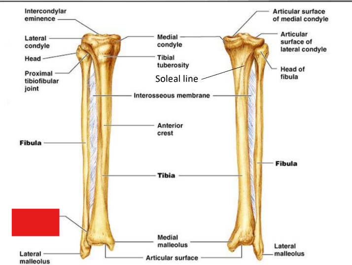
fill in the red box:
Lateral malleolus
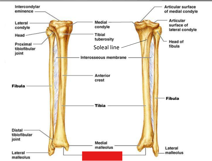
fill in the red box:
Distal tibiofibular joint
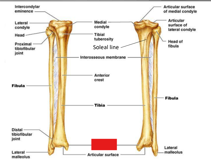
fill in the red box:
Articular surface
fill in the red box:
Medial malleolus
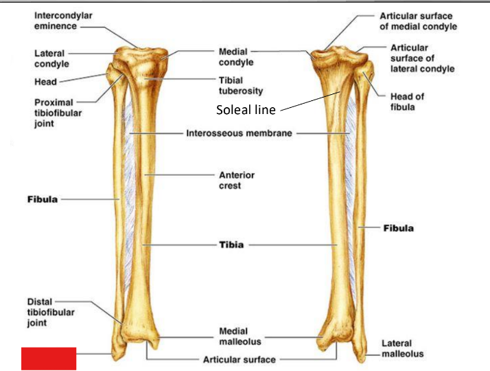
fill in the red box:
Anterior crest
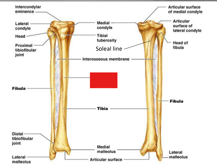
fill in the red box:
Soleal line
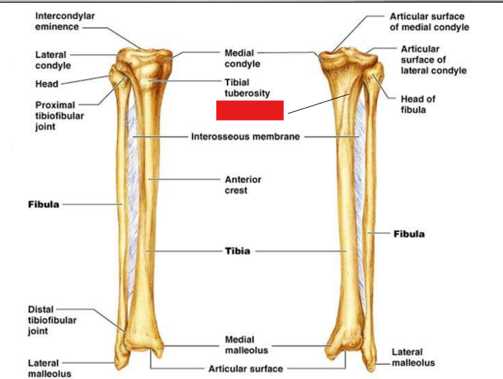
fill in the red box:
Tibial tuberosity
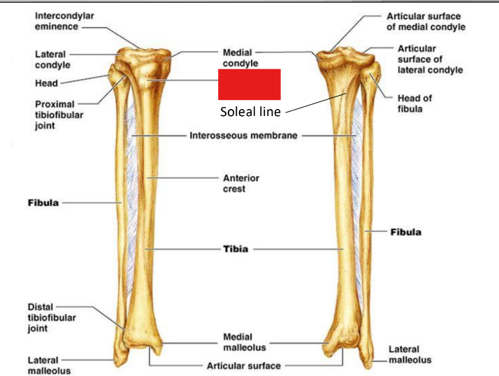
fill in the red box:
Medial condyle
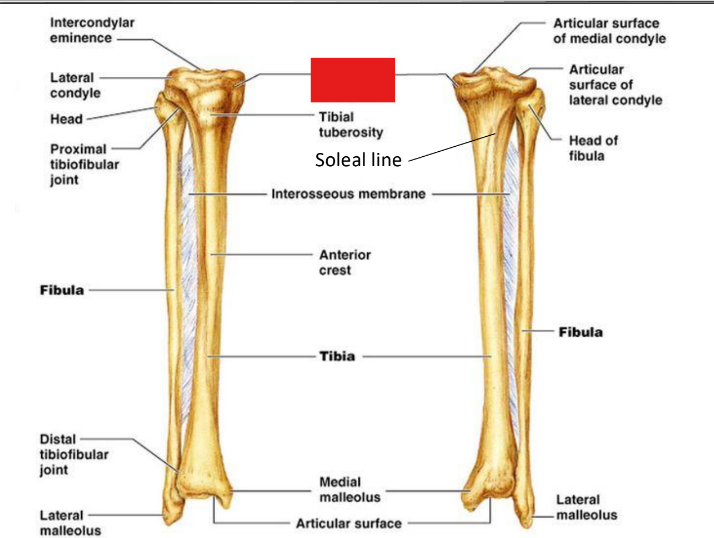
fill in the red box:
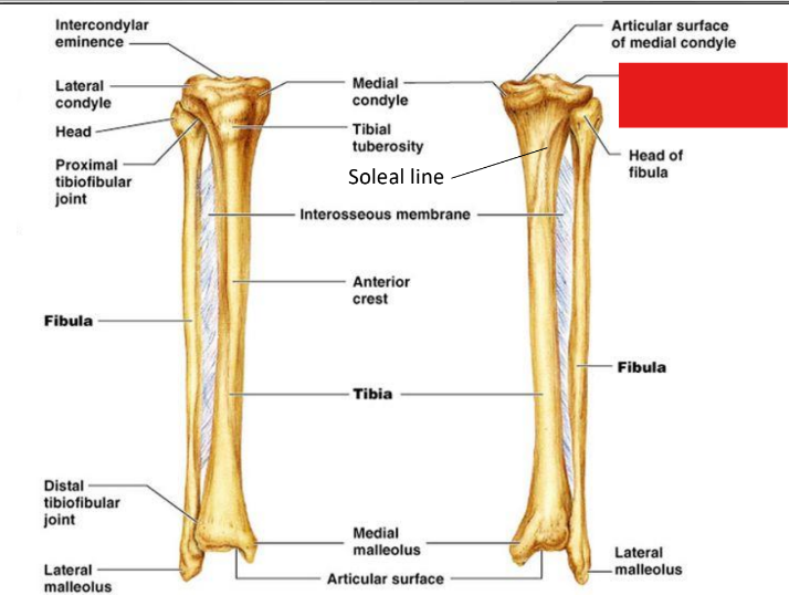
Articular surface of medial condyle
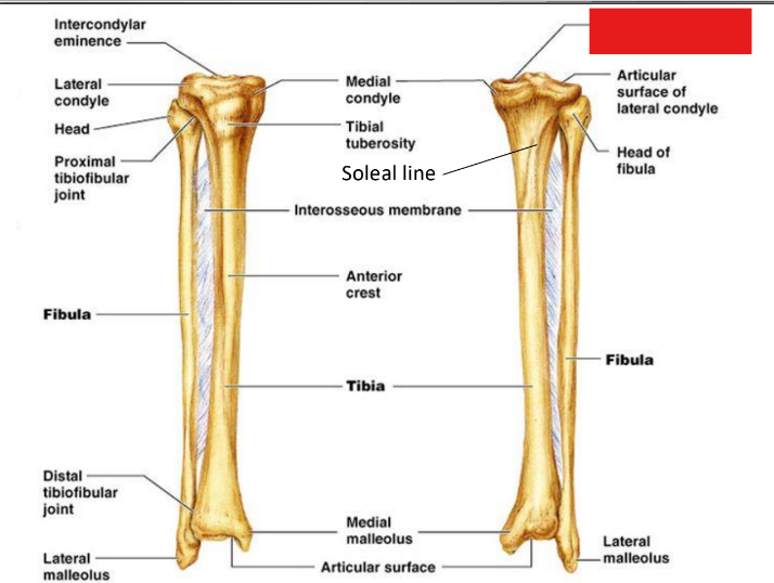
fill in the red box:
Articular surface of lateral condyle
fill in the red box:
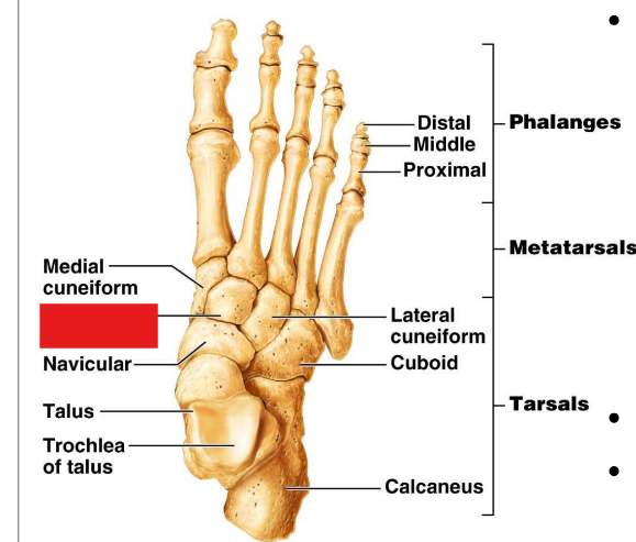
Medial cuneiform
fill in the red box:
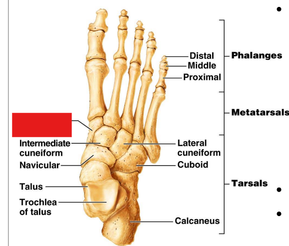
Intermediate cuneiform
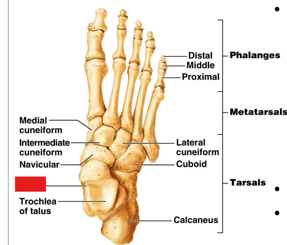
fill in the red box:
Navicular
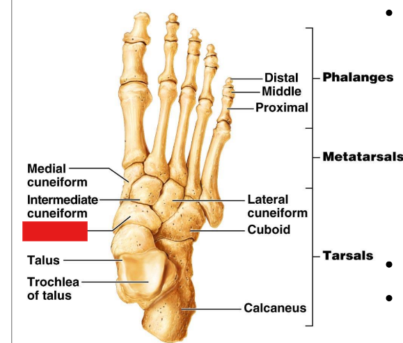
fill in the red box:
Talus
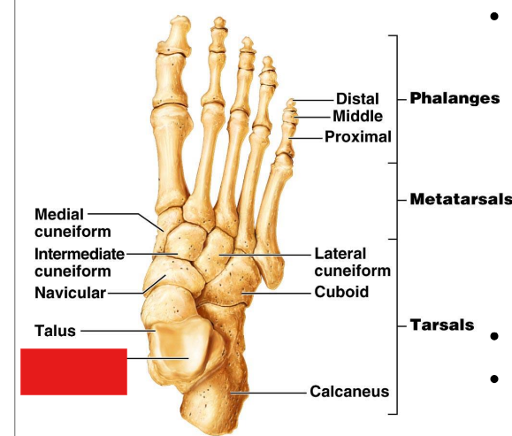
fill in the red box:
Trochlea of talus
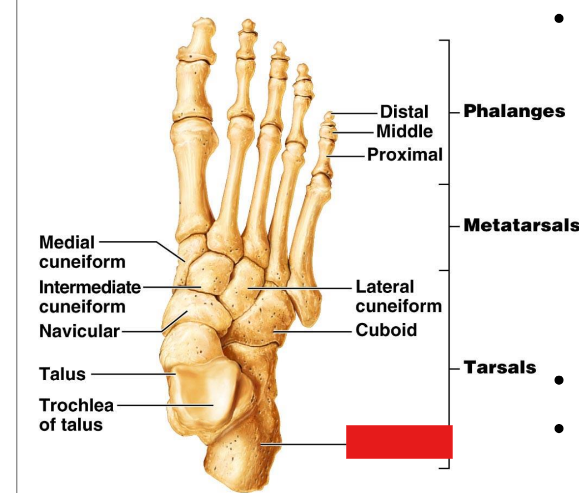
fill in the red box:
Calcaneus
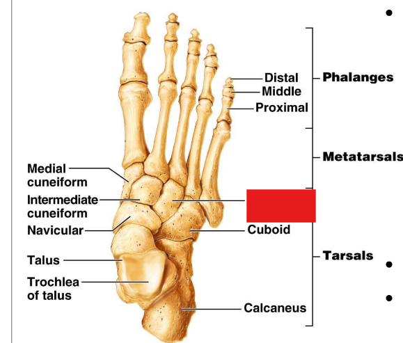
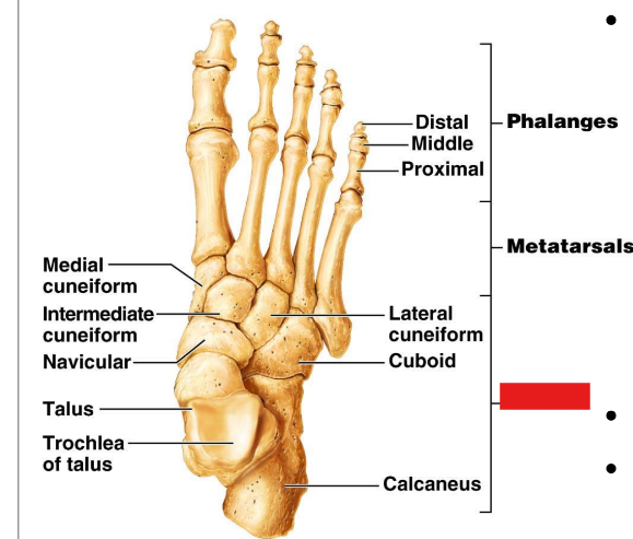
fill in the red box:
Cuboid
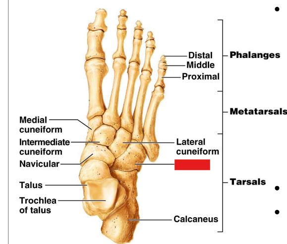
fill in the red box:
Lateral cuneiform
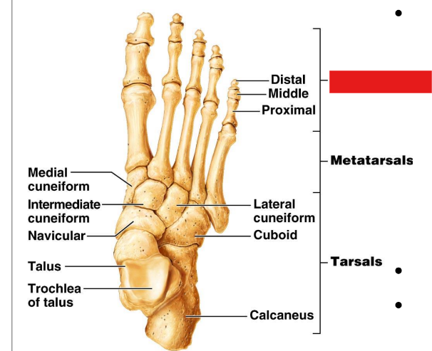
fill in the red box:
Phalanges
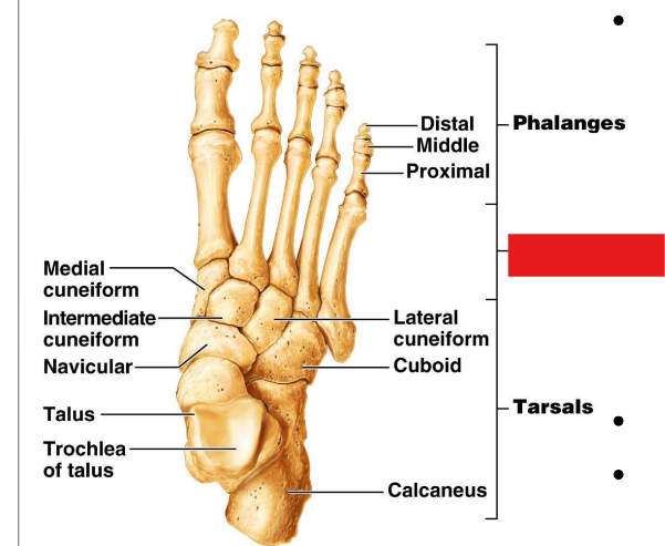
fill in the red box:
Metatarsals
fill in the red box:
Tarsals
The hip or coxal joint articulates the ________ ______ at the ______
hip bone; femur
The hip or coxal joint is a ______ ______ and ______ joint
synovial; ball; socket
The hip joint allows for ______ movement including …
diarthrotic; flexion, extension, abduction, adduction, rotation, circumduction of thigh
The knee joint articulates the ______ and ______
femur; tibia
The knee joint is a ______ ______ ______
synovial modified hinge
The knee joint is ______ allowing for …
diarthrotic; flexion and extension of the leg, some rotation
The knee cap joint articulates the ______ and ______
femur; patella
The knee cap joint is a ______ ______ joint
synovial plane
The knee cap joint is ______ allowing for … movement
diarthrotic; gliding of the patella
The ankle joint articulates the ______ and ______ with the ______
Tibia; fibula; talus
The ankle joint is a ______ ______ joint
synovial hinge
The ankle joint is ______ allowing … movement
diarthrotic; dorsiflexion and plantar flexion of foot
The toe joint articulates ______ ______
adjacent phalanges
The toe joints are ______ ______ joints
synovial hinge
The toe joints are ______ allowing for … movement
diarthrotic; flexion and extension of the toes
The patellar ligament connects the ______ to the ______ ______
patella; tibial tuberosity
The medial tibial collateral ligament connects the ______ ______ of the femur to the ______ ______ of the tibia
medial epicondyle; medial condyle
The lateral fibular collateral ligament connects the lateral epicondyle of the ______ to the head of the ______
femur; fibula
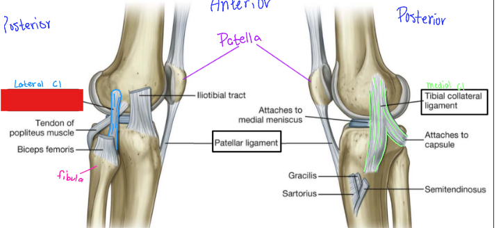
fill in the red box:
Fibular collateral ligament