BY 124L - Exam 3 Jars and Models
 Call Kai
 Learn
Learn Practice Test
 Spaced Repetition
 Match
Match Flashcards
 Knowt Play
1/117

There's no tags or description
Looks like no tags are added yet.
Name | Mastery | Learn | Test | Matching | Spaced | Call with Kai | Chat |
|---|
No analytics yet
Send a link to your students to track their progress
118 Terms
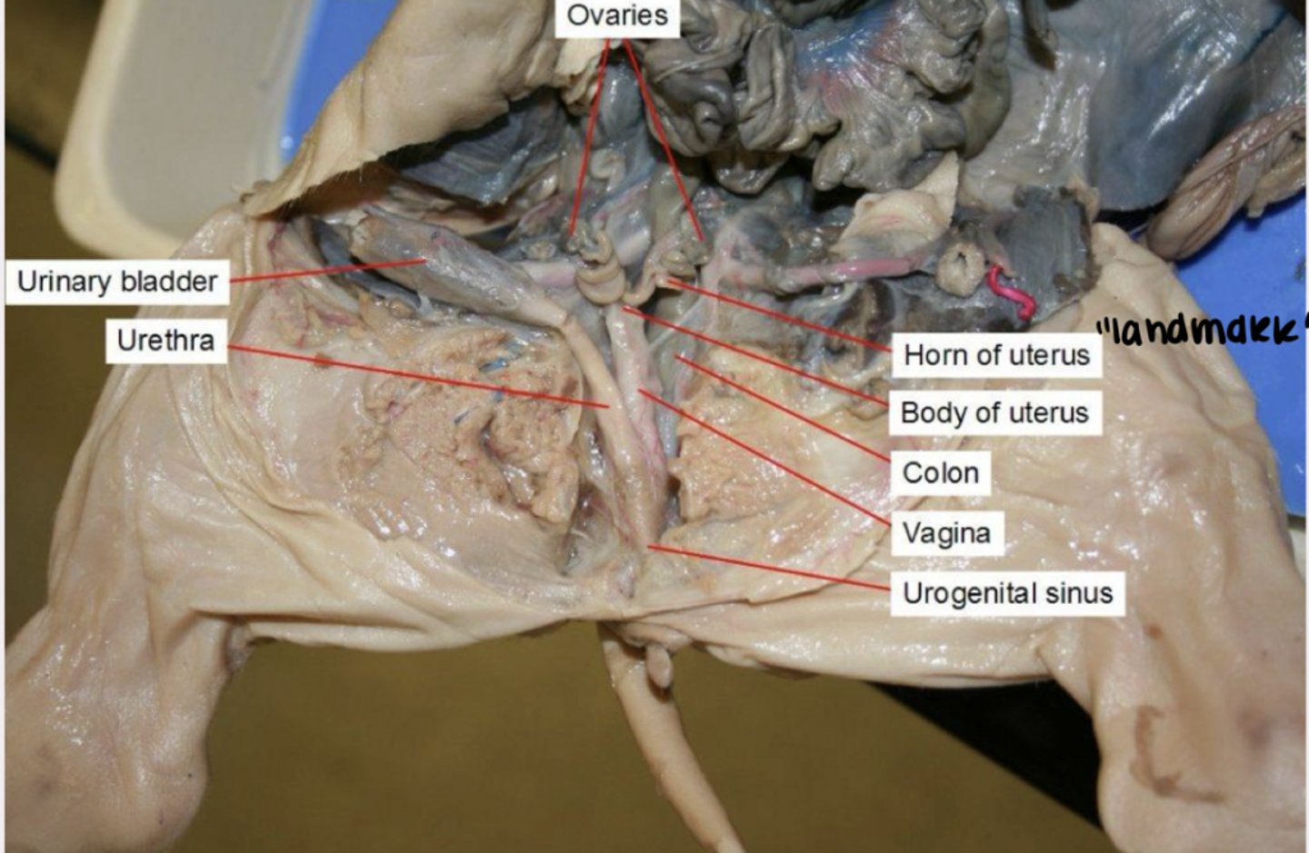
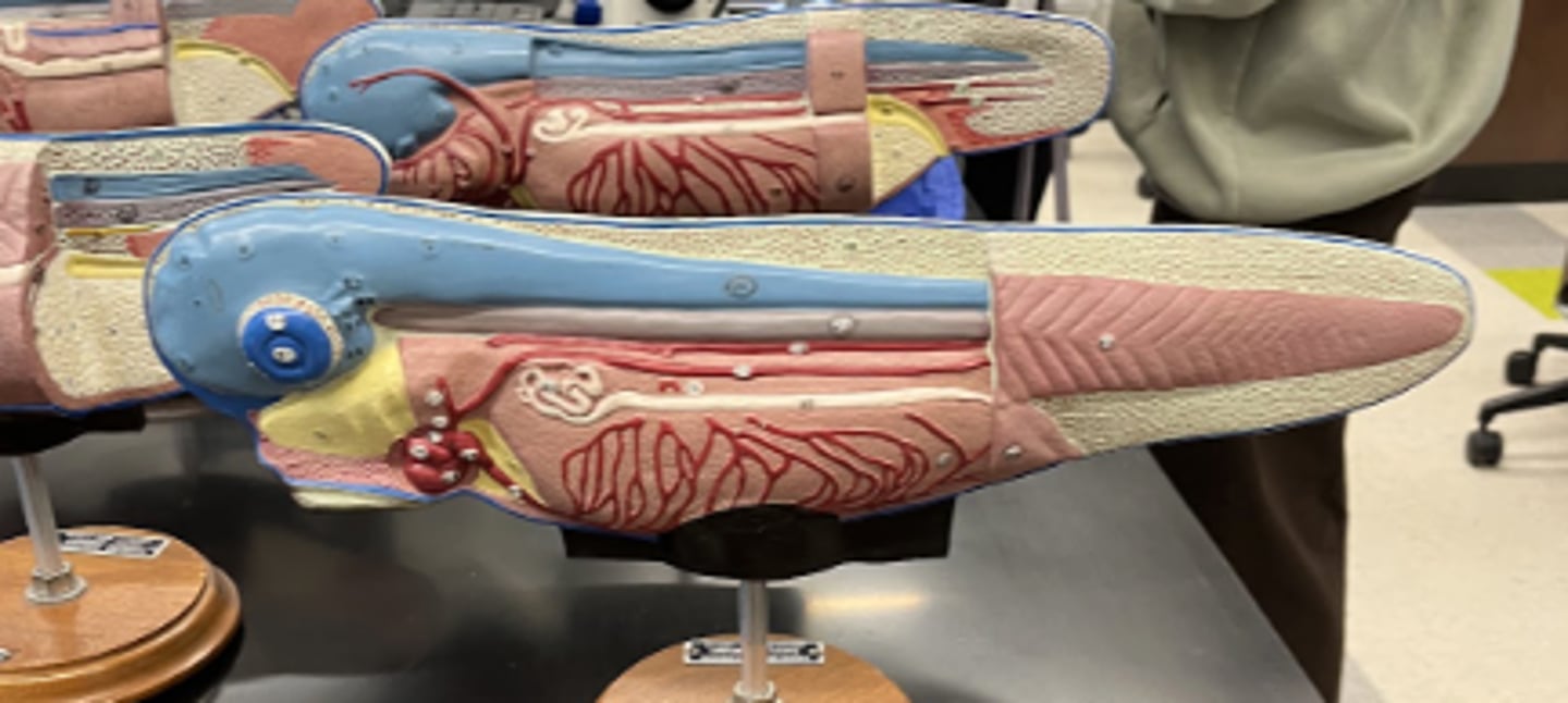
Female Pig Reproductive System
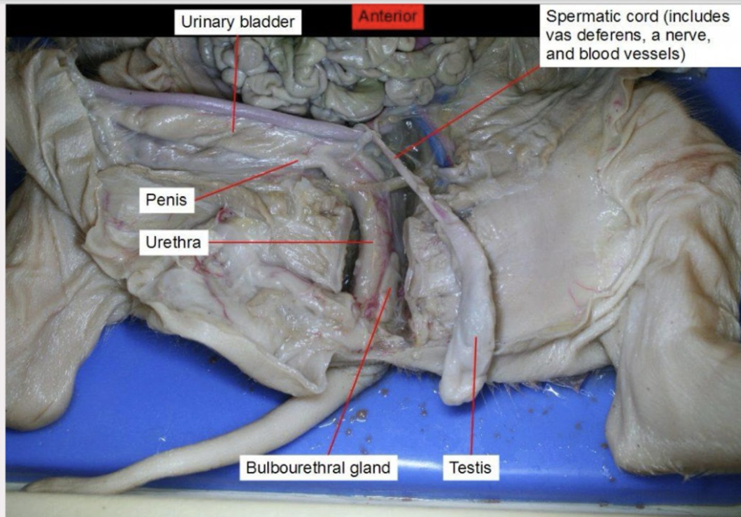
Pig Male Productive System
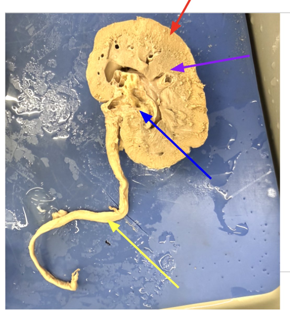
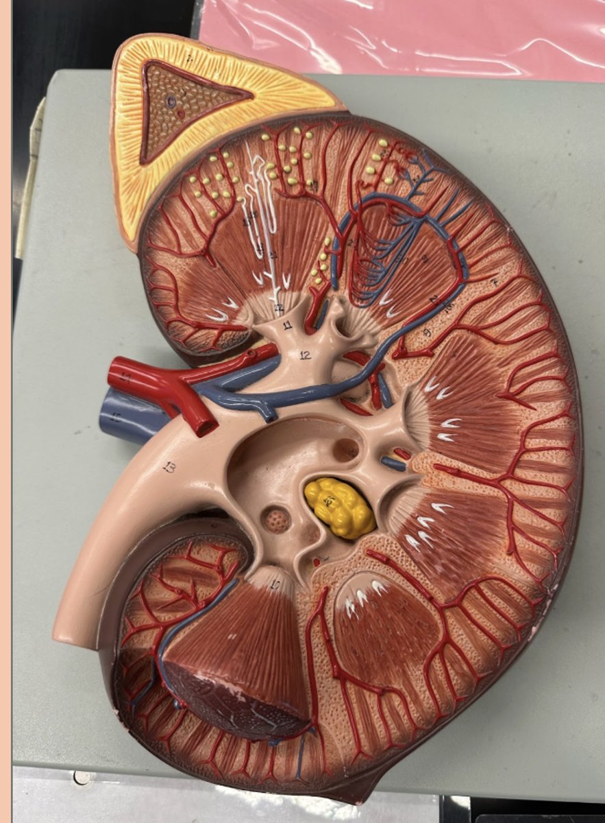
Kidney
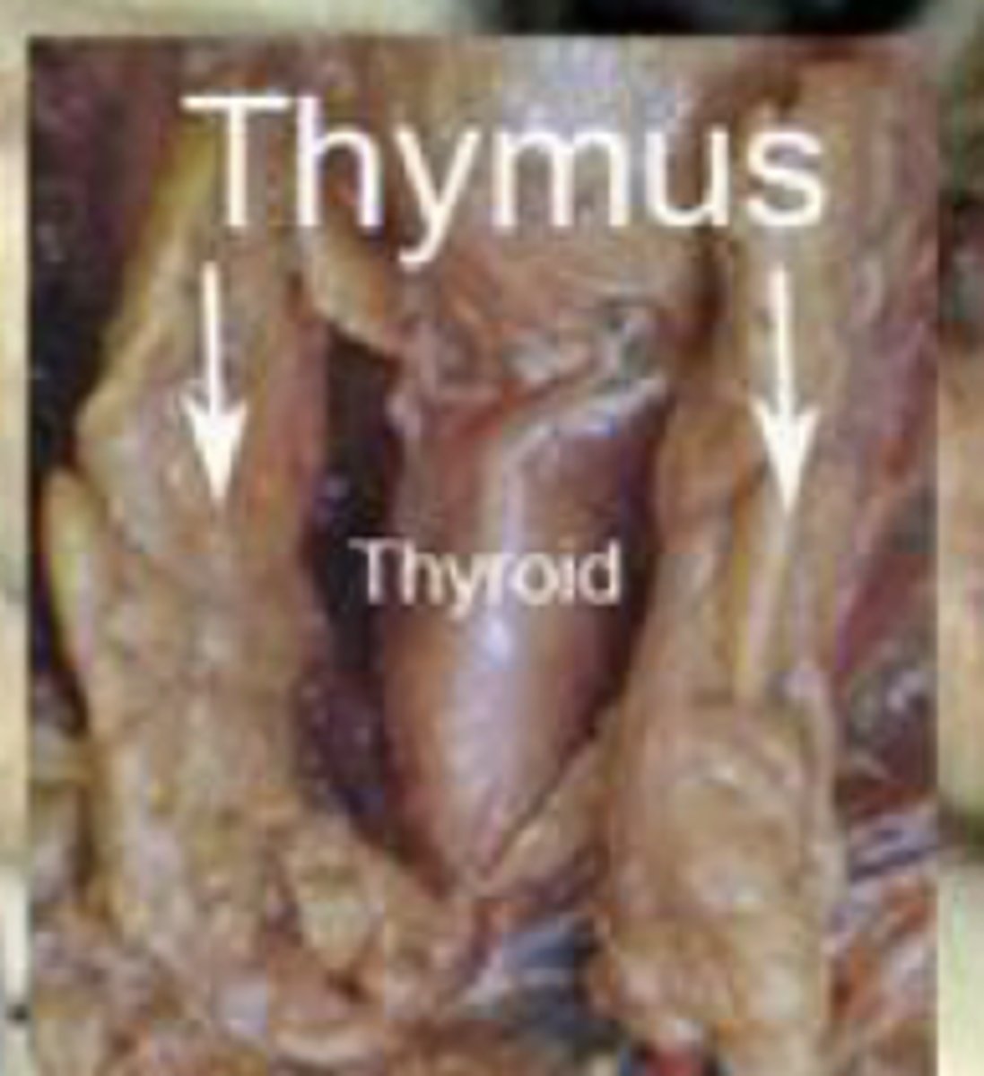
thymus
• produces thymosin
• thymosin stimulates development of lymphocytes/T cells
Hypophysis
green thing
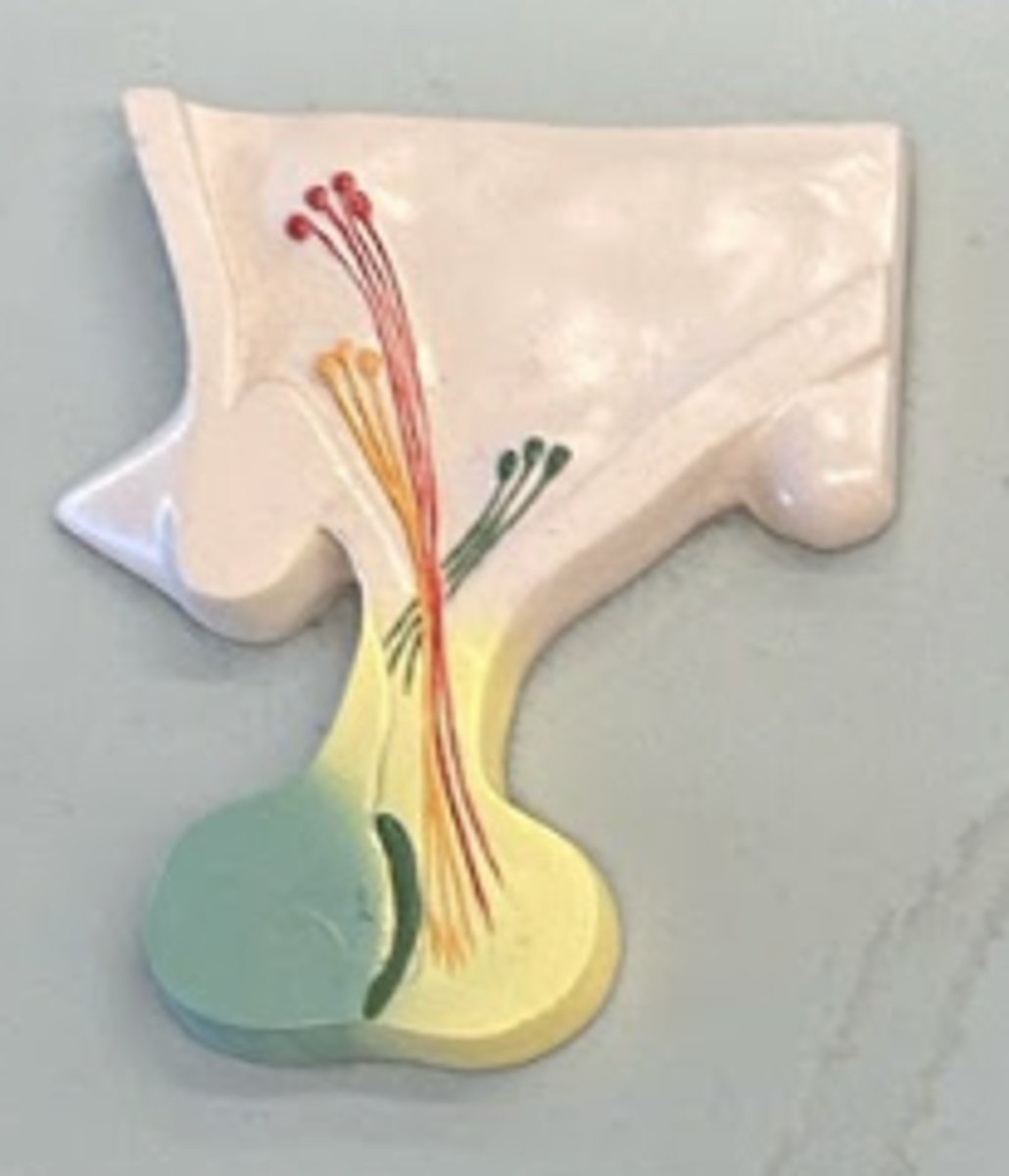
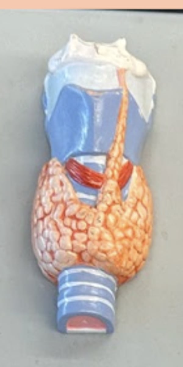
thyroid gland
cuboidal cells
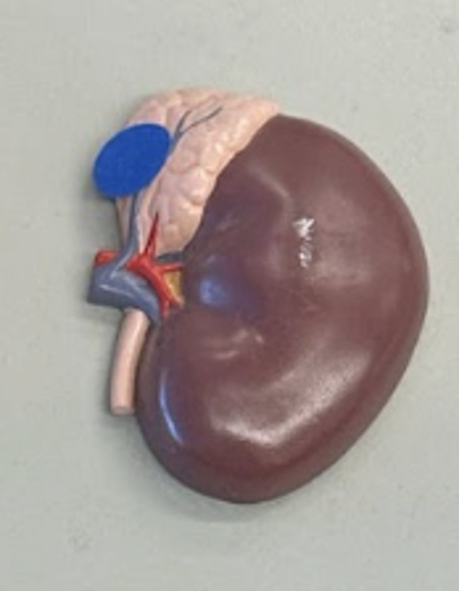
adrenal glands
on top of kidney
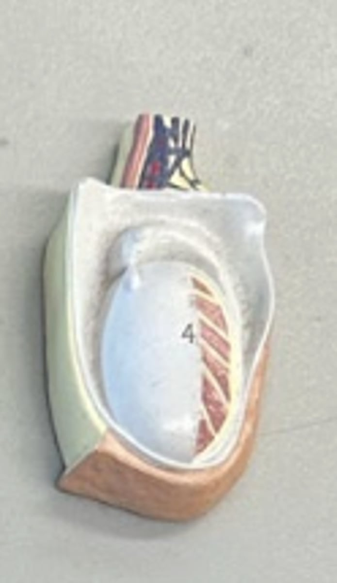
testis
• sperm production
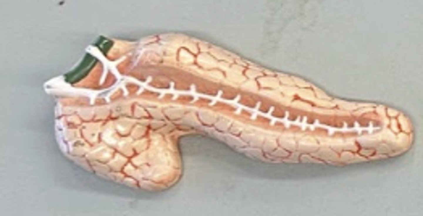
Pancreas
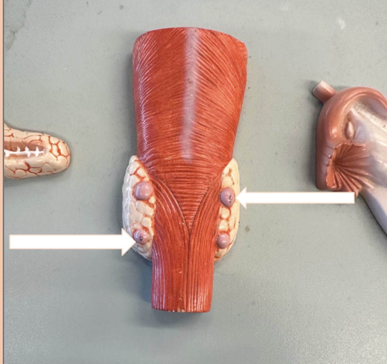
Parathyroid gland
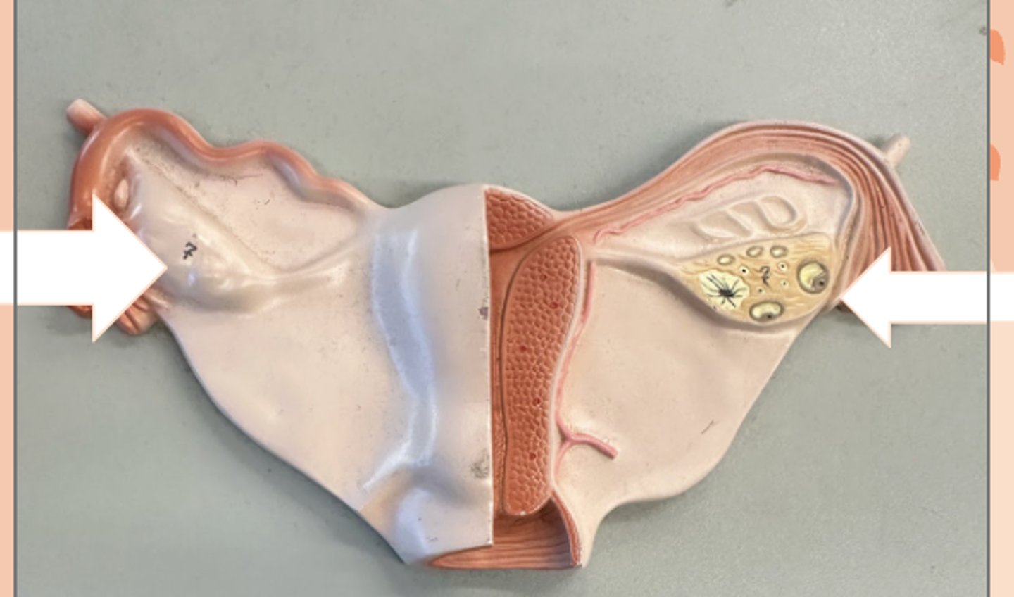
Ovary
Bull testicle
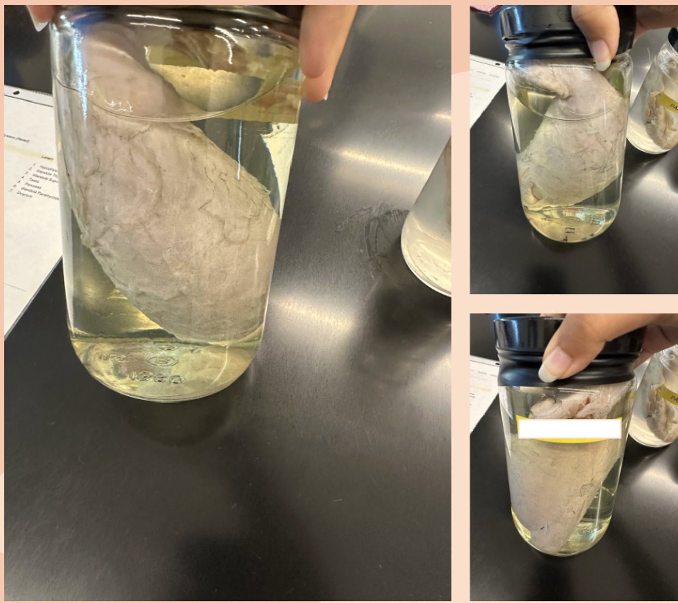
Pancreas
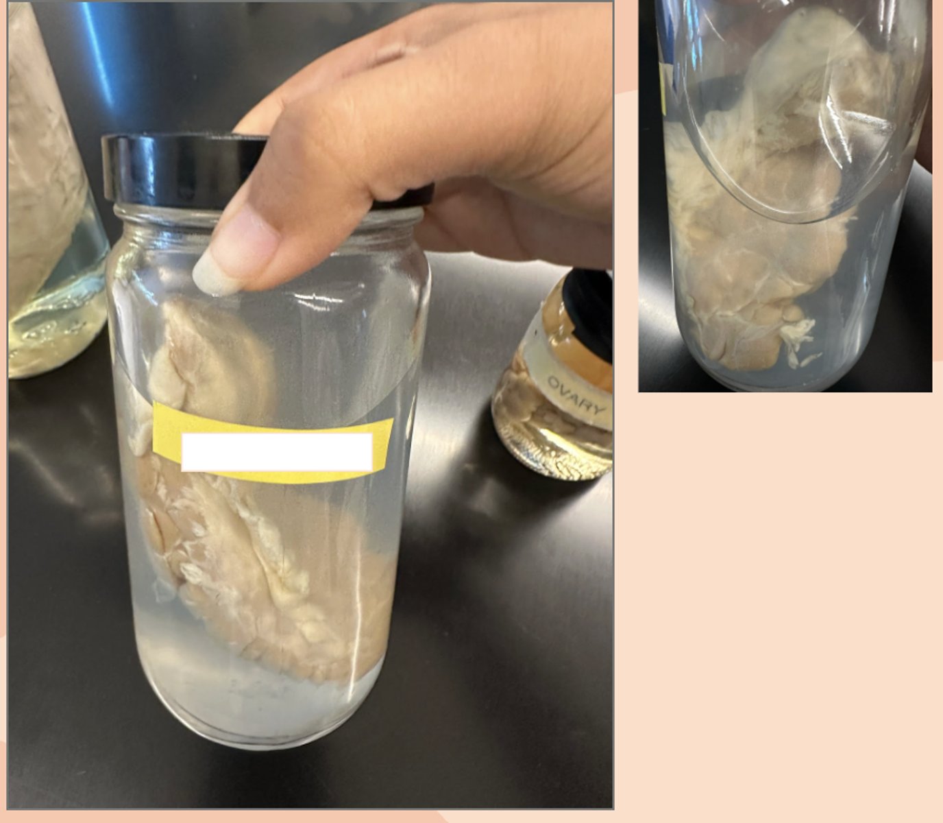
Ovary
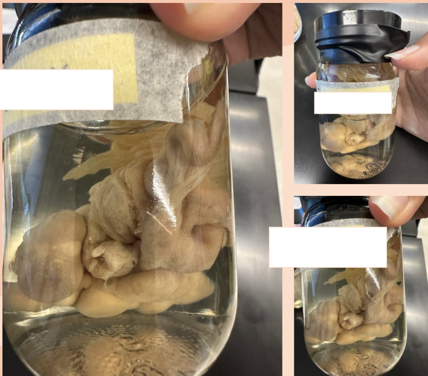
Uterus
• site of implantation
fimbriae
#1
• finger-like projections at the lateral end which create sweeping movements to draw eggs into oviduct
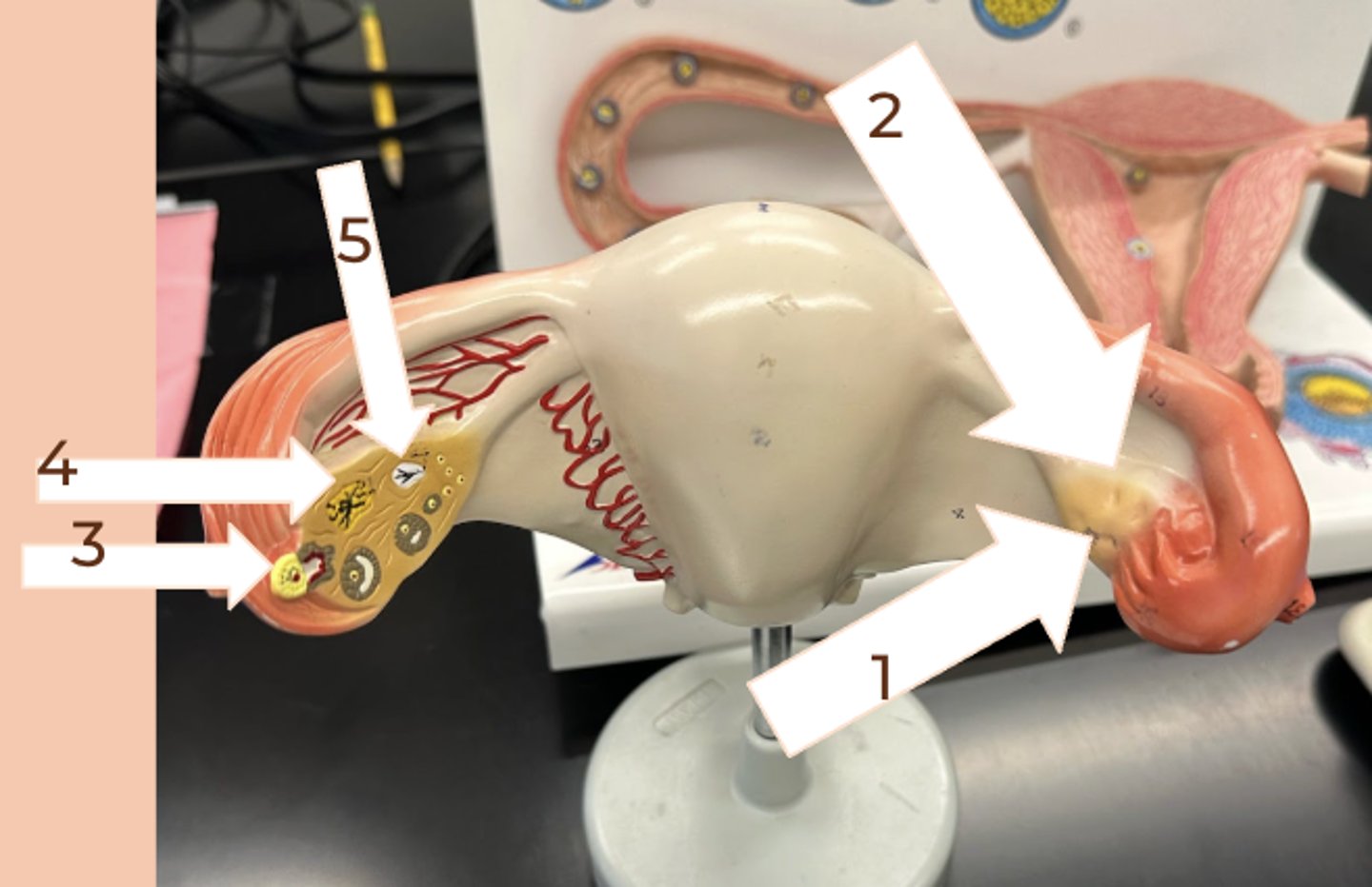
Ovary
#2
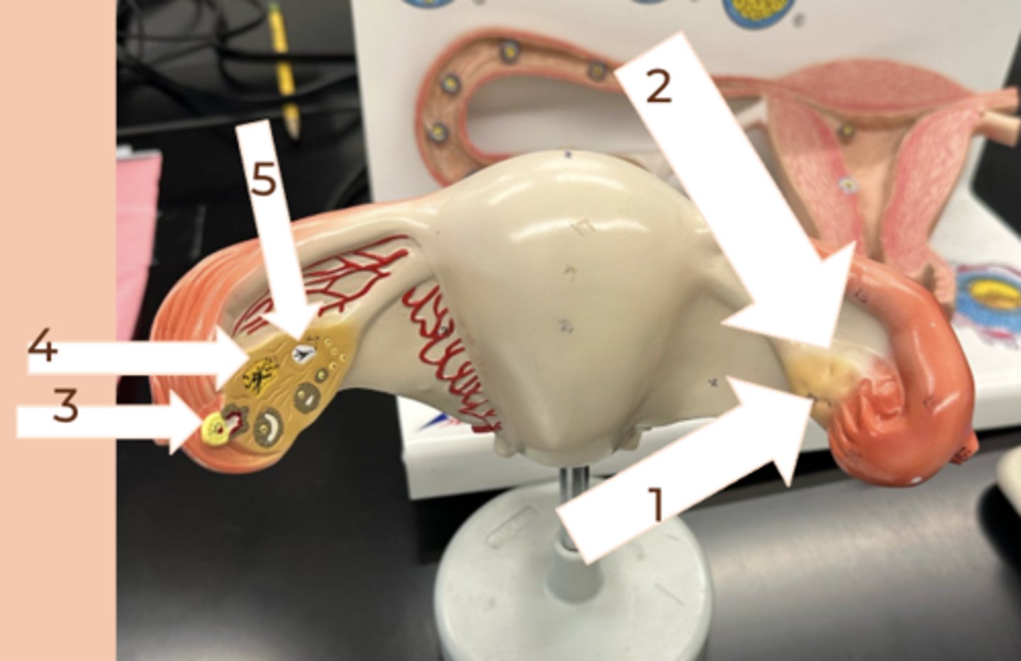
Mature Follicle
#3
• released during ovulation
• luteinizing hormone stimulates rupture of follice to release egg
• Follicle Stimulating hormone stimulates the maturation of follicle
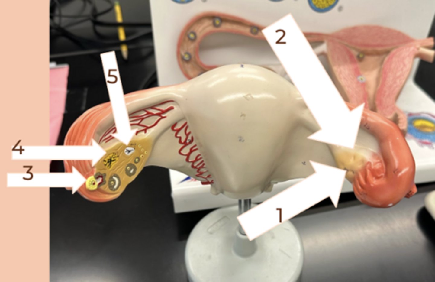
corpus luteum
#4
• temporary collection of cells that forms in ovaries after the egg is released and it releases progesterone which helps protect the uterine lining for implantation
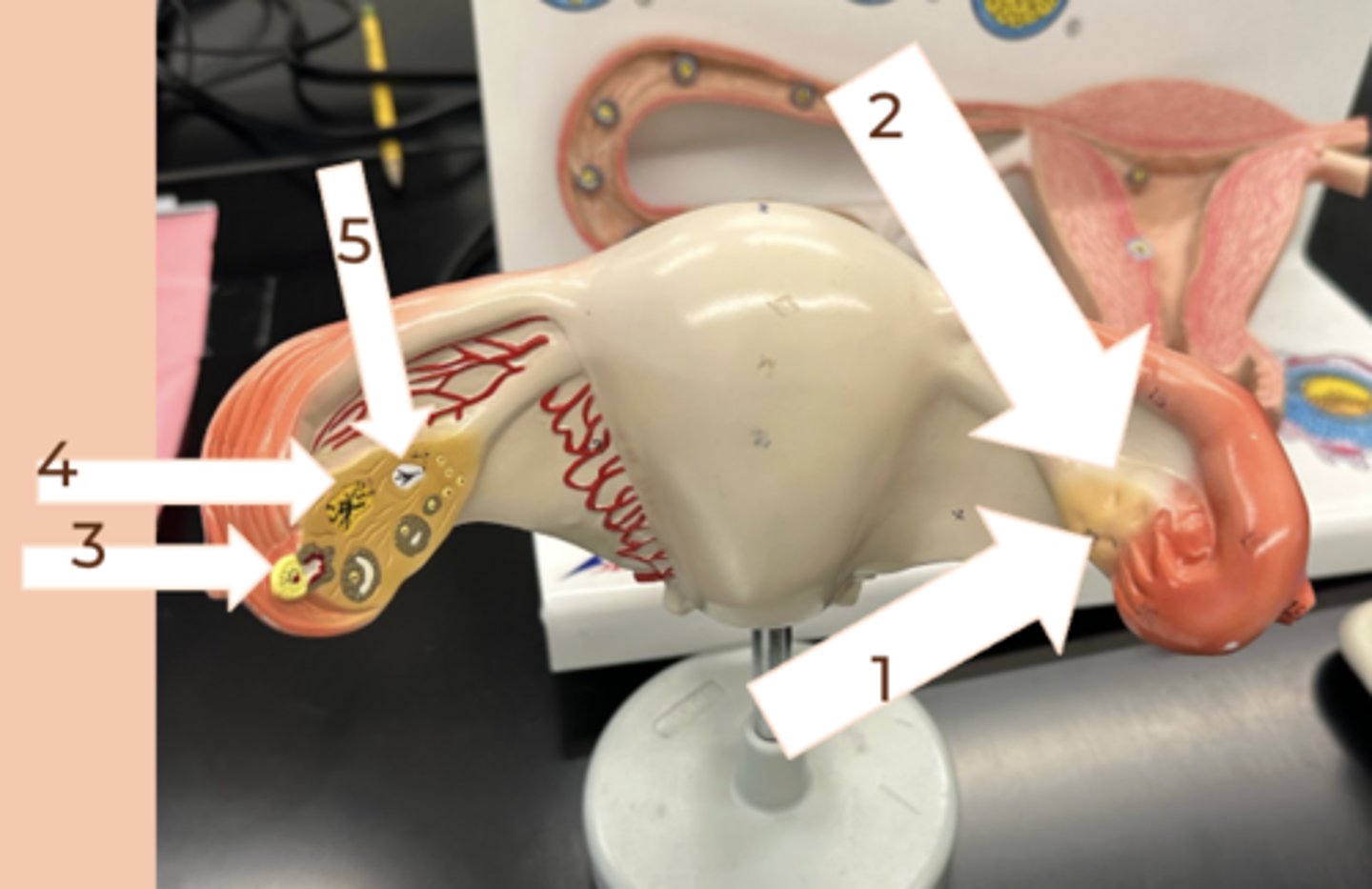
corpus albicans
#5
• scar tissue that forms after ovulation; created via the breakdown of the corpus luteum
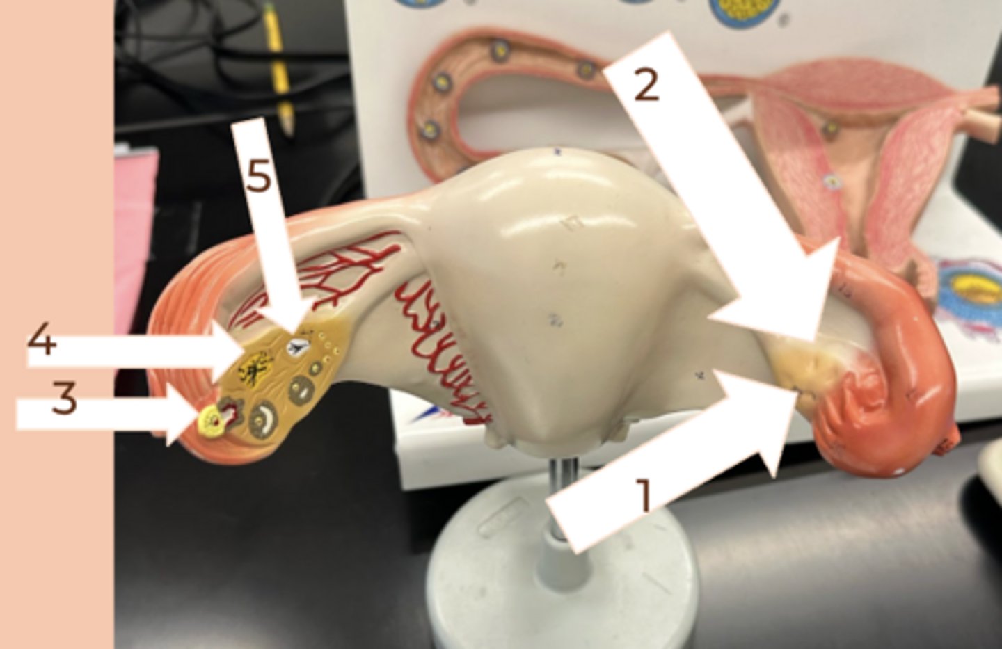
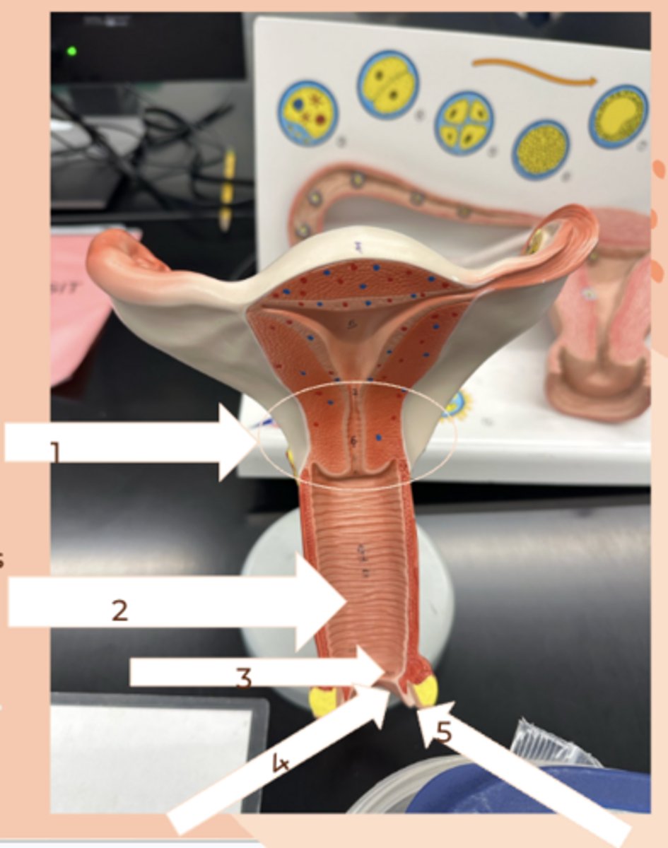
cervix
#1
• semi-cartilaginous ring of tissue separating the body of the uterus from the vagina
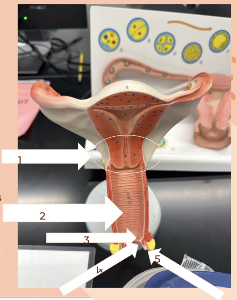
Vagina
#2
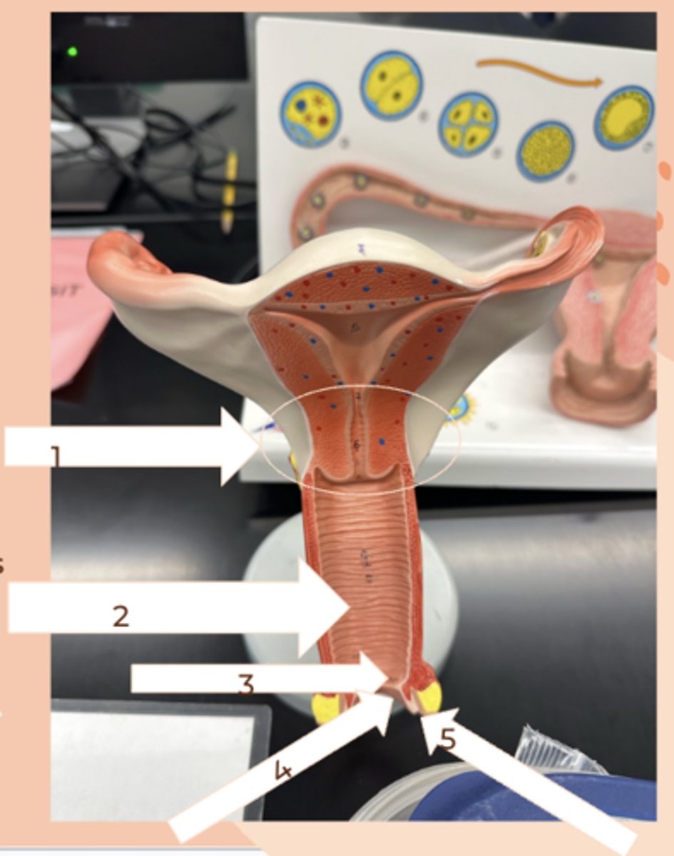
Hymen
#3
• thin layer of tissue that partially covers the vaginal opening in some women
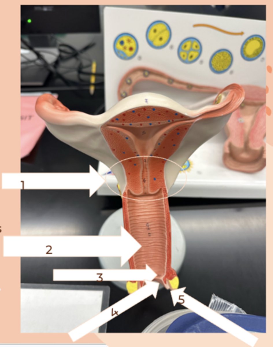
Labium minus
#4
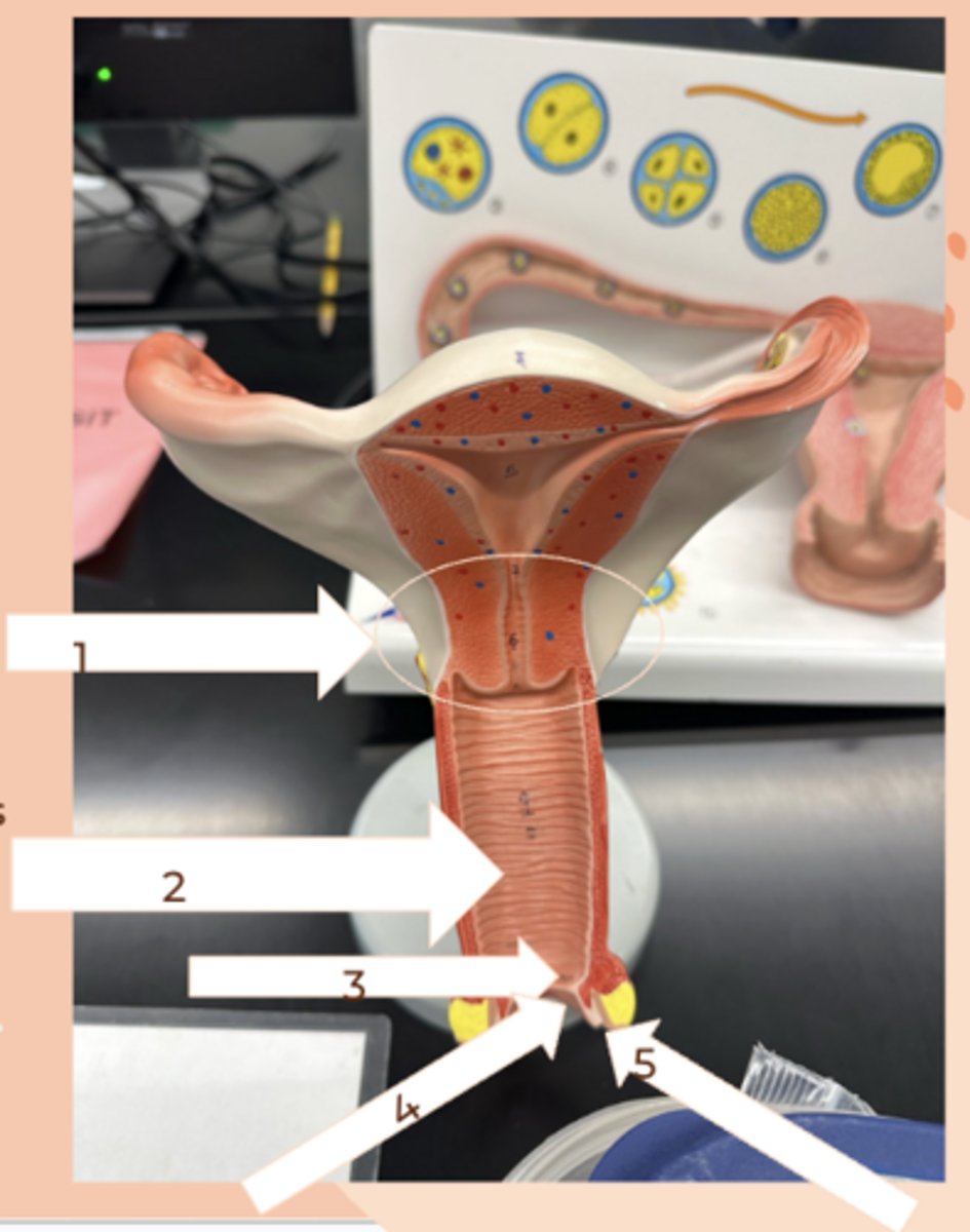
Labium majus
#5 (outside)
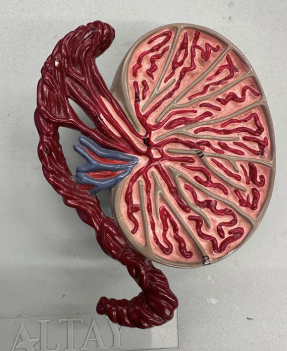
Testis
• sperm produced via meiosis here
• have leydig cells and seminiferous tubules
• surrounded by the scrotum
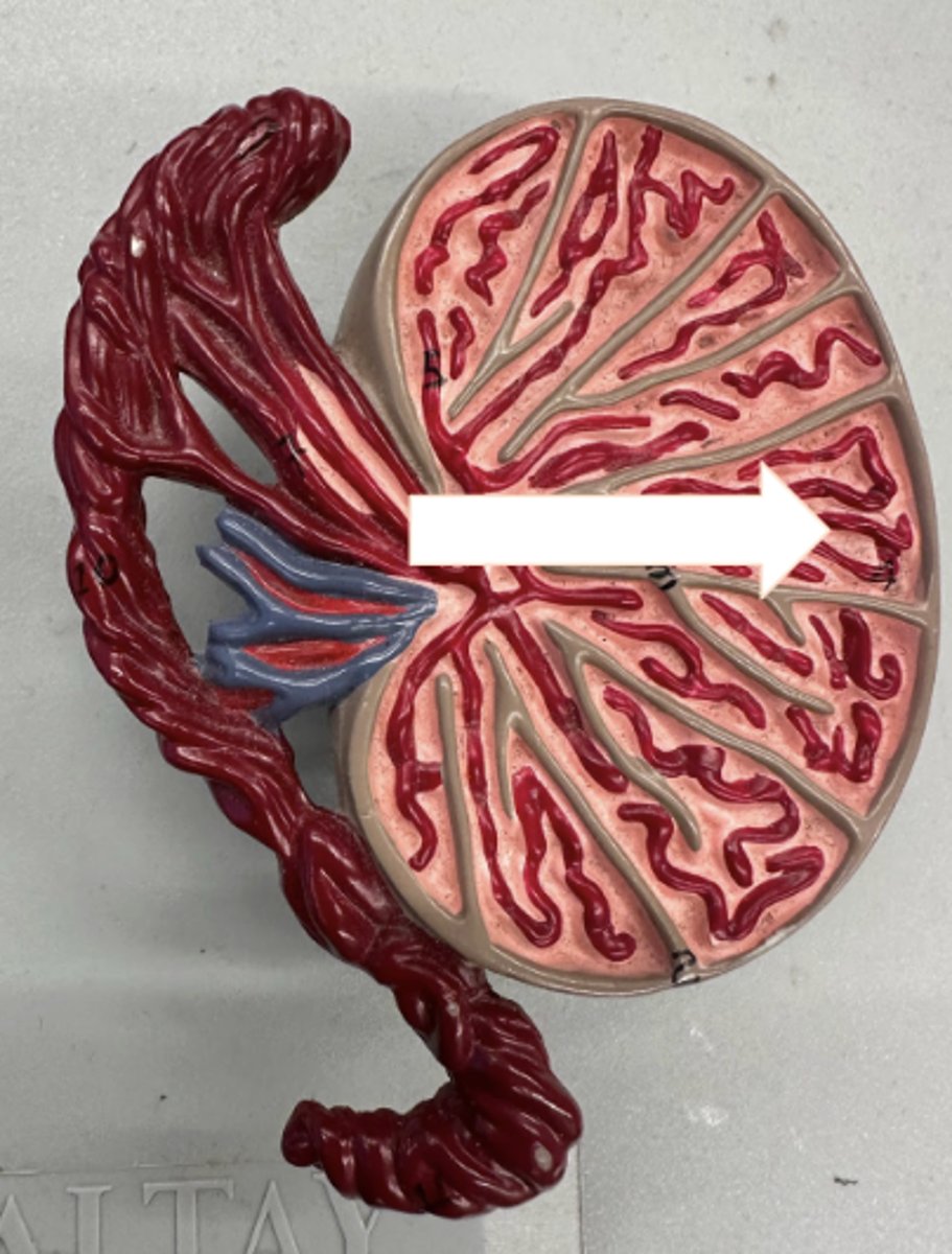
Seminiferous Tubules
• microscopic ducts for sperm cells
• Leydig cells are OUTSIDE these tubules and produce testosterone
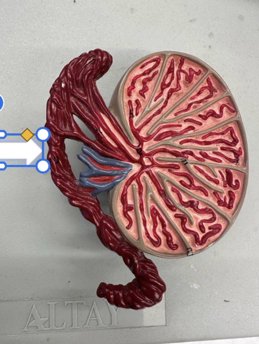
Epididymis
• Sperm mature here
• younger sperm at the head, older sperm towards the tail
Testis
• has spermatic cord
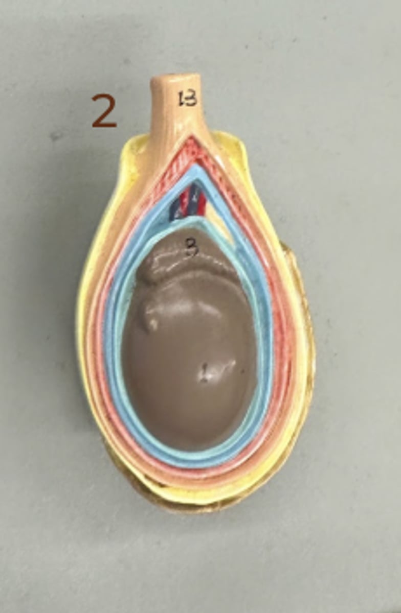
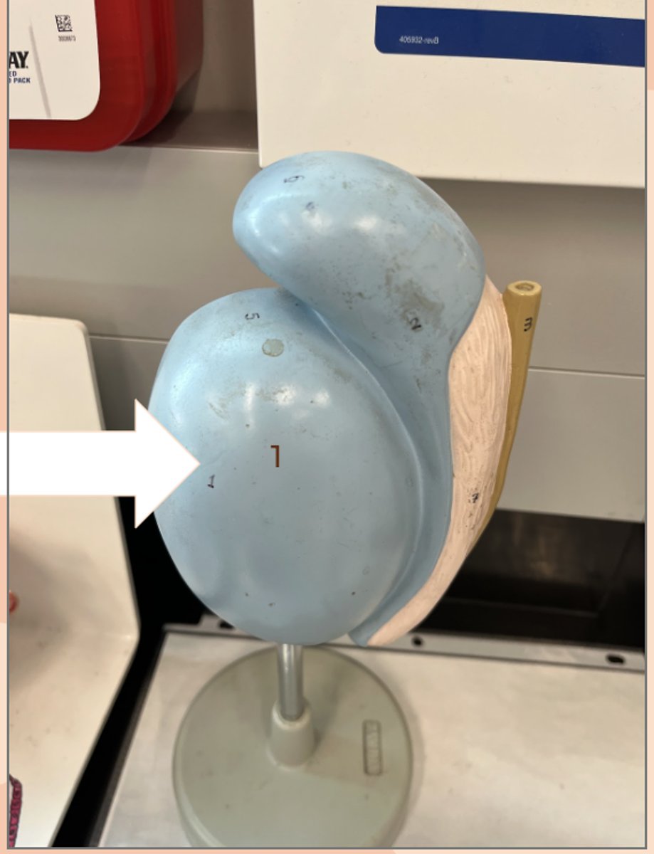
Testis
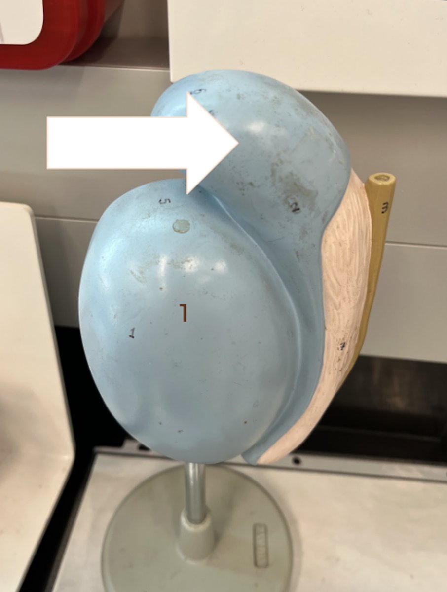
Epididymis
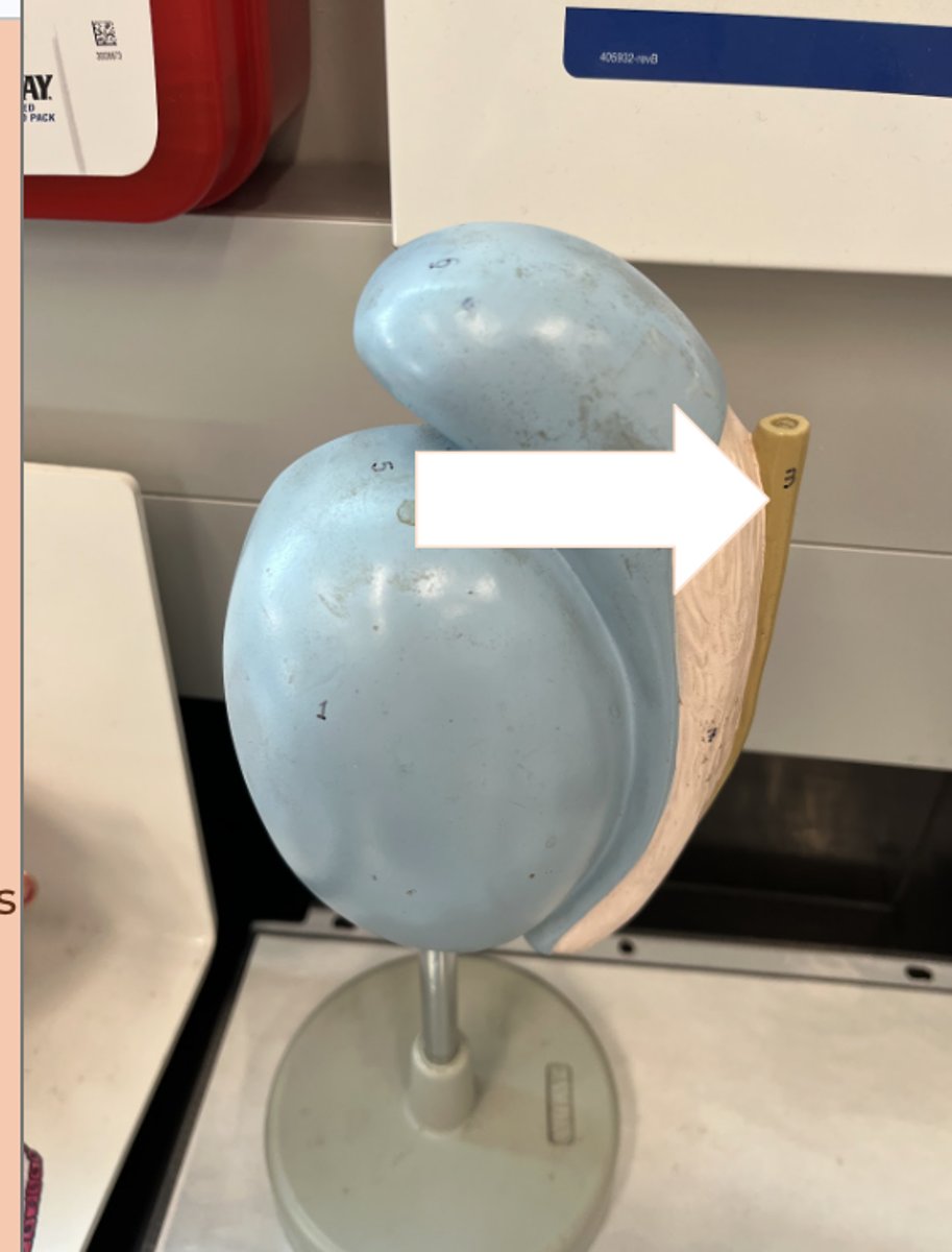
ductus deferens (vas deferens)
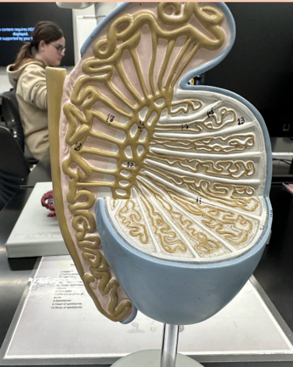
Body of Epididymis
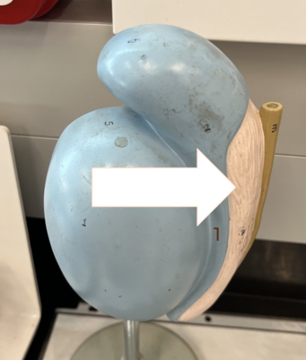
Testis
(AGAIN!)
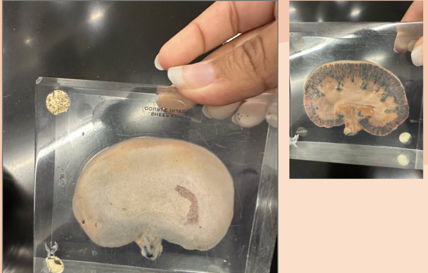
Kidney
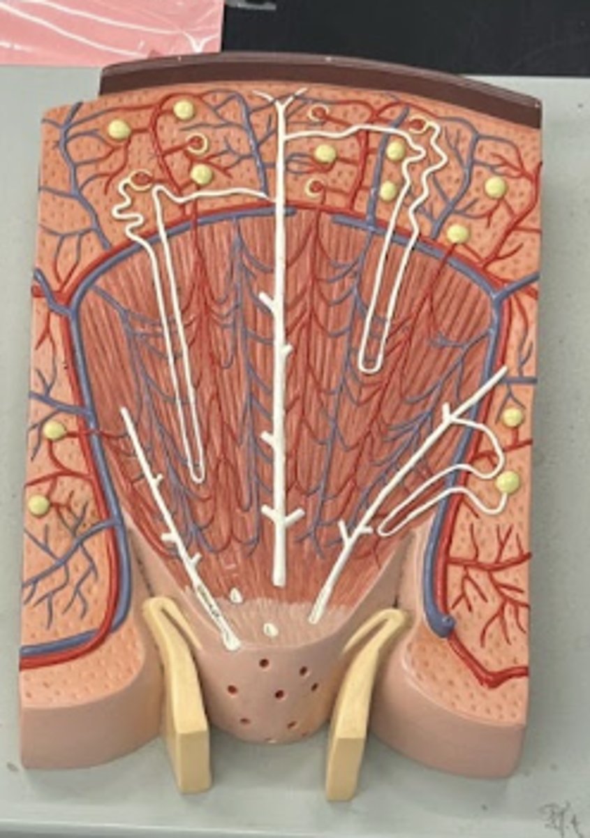
Kidney Nephron
• proximal convoluted tubule
• descending limb
• loop of henle
• ascneding limb
• collecting duct (middle long white thing)
• distal convoluted tubule
Kidney
cortex (outer)
renal artery (red)
Renal vein (blue)
pelvis (inner white thing)
medulla with pyramid that thing
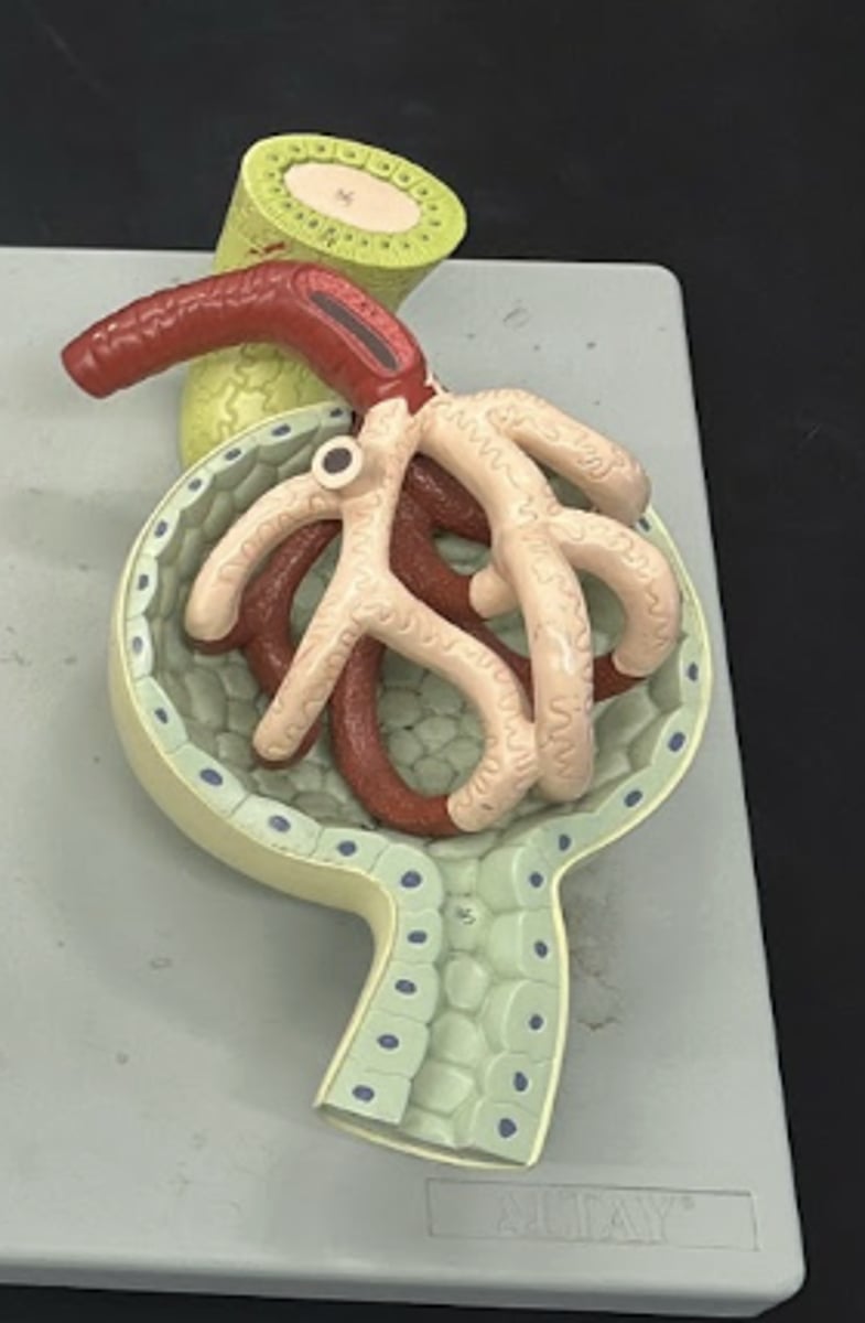
glomerulus and bowmans capsule
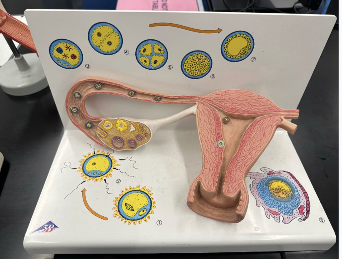
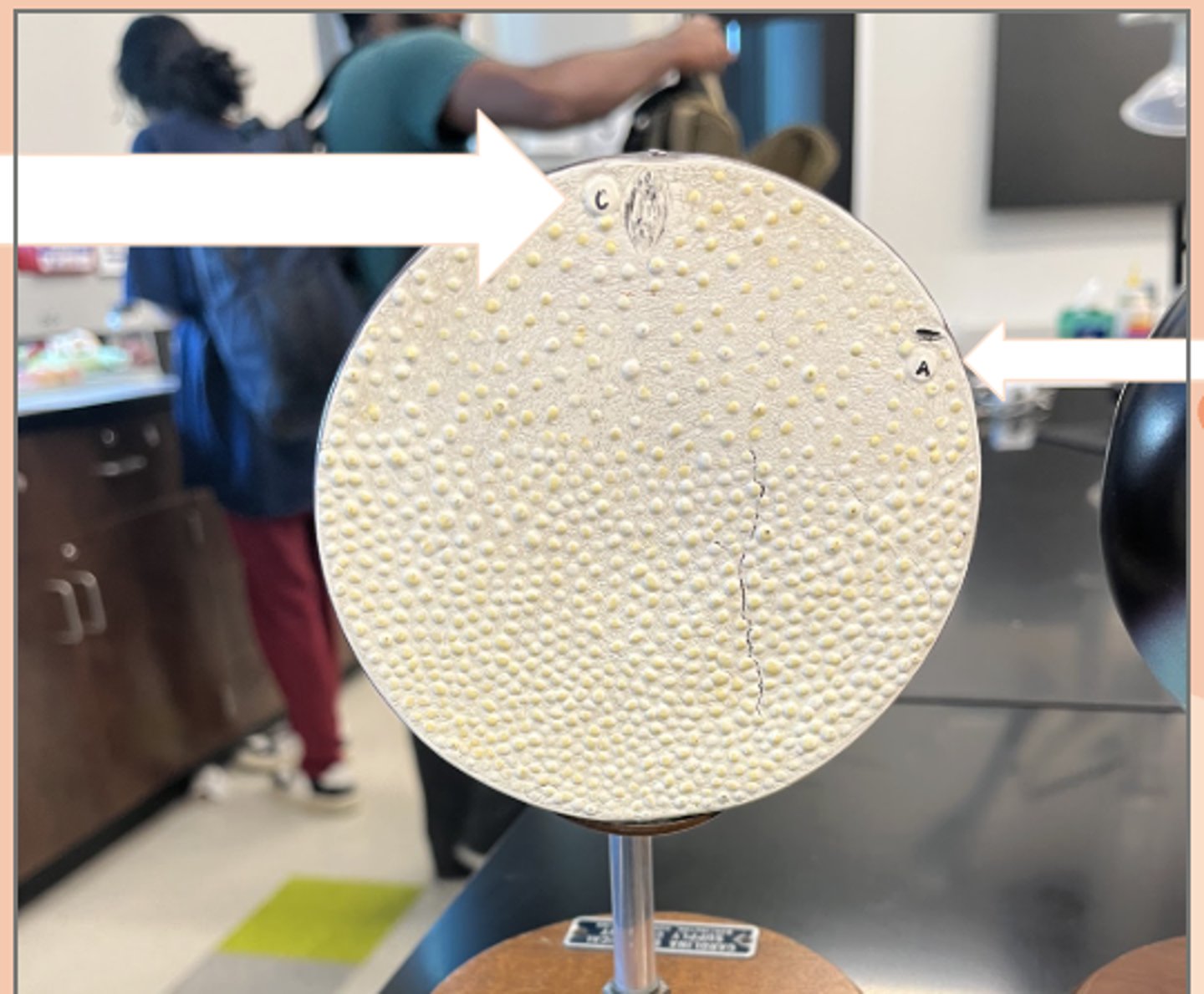
fertilization
C) egg nucleus
A) Sperm nucleus has penetrated egg
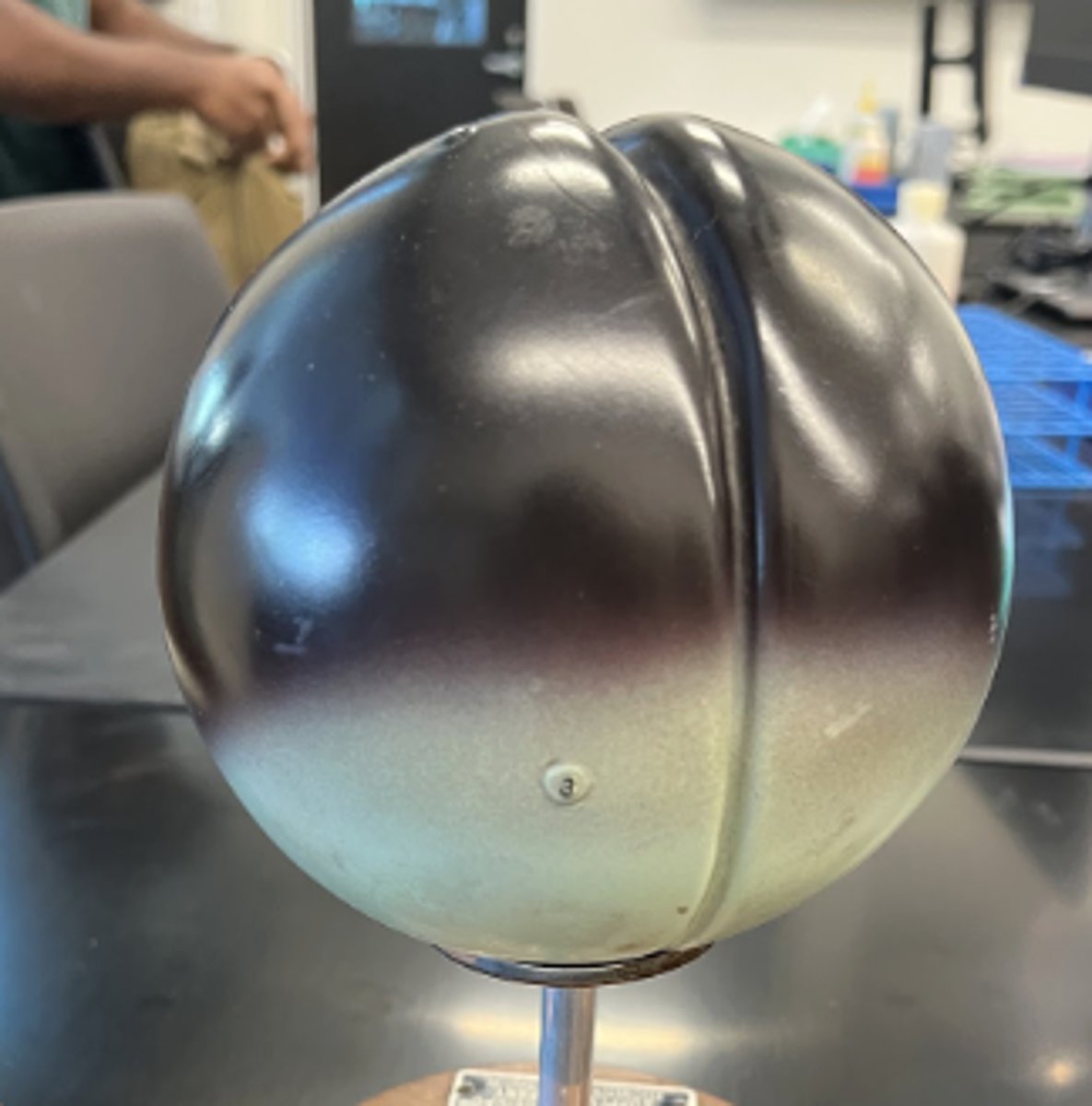
First Cleavage Event
• completed meiosis II
• longitudinal/verticle
• darker pole: animal pole
• lighter pole: vegetal pole
second cleavage event
• longitudinal
• results in 4 blastomeres
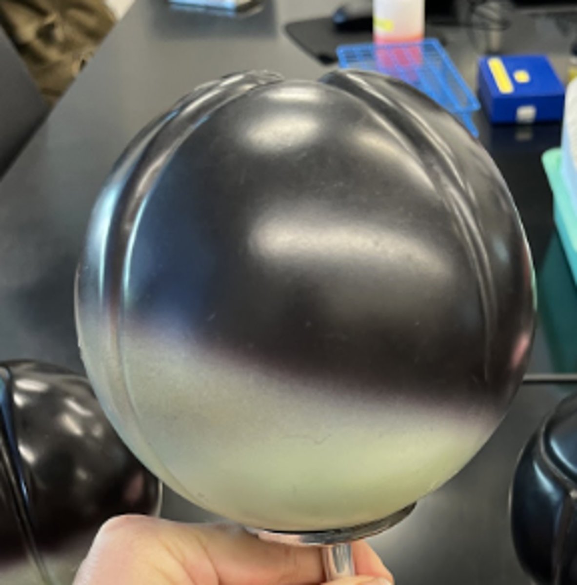
Third Cleavage event
• Horizontal cleavage
• 8 cells
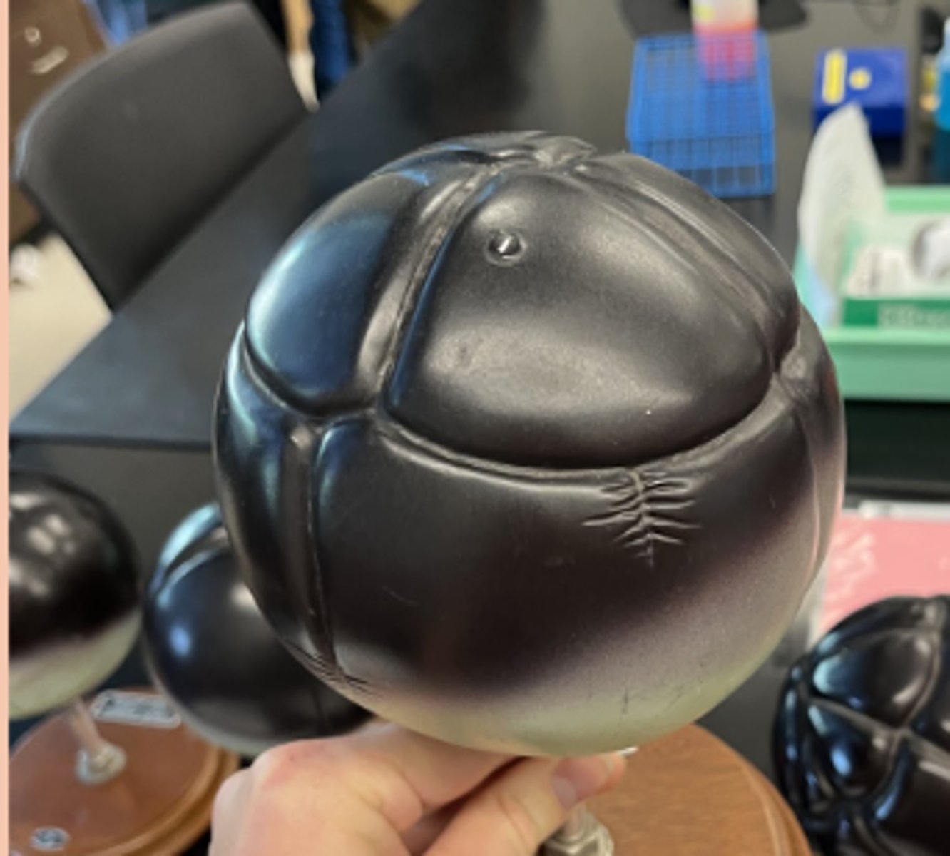
Early Morula
• solid ball of cells
• blastocoel begins to form
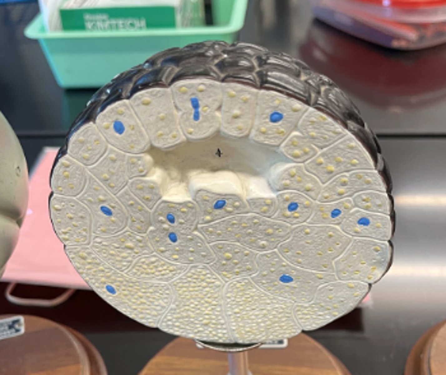
Late Morula
• blastocoel continues forming
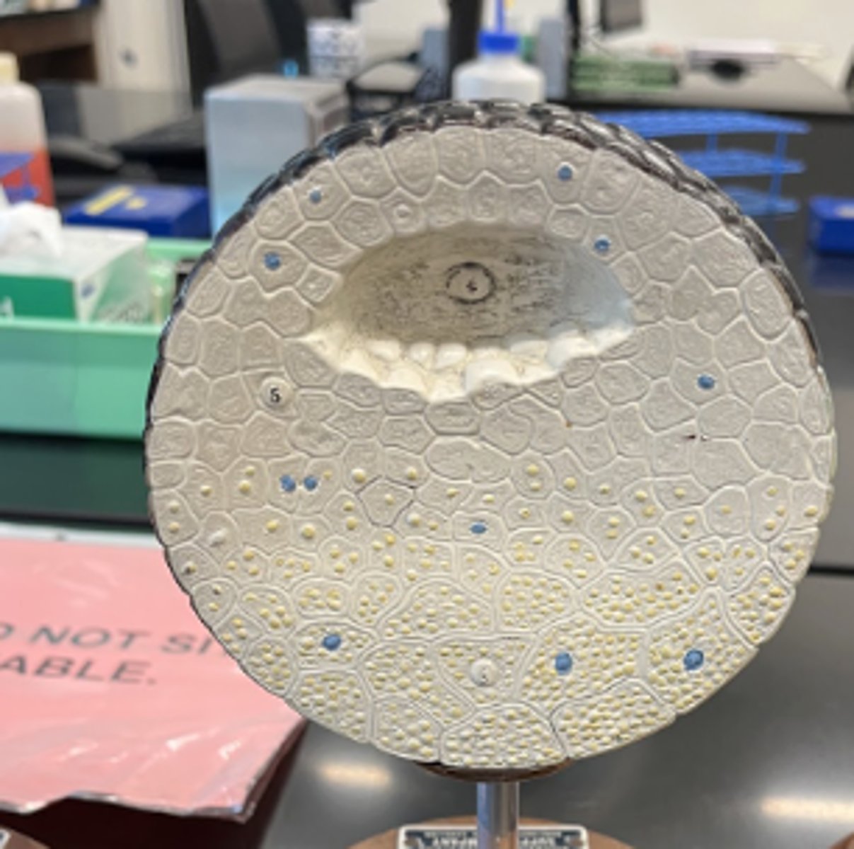
Blatsula
• fluid filled blastocoel is completed
• ectoderm (blue) is present
• arrow pointing to dorsal lip, where cells will invaginated during gastrulation
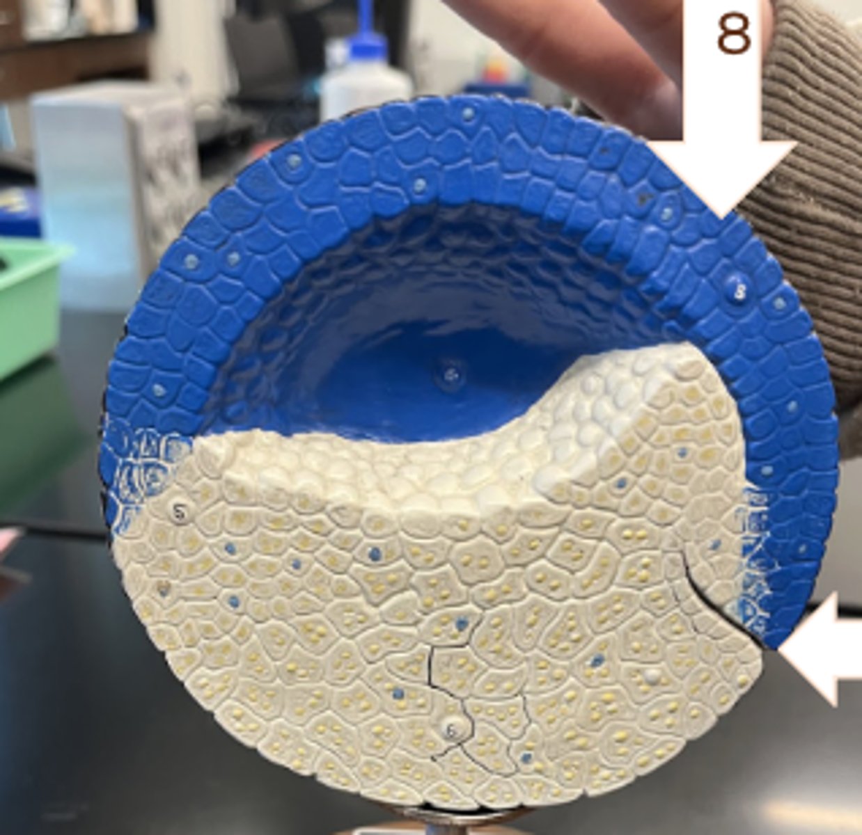
Gastrulation
• cells migrate inward
• archeteron obliterates blastocoel
• undifferentiated mesoderm arises
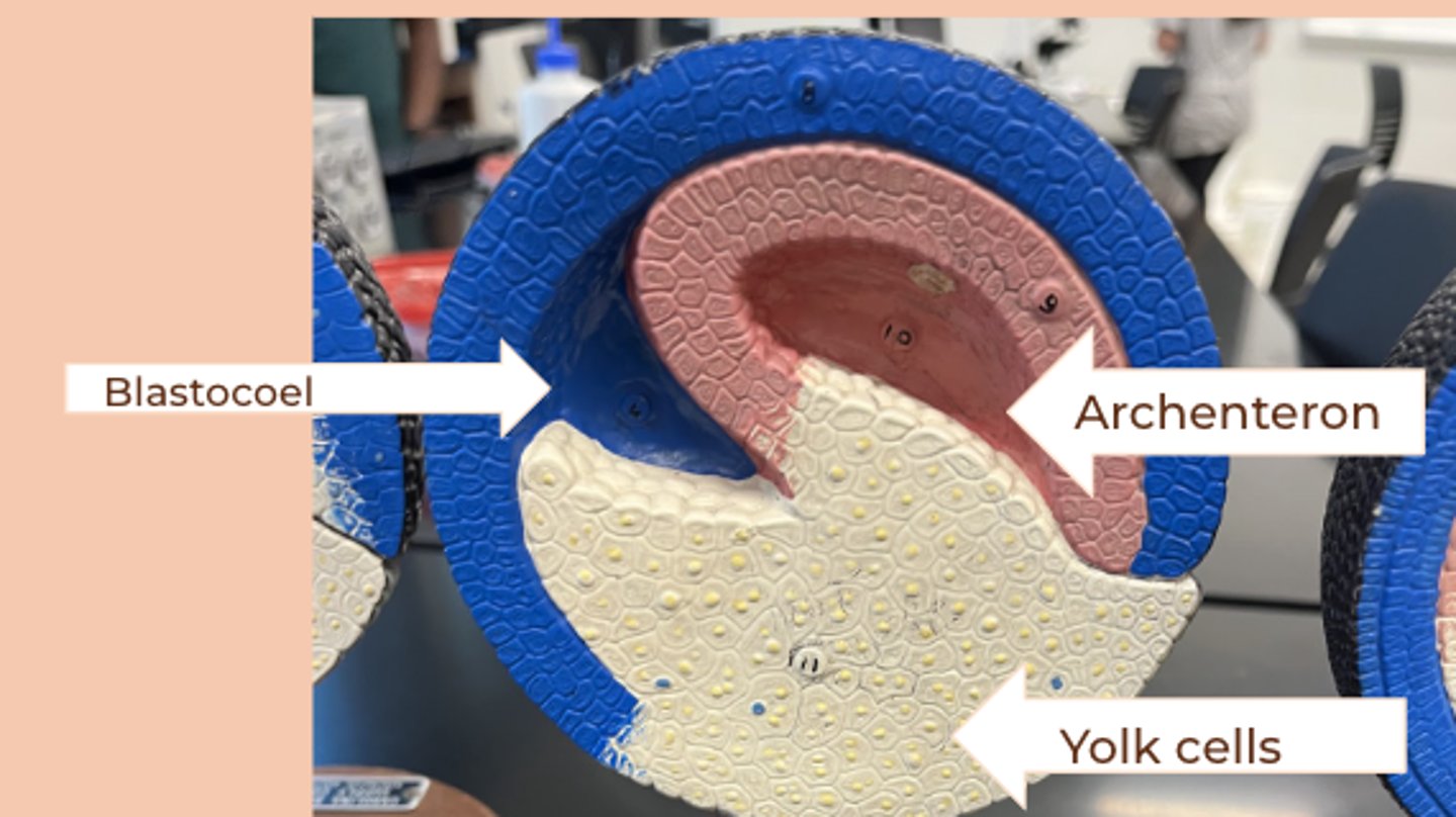
End of Gastrulation
• three germ layers present
• complete archenteron
• yolk plug occupies what will become the blastospore
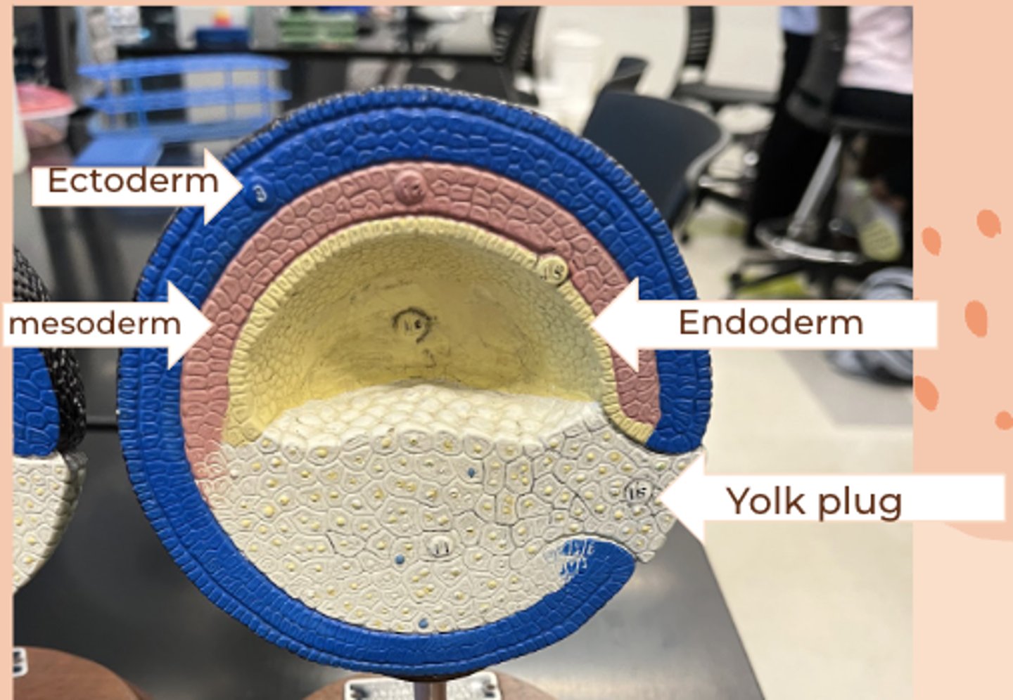
first stage of neurulation
• neural plate is forming at the top of the embryo
• blastopore forming as result of shrinking yolk plug
• neural groove formed by ectodermal cells flattening out and sinking
• notochord forms from purple mesodorm
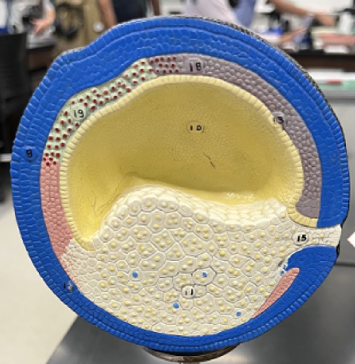
neurulation
• neural crest and neural groove are forming at top of embryo
• blastopore is nearly free of yolk
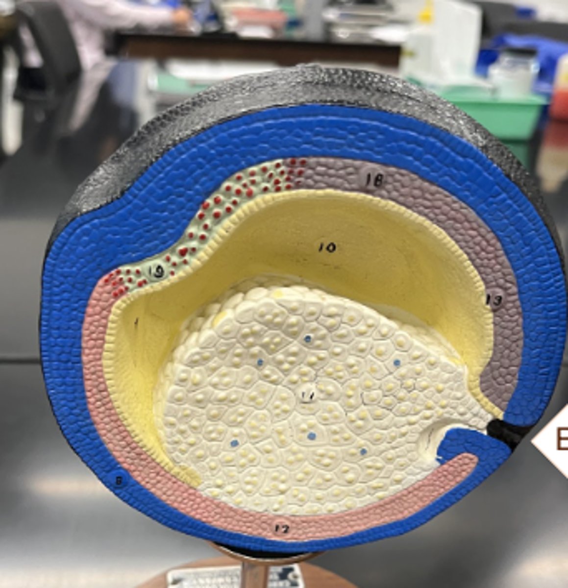
neurulation: two neural crest fuse to form hollow tube
• two neural crests are almost completely fused together forming a hollow tube lined with ectoderm
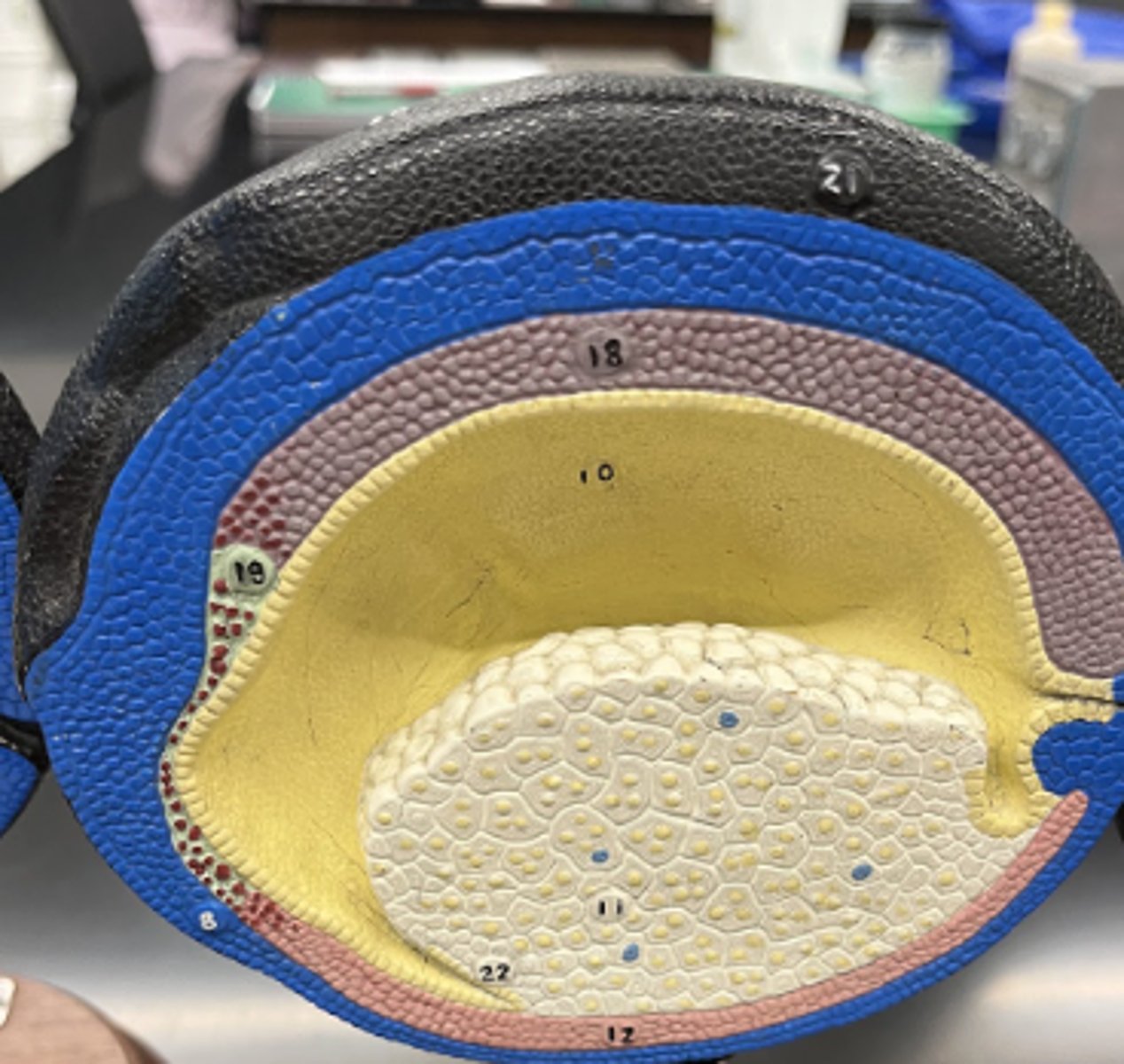
neurulation: fusing of ectoderm and endoderm
• oral plate
• ectoderm and endoderm fuse, forming the oral cavity
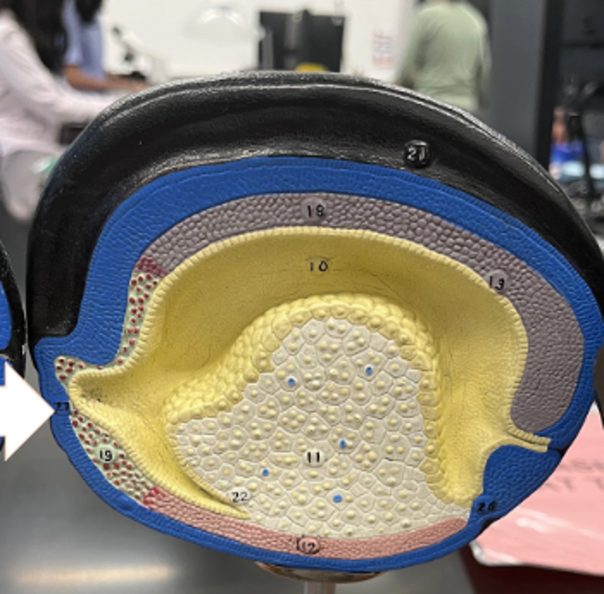
neurulation: specialization of derm tissues
• brain and spinal cord from ectoderm
• notochord is fully formed
• hypophysis is the pituitary gland
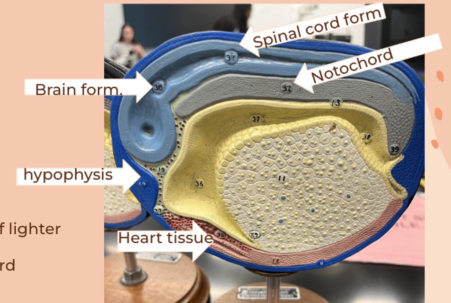
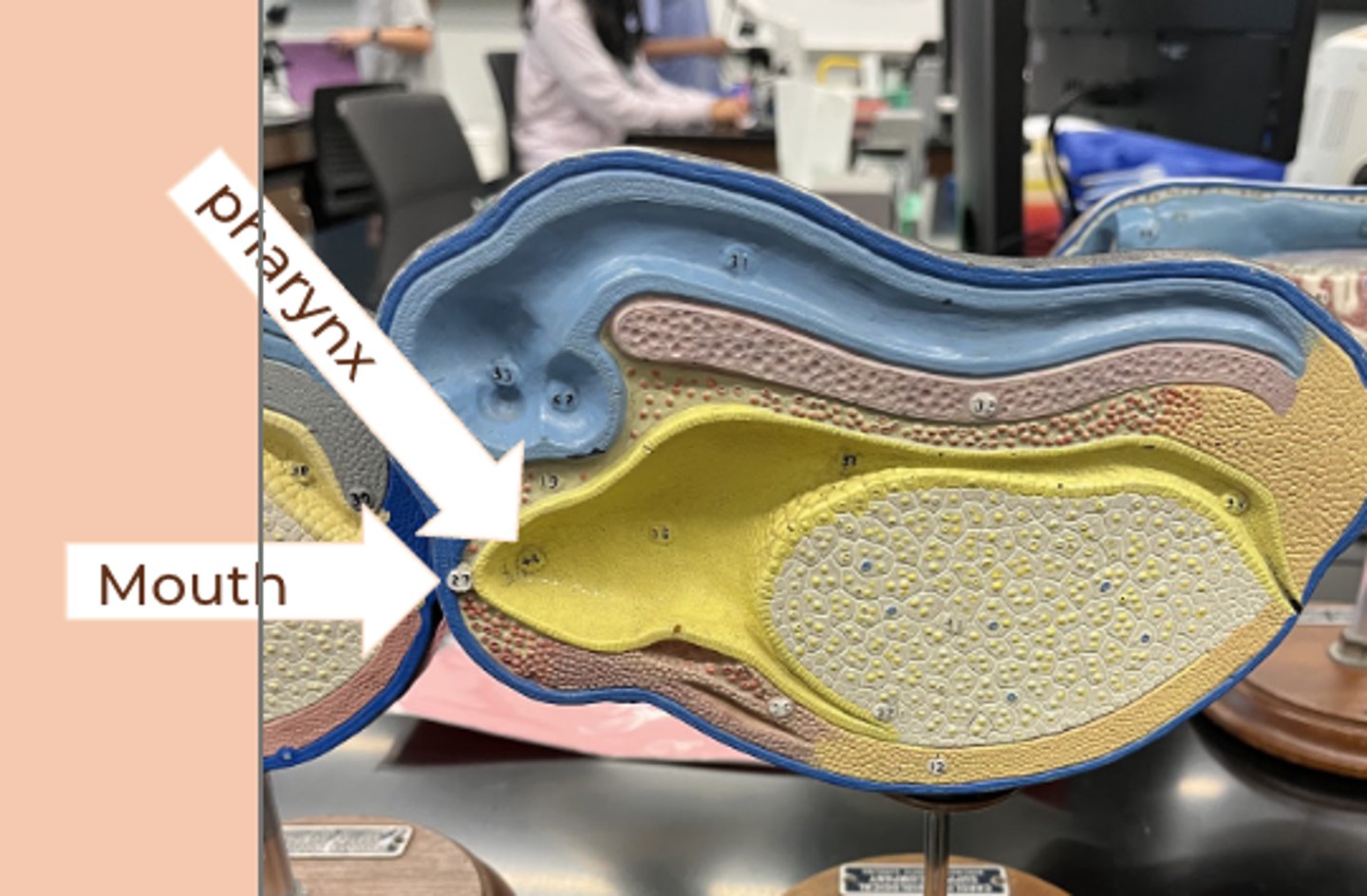
neurulation: mouth and anus formation
• ectoderm and endoderm fuse to form the mouth
• anal opening from blastopore
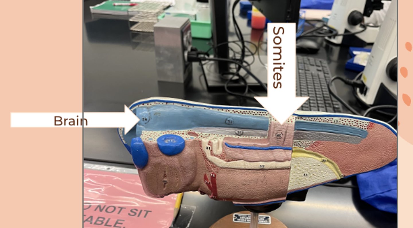
neurulation: formation of somites and brain
• brain has formed
• somites: condensed mesoderm that give rise to vertebrae and back muscles
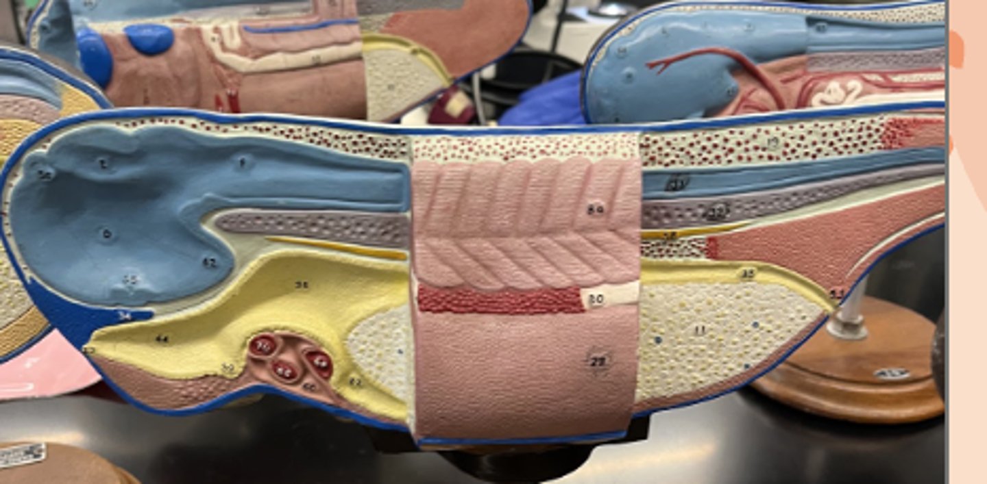
neurulation: elongation of embryo
neurulation: elongation of embryo
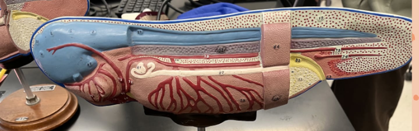
neurulation: final elongation of embryo
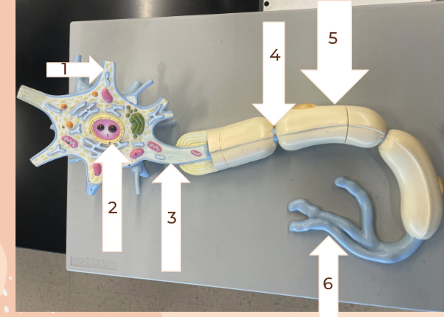
neuron model
Dendrite
soma/body
Axon
Nodes of ranvier
Myelinated
Terminal branches
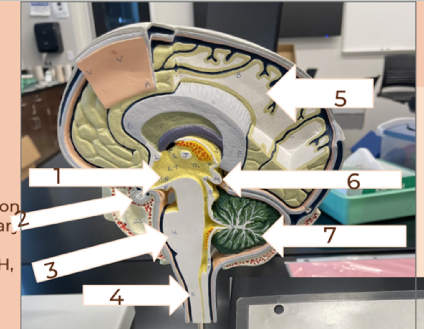
Hypothalamus
#1
• contains areas which govern emotions
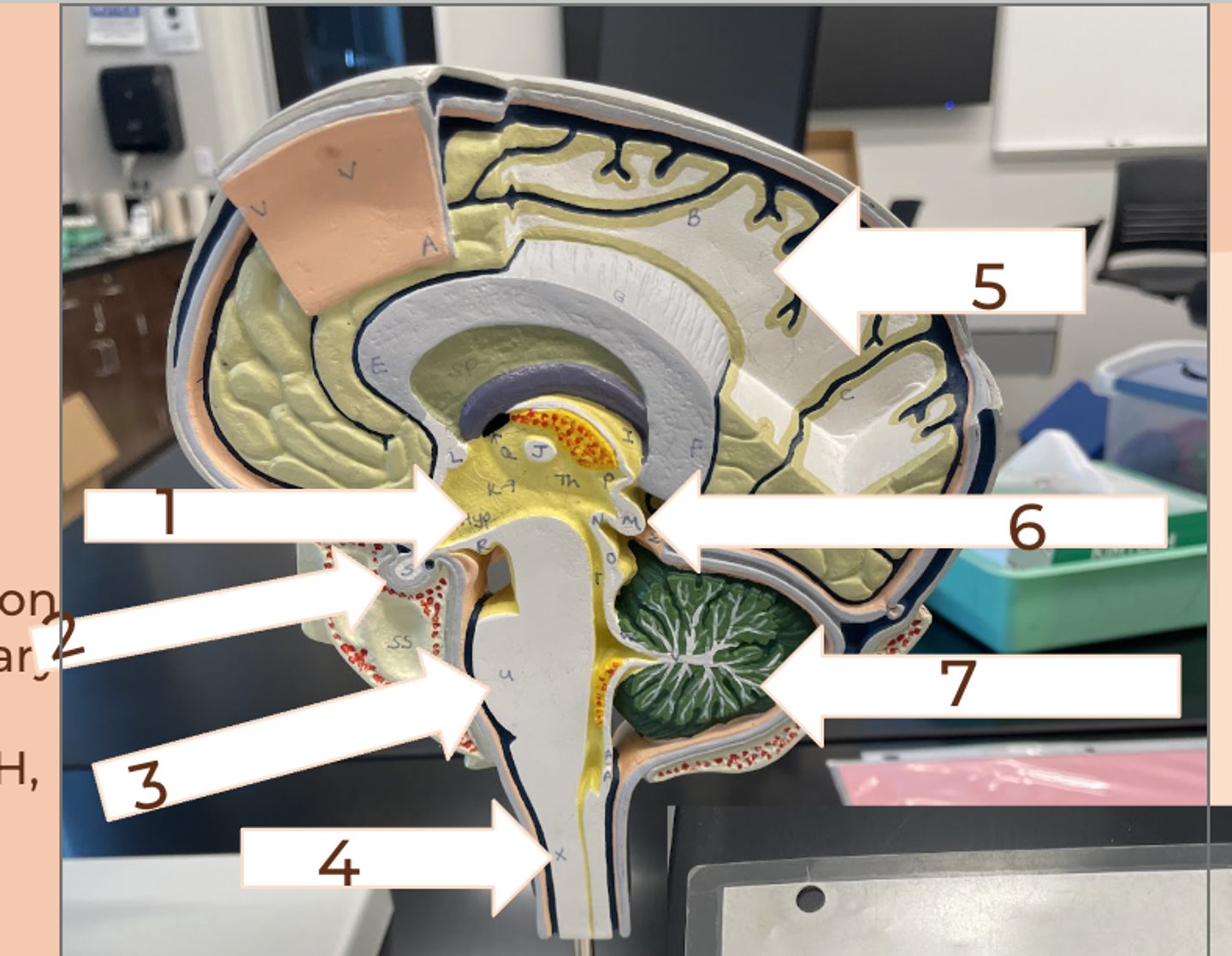
Pituitary gland
#2
• release many hormones
Pons
#3
• helps medulla oblongata with its function
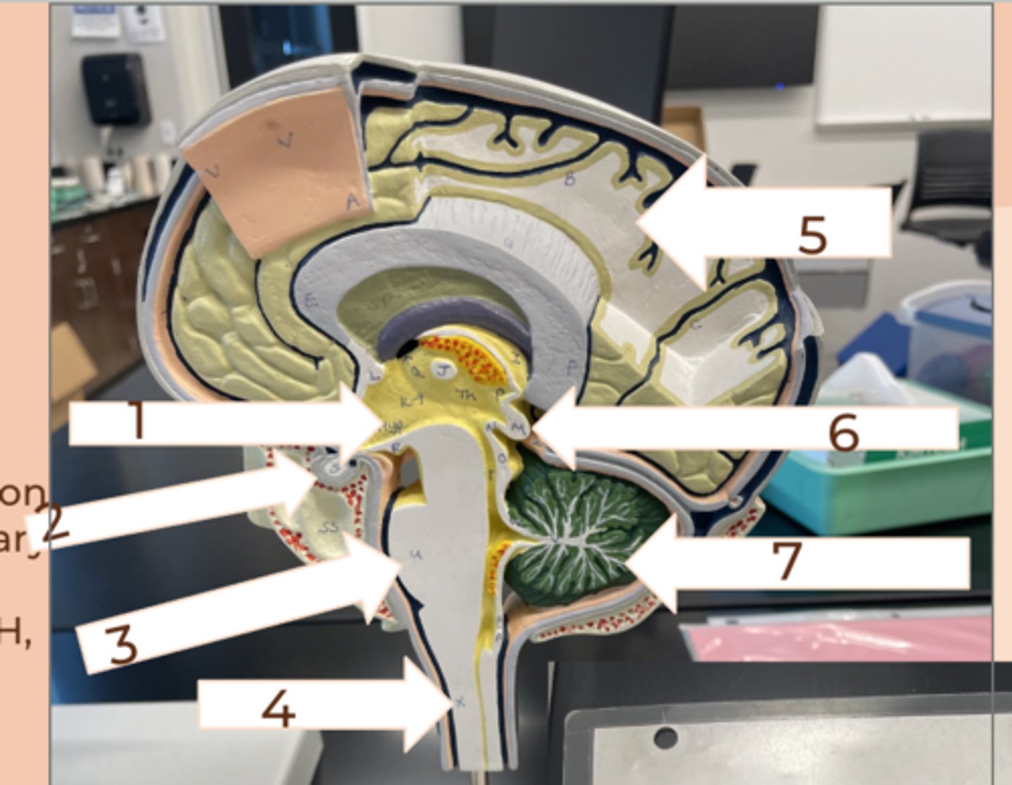
medulla oblongata
#4
• autonomic function: swallowing, breathing, digestion, heart rate
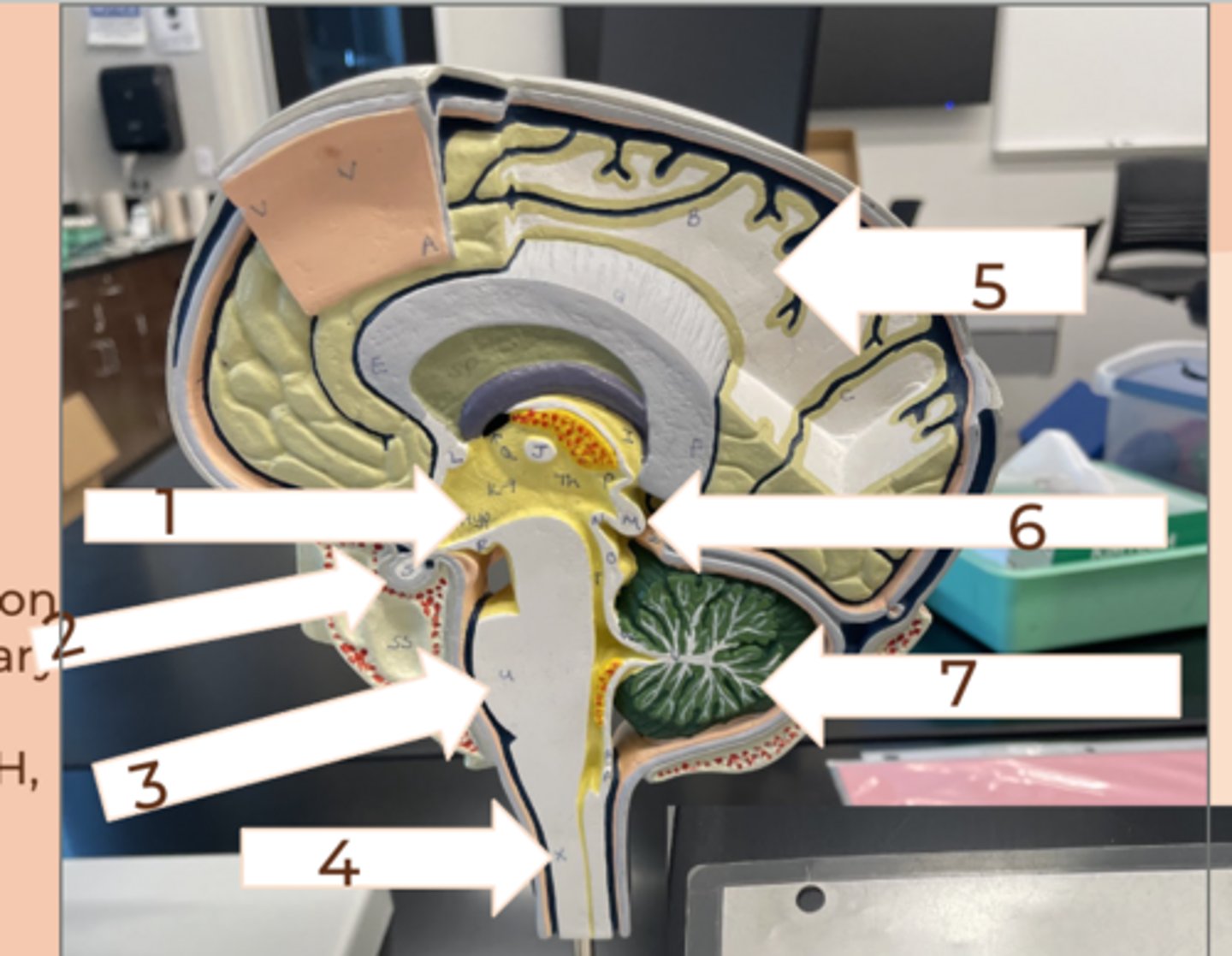
cerebrun
#5
• largest region of brain divided into lobes
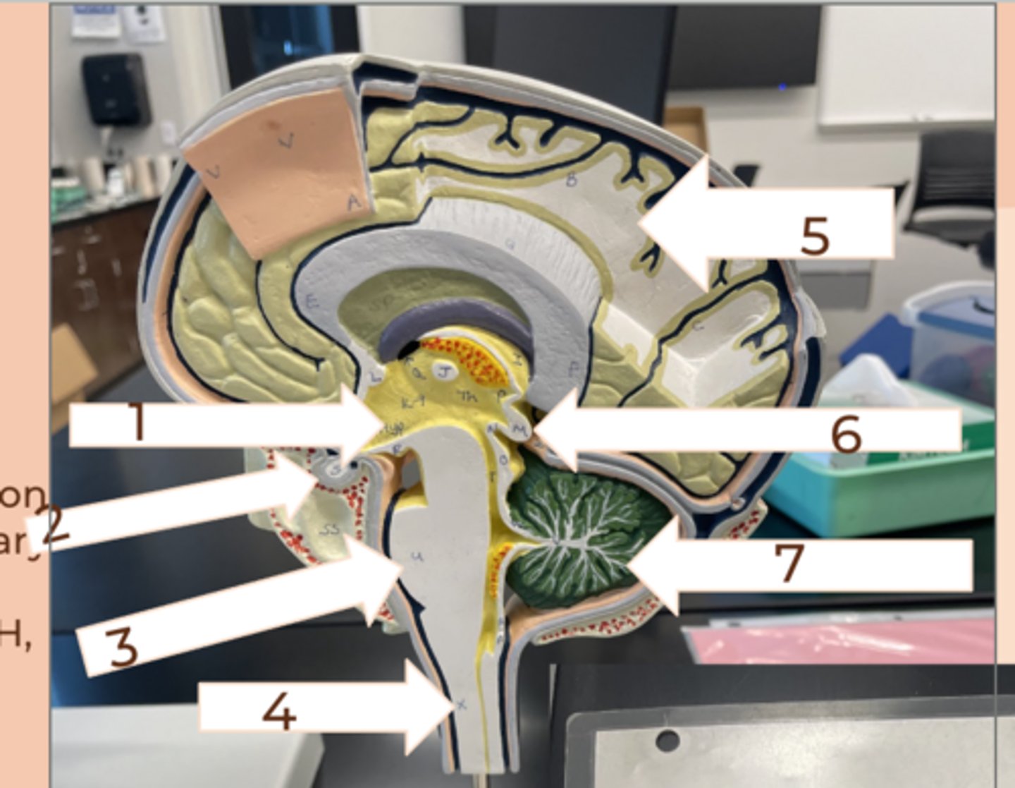
pineal gland
#6
• releases melatonin
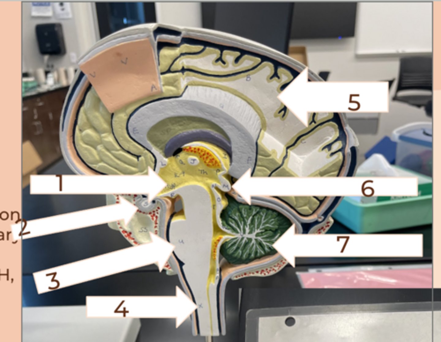
cerebellum
#7
• coordinates movement and balance
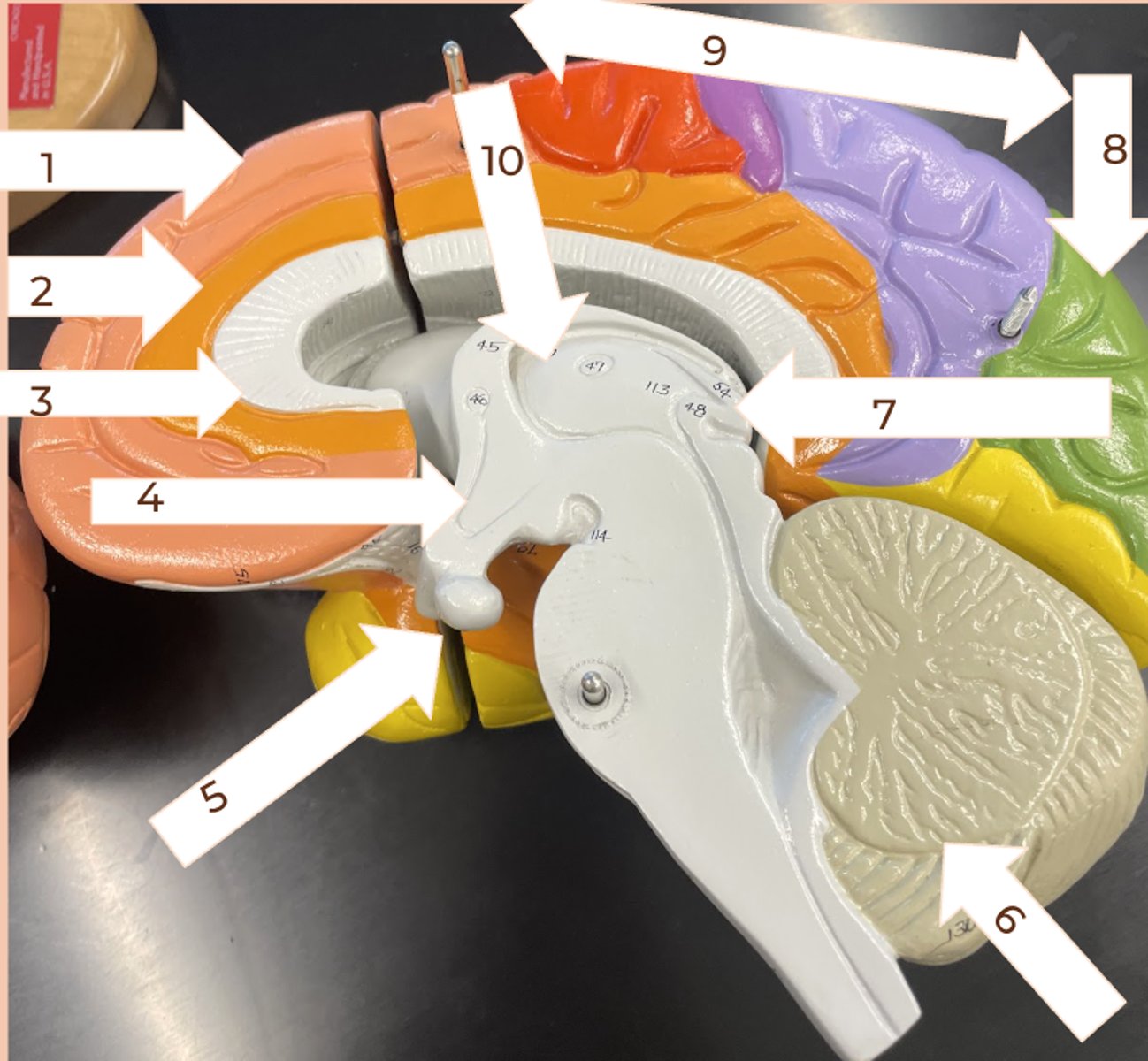
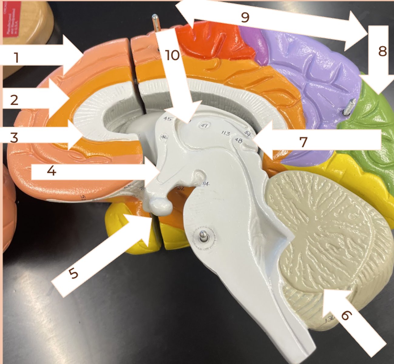
frontal lobe
#1
• primary motor area and though center
• speech
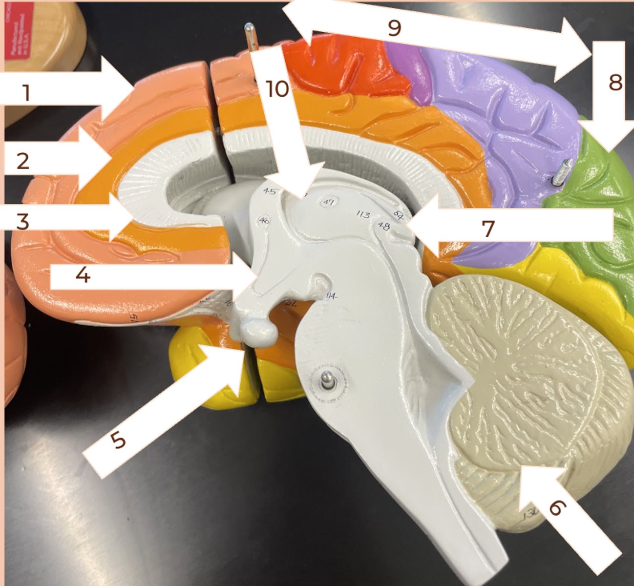
Limbic Lobe
#2
• governs emotions
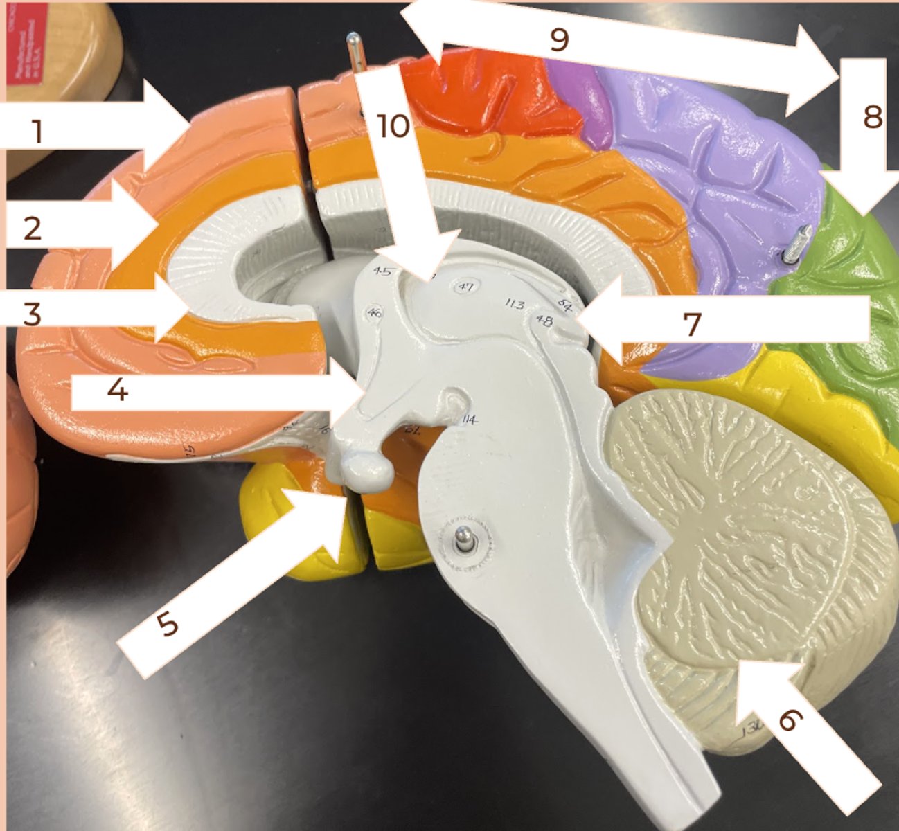
Corpus collosum
#3
• along internal midline of cerebrum where 2 hemispheres connect; relay center
hypothalamus
#4
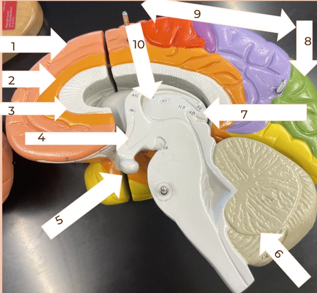
Pituitary gland
#5
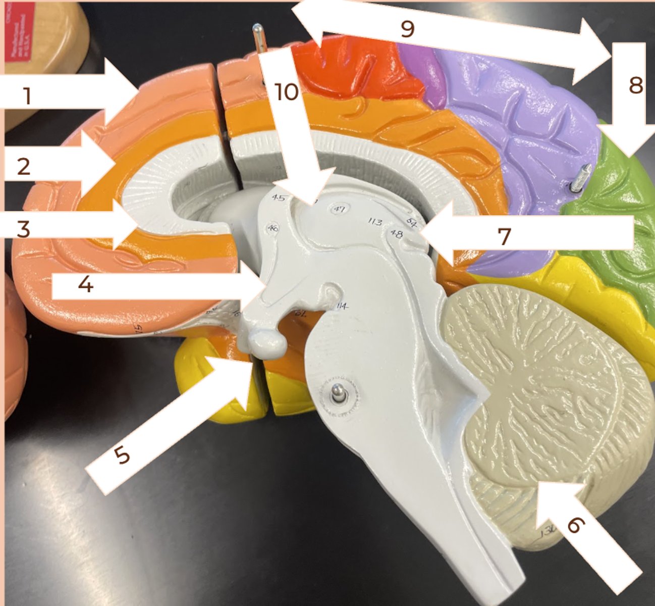
Cerebellum
#6
• coordinates movement and balance
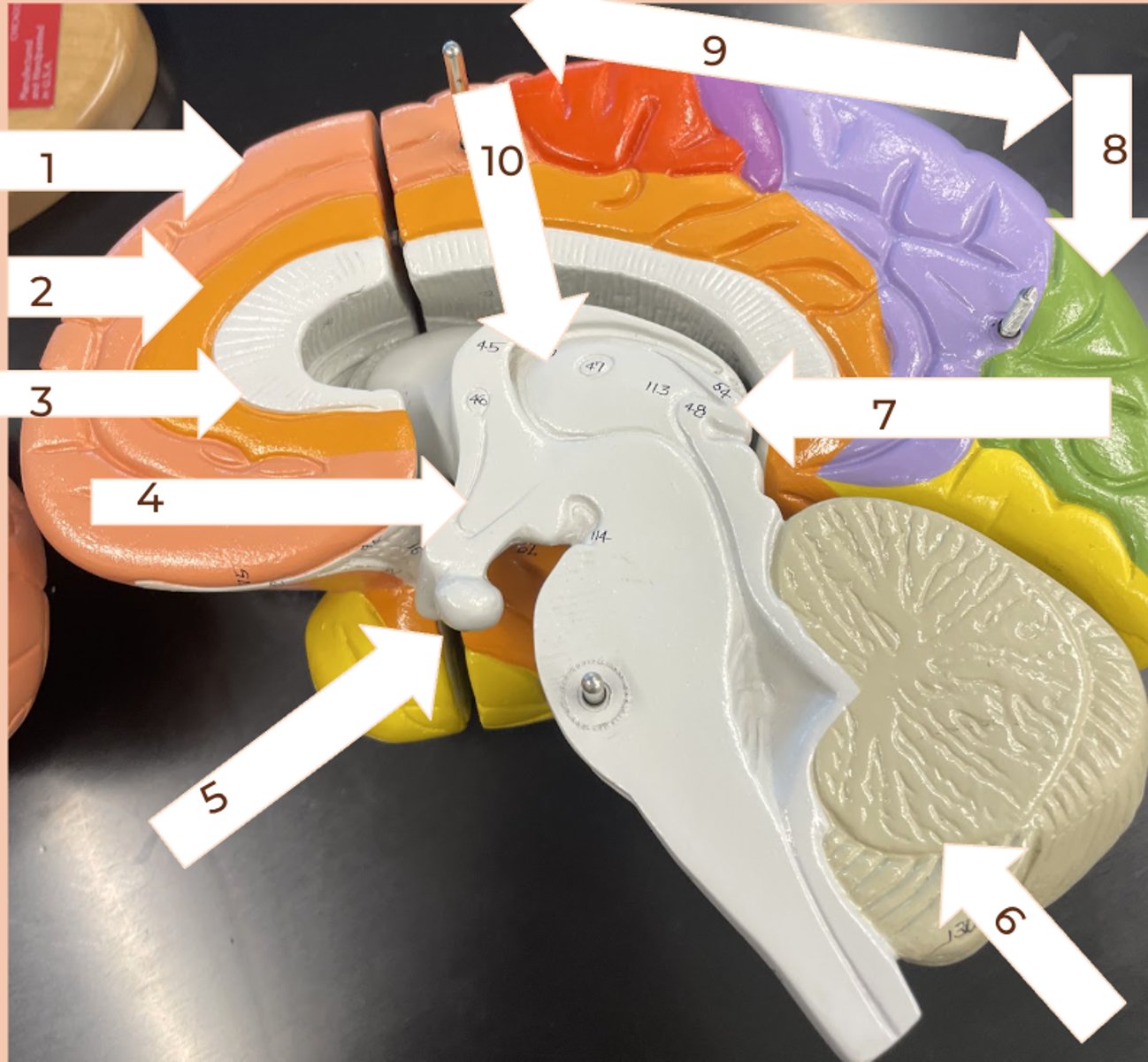
Pineal Gland
#7
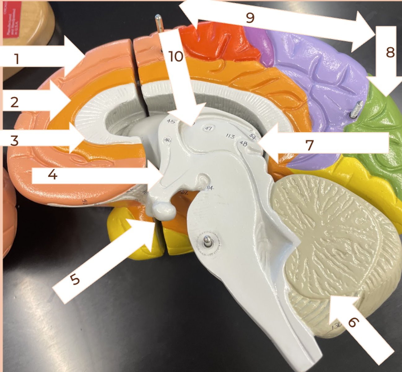
Occipital lobe
#8
• vision
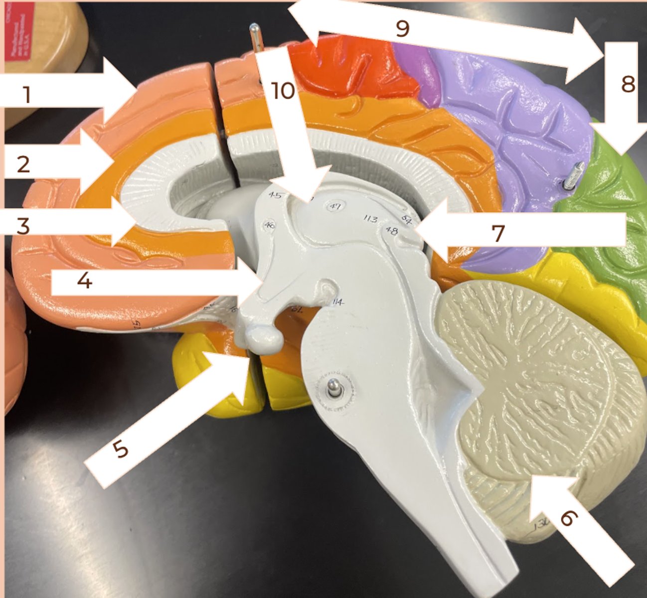
Parietal Lobe
#9
• primary sensory area for speech, reading, taste, touch
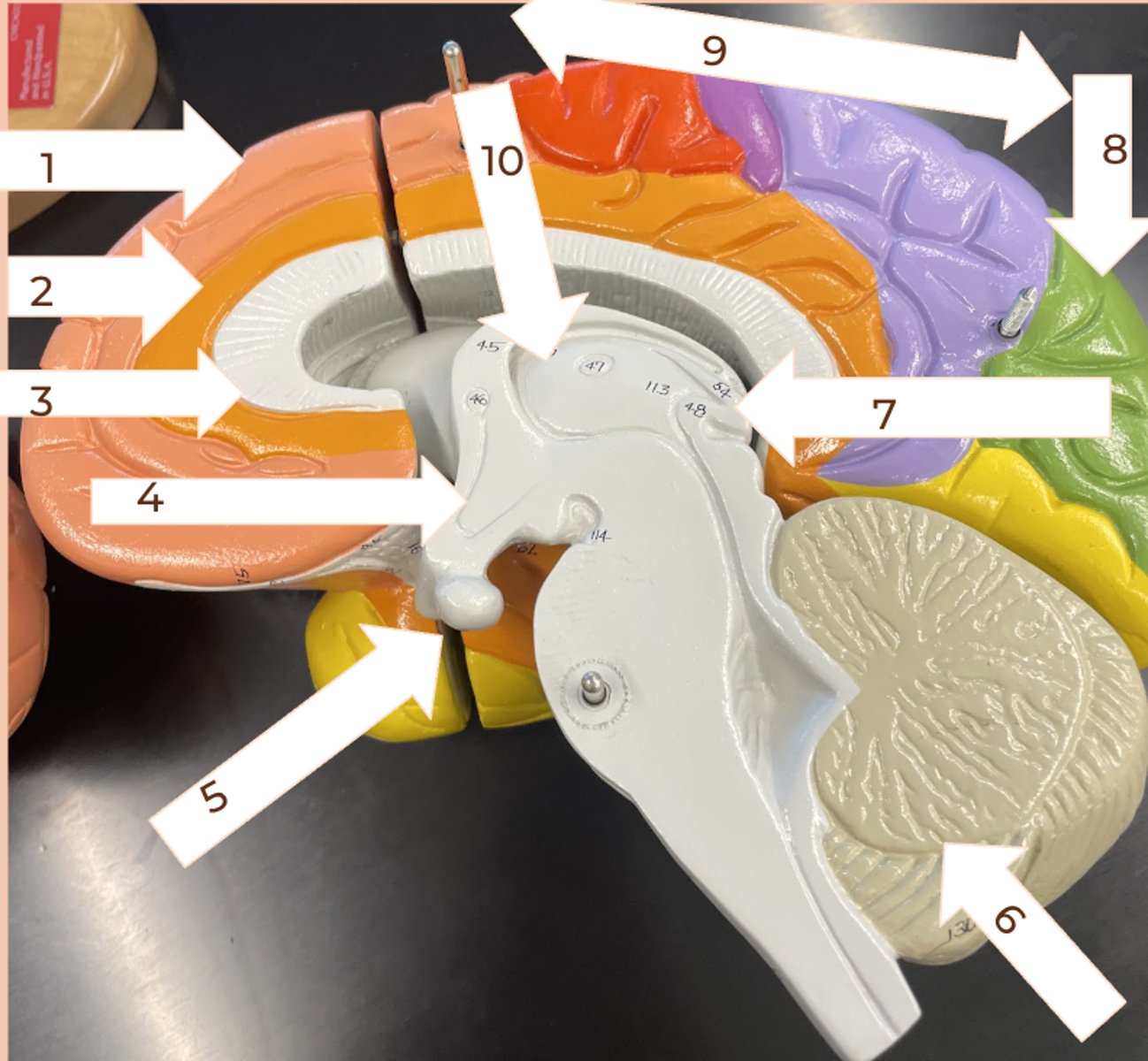
Thalamus
#10
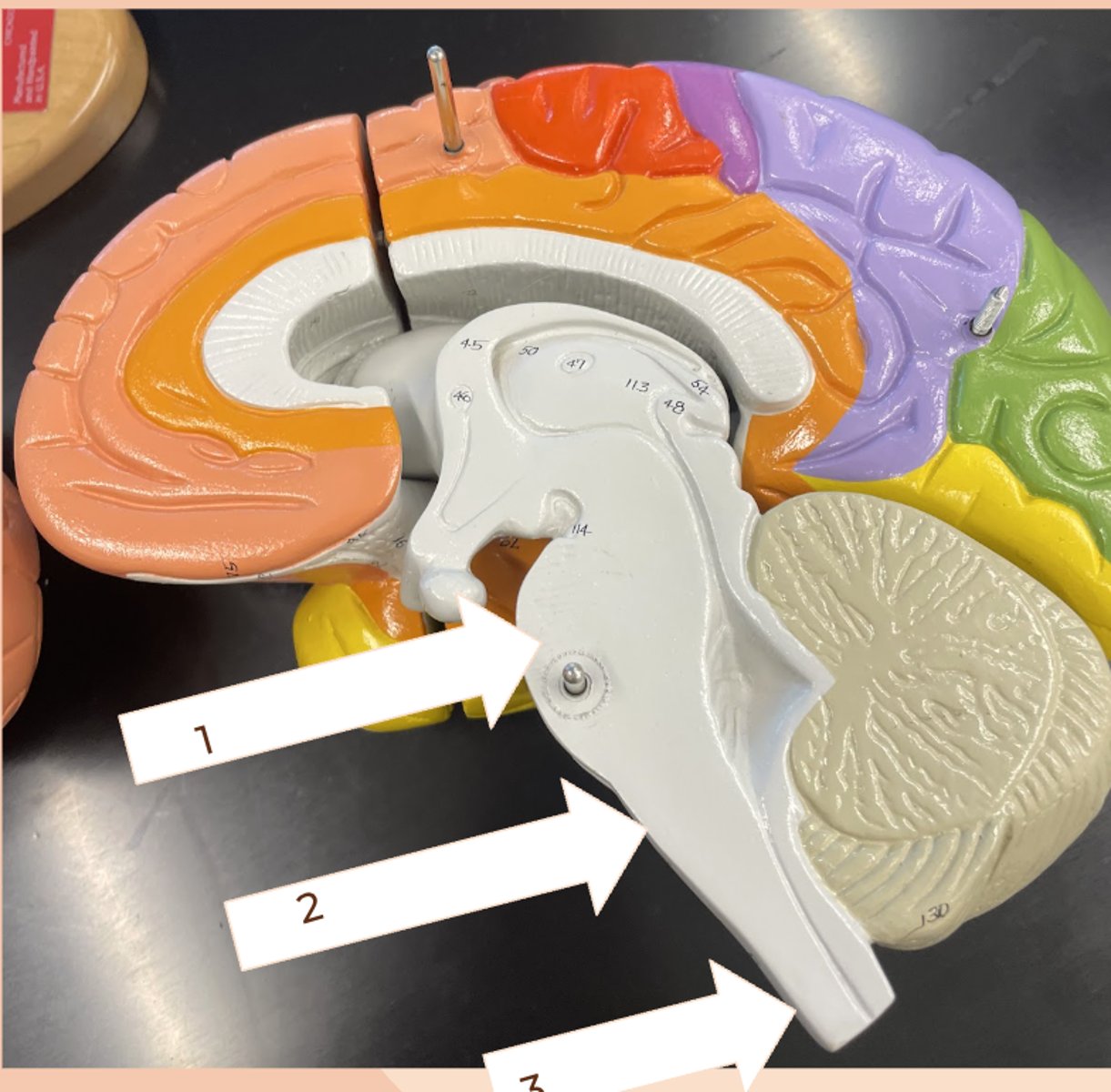
Brainstem
1) pons
2) medulla oblongata
3) spinal cord
fovea
#1
• center of visual field
• densest concentration of cones
• see the sharpest
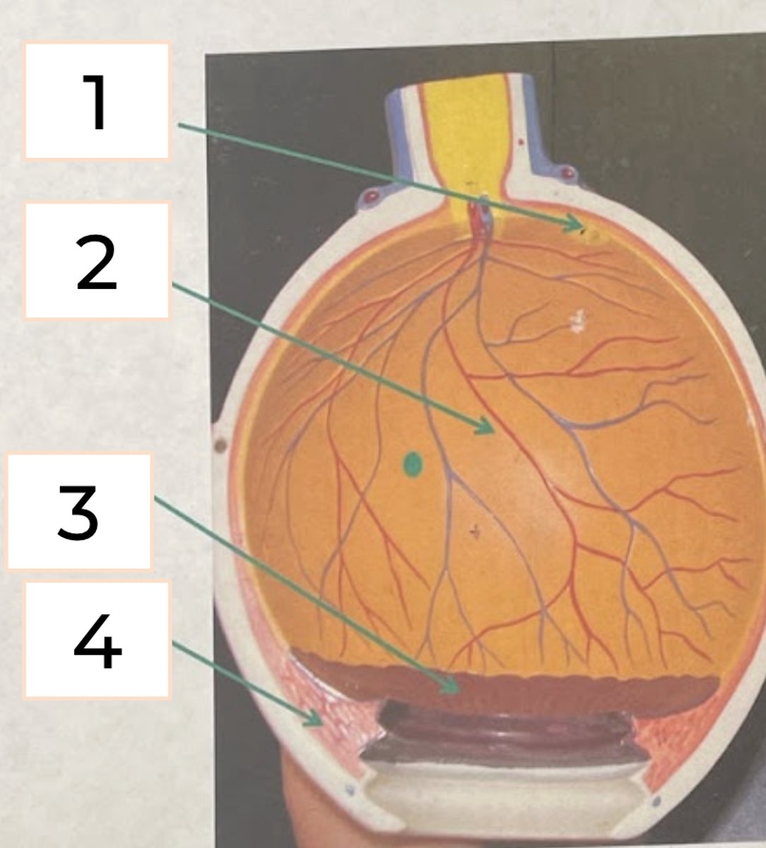
retina
#2
• innermost layer of eyeball
• has cones and rods
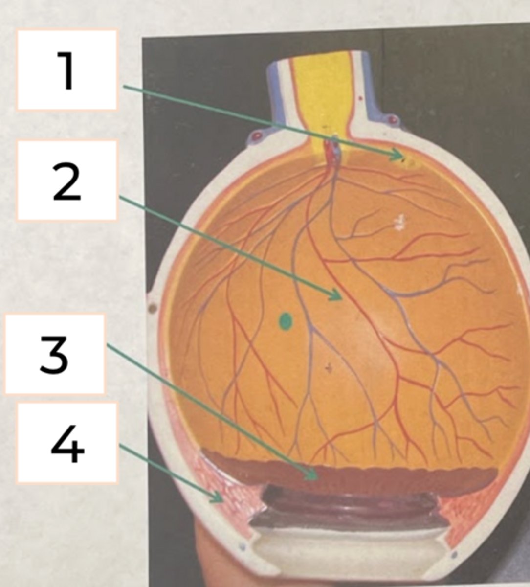
ciliary bodies
#3
• produces clear water fluid that fills the space between the cornea and the lens
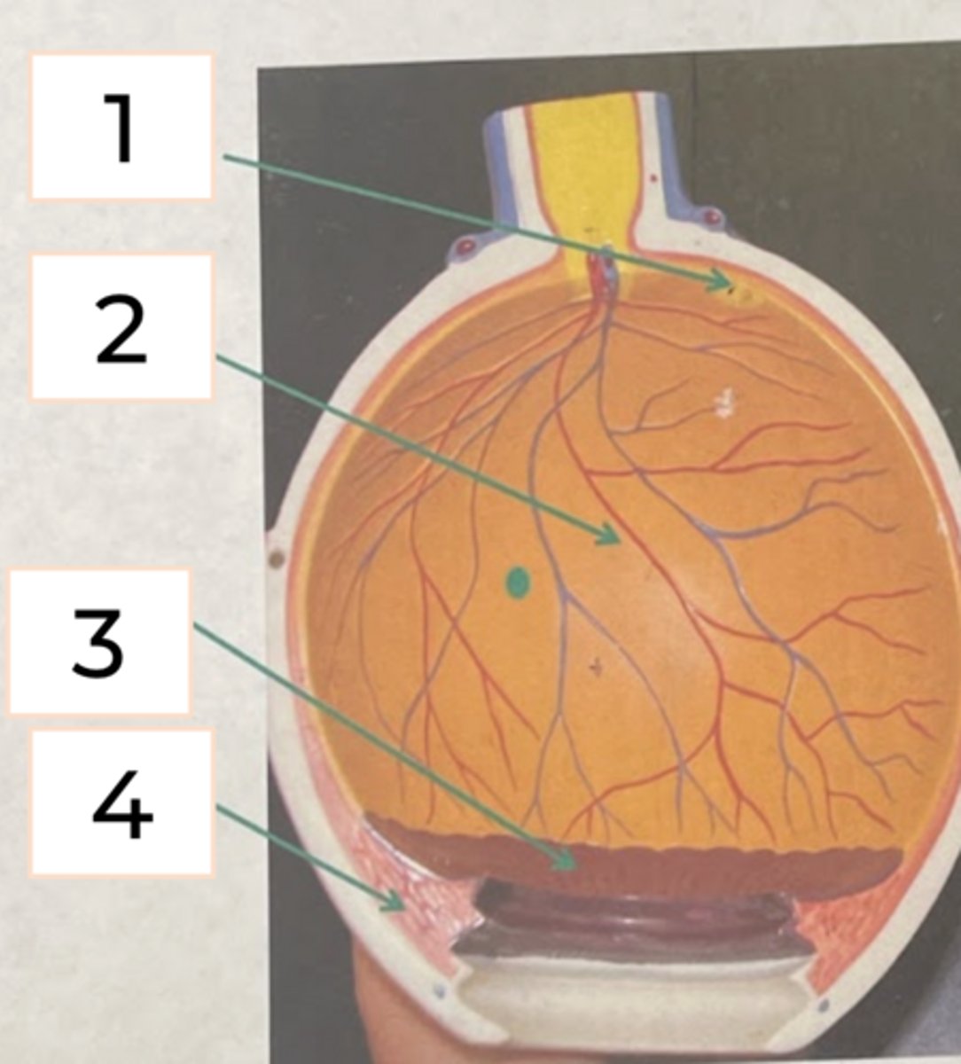
Choroid
• Pigmented region of the eye between the retina and lens
• responsible for "night shine" in nocturnal mammals
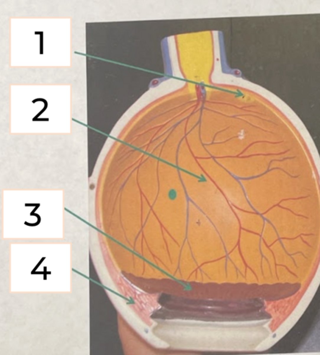
Optic nerve
#5
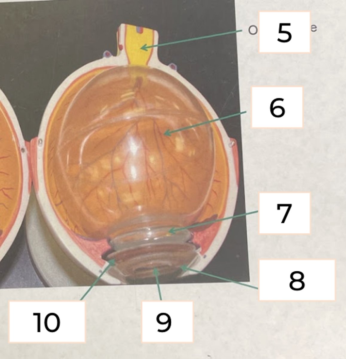
Vitreous Humor
#6
• jellylike material that occupies the area behindthe lens; fills up most of eye
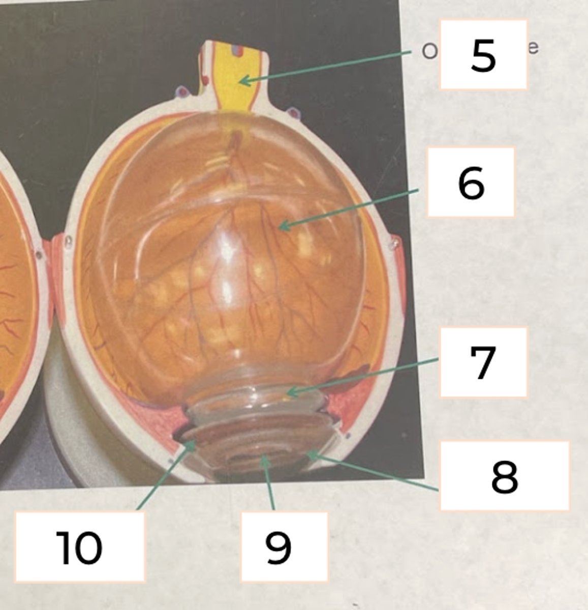
Lens
#7
• hardened structure located behid the cornea that helps focus light
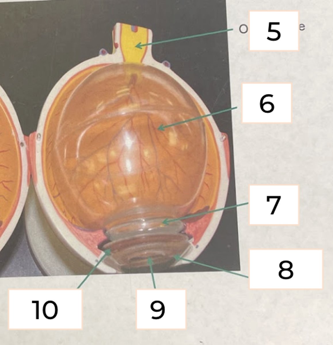
Cornea
#8
• front of the eye where sclera is transparent
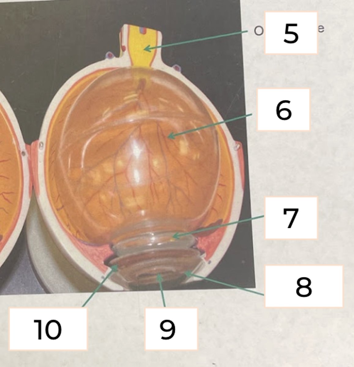
Pupil
#9
• hole in the center of iris
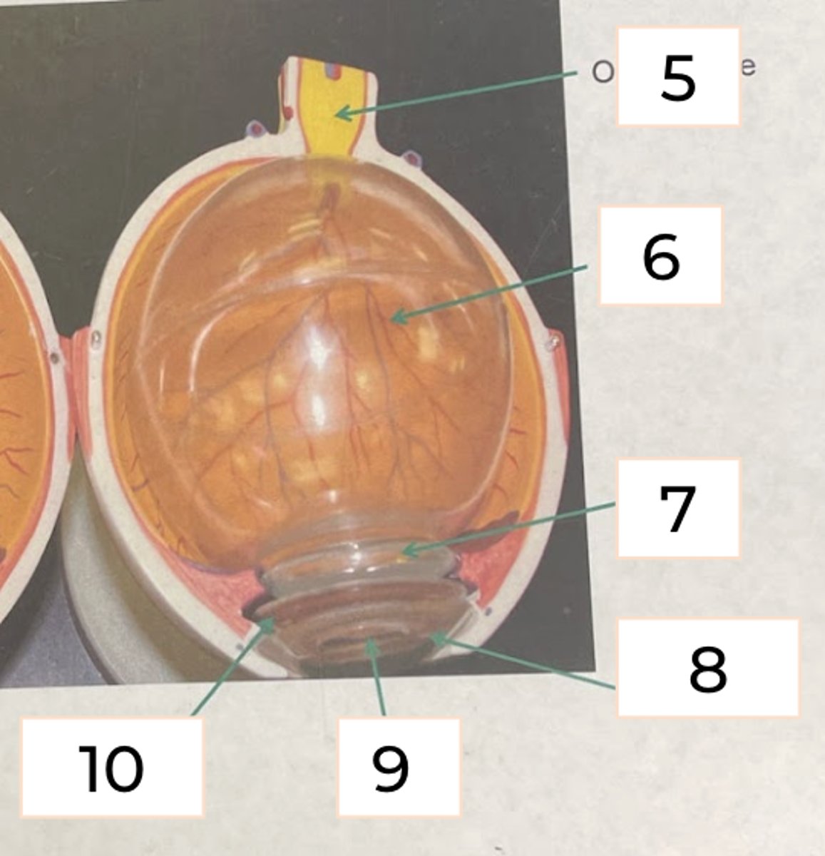
Iris
#10
• Pigmented outer layer of the eye which surrounds the pupil
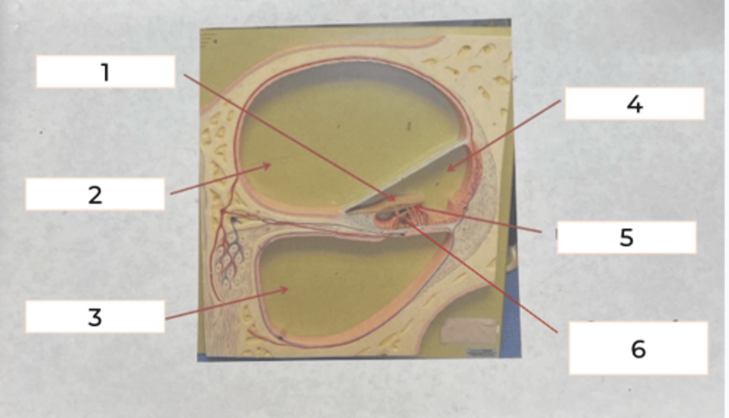
Cochlea
• coiled, snail-shaped structure which contains hair cells that are used for the detection of sound
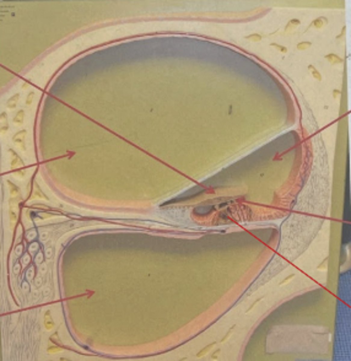
tectorial membrane
#1
• plays a role in how we receive sound
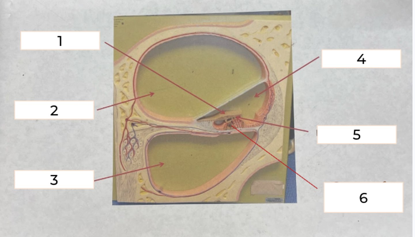
vestibular canal
#2
• upper canal of the cochlea
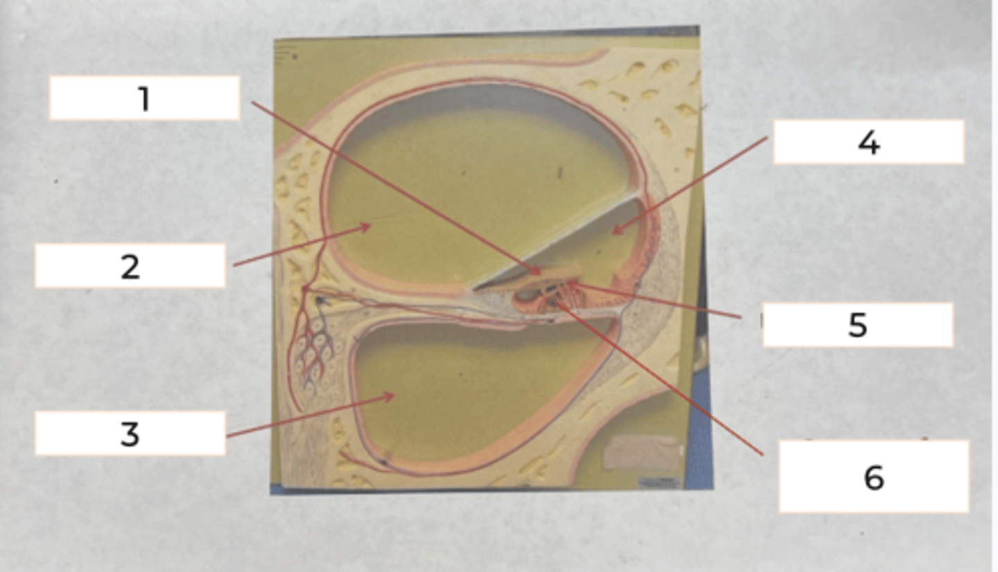
tympanic canal
#3
• lower canal
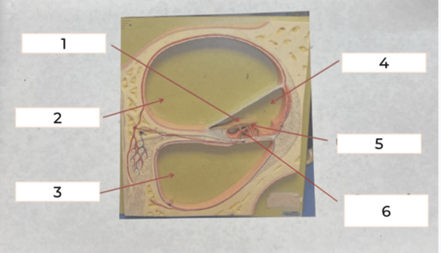
cochlear duct
#4
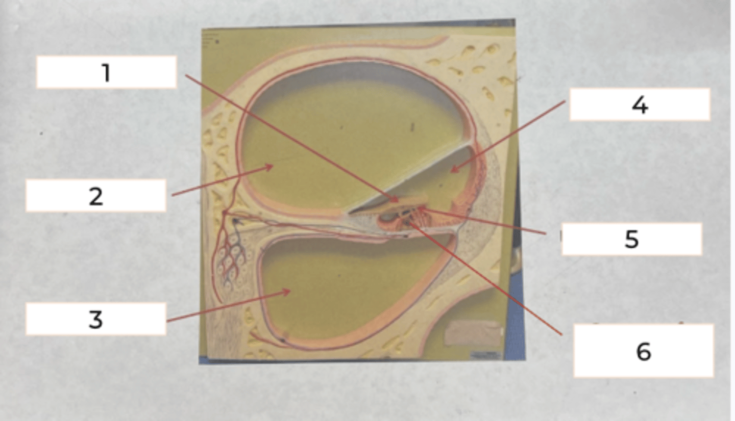
hair cells
#5
long hairs = low pitch
short hairs = high pitch
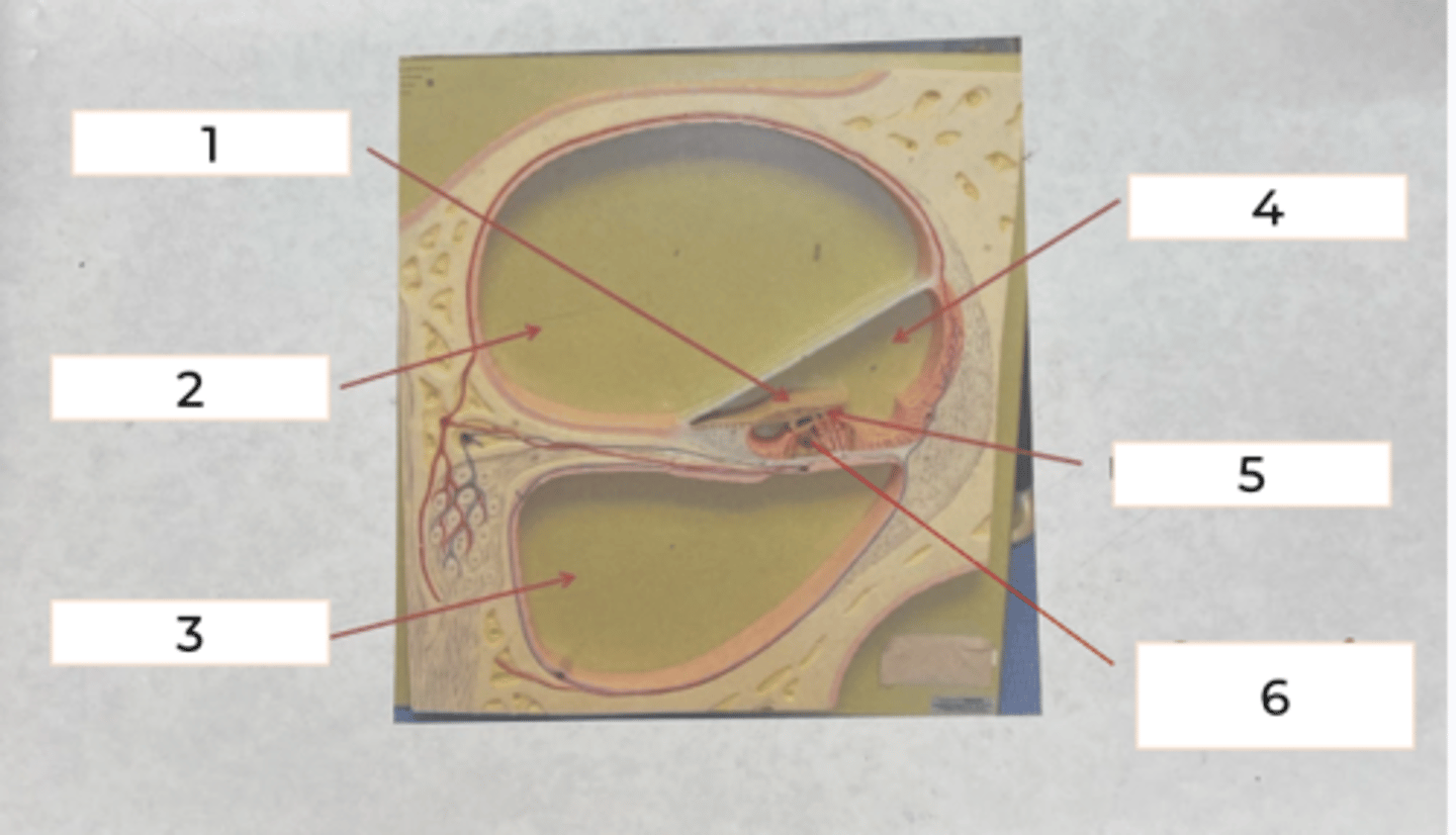
organ of corti
• #6
contains receptor cells and hair cels
ear
outer, middle, inner
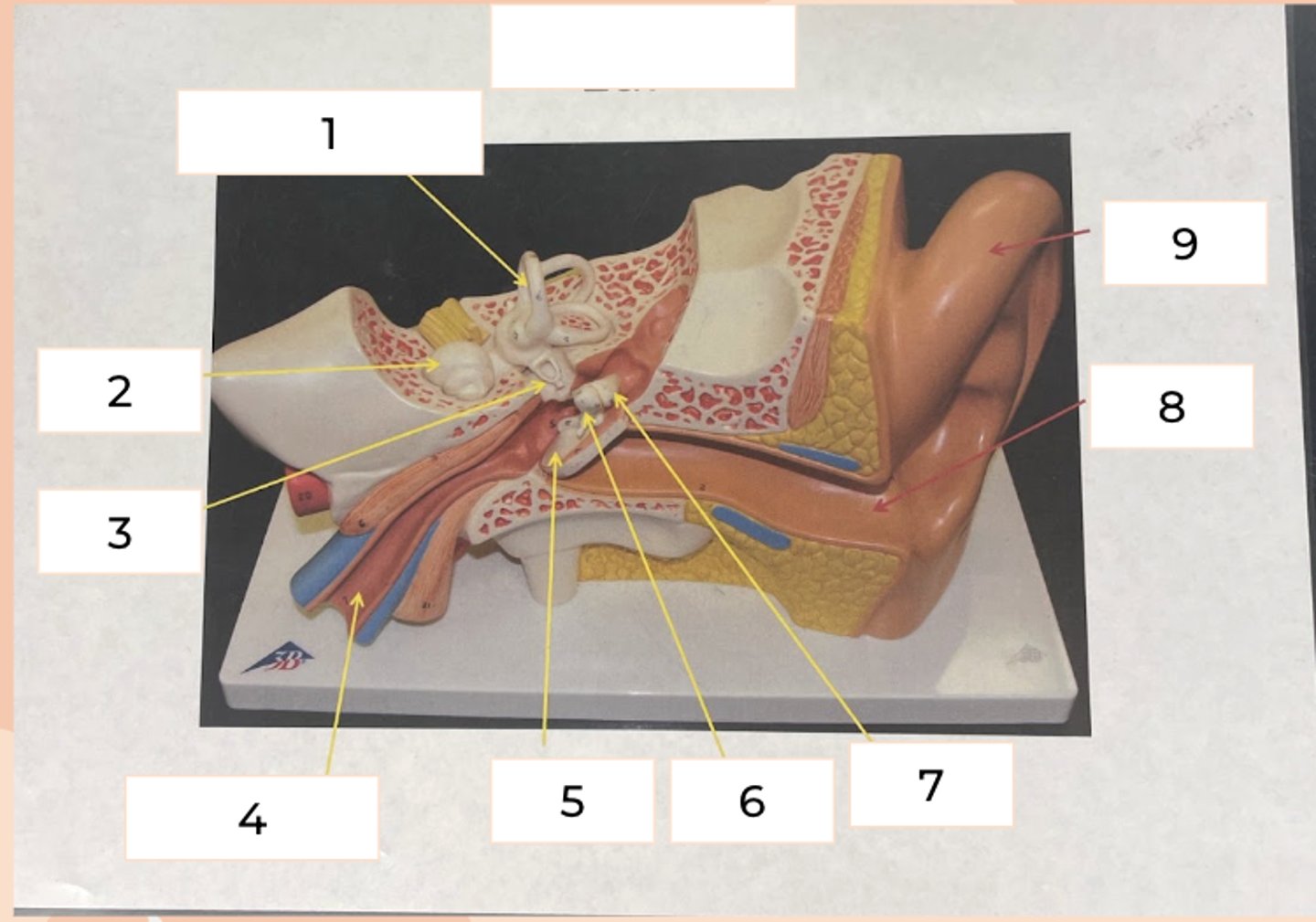
semicircular canals
#1
series of three structures in 3 spatial planes and detect position of head
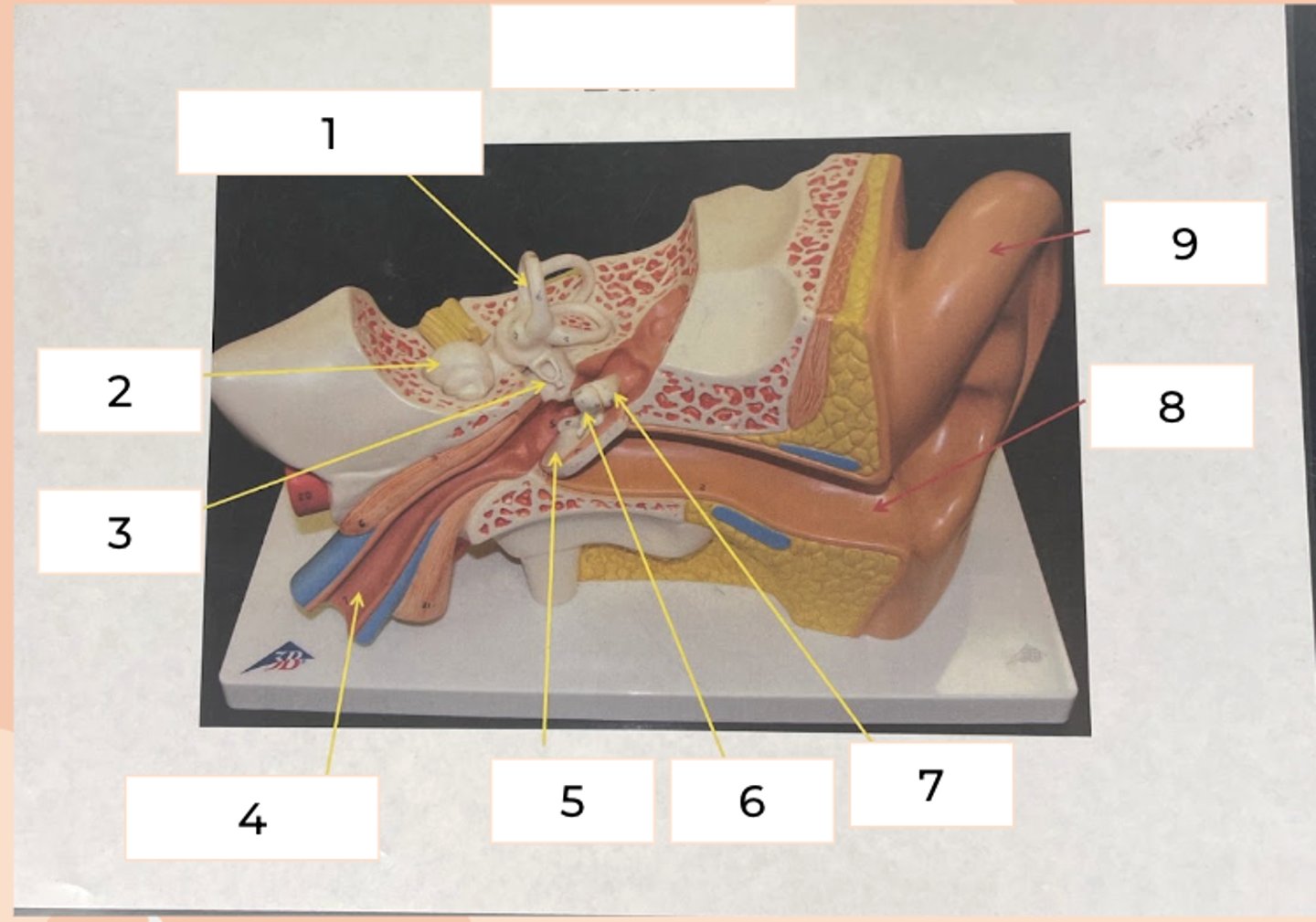
Cochlea
#2
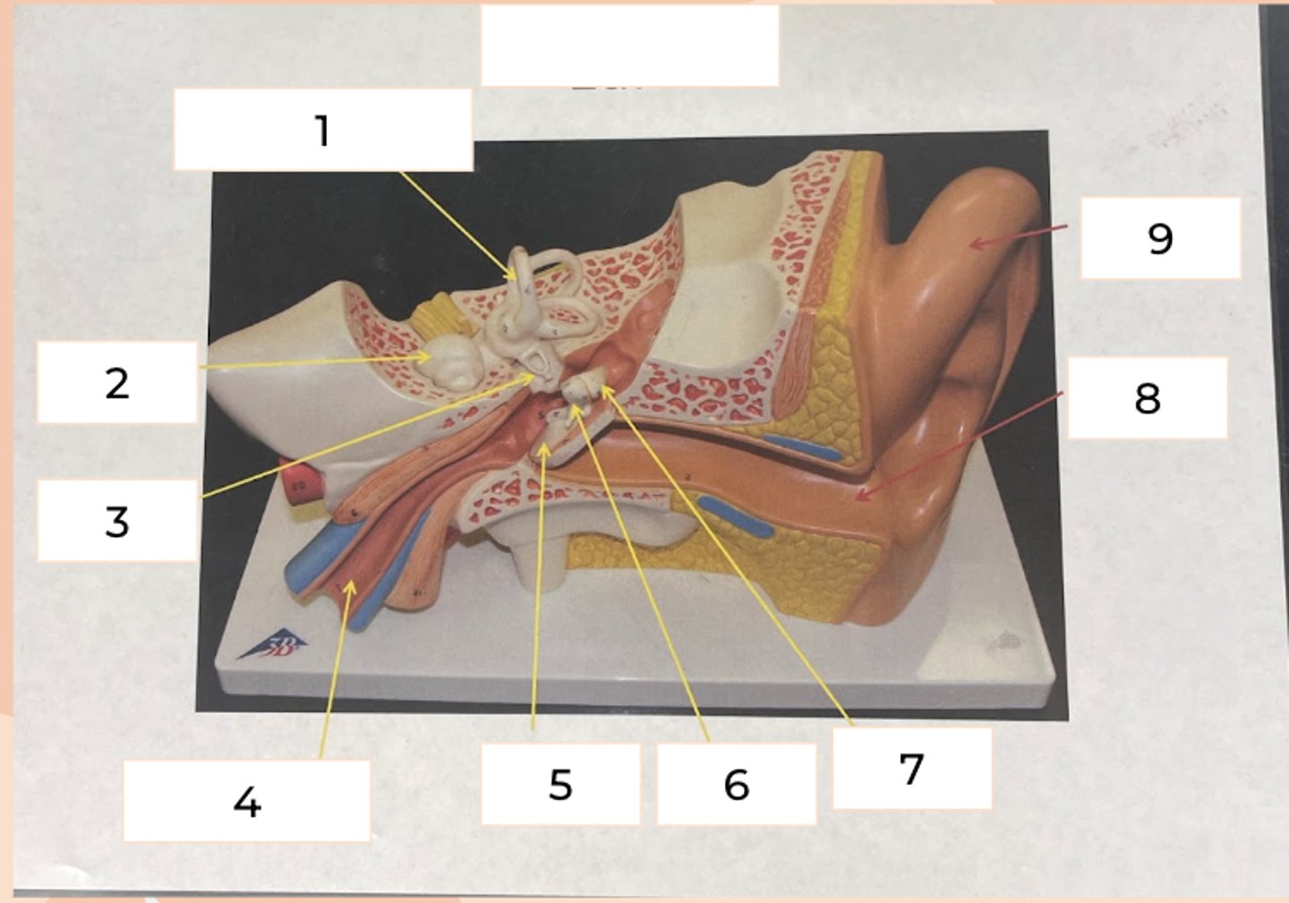
Stirrup
#3
• last ossicle; attaches to oval window
eustachian tube
#4
• connects middle ear to back of throat
• when open, equalizes air pressure of middle ear with atmosphere (popping)
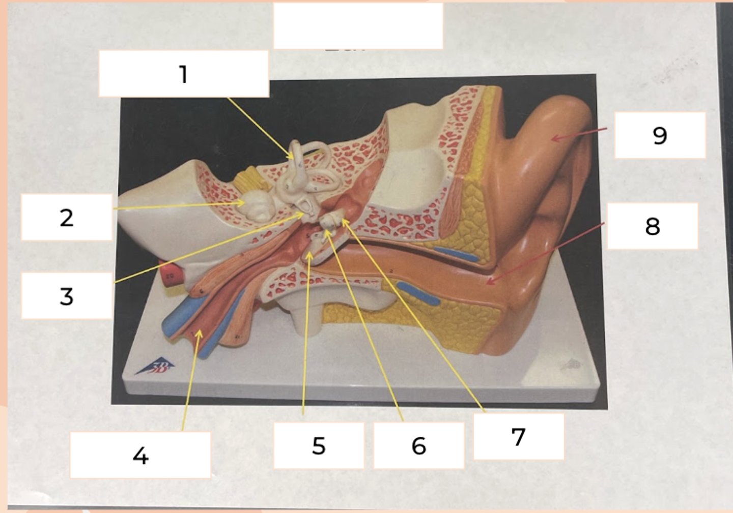
Eardrum
#5
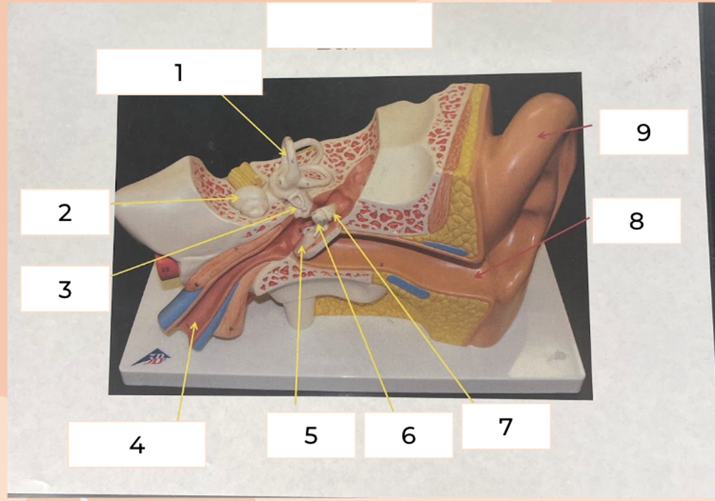
hammer
#6
first ossicle
directly attaches to eardrum