J CM Derm pt 2
 Call Kai
 Learn
Learn Practice Test
 Spaced Repetition
 Match
Match Flashcards
 Knowt Play
1/177

There's no tags or description
Looks like no tags are added yet.
Name | Mastery | Learn | Test | Matching | Spaced | Call with Kai | Chat |
|---|
No analytics yet
Send a link to your students to track their progress
178 Terms
6 classic rashes of childhood
rubeola 9measles)
scarlet fever
rubella (german measles)
4th disease (duke’s disease)
5th disease (erythema infectiosum)
roseola infantum
descending rash with a prodrome of cough, coryza, and conjunctivitis 2n1s
rubeola (measles)
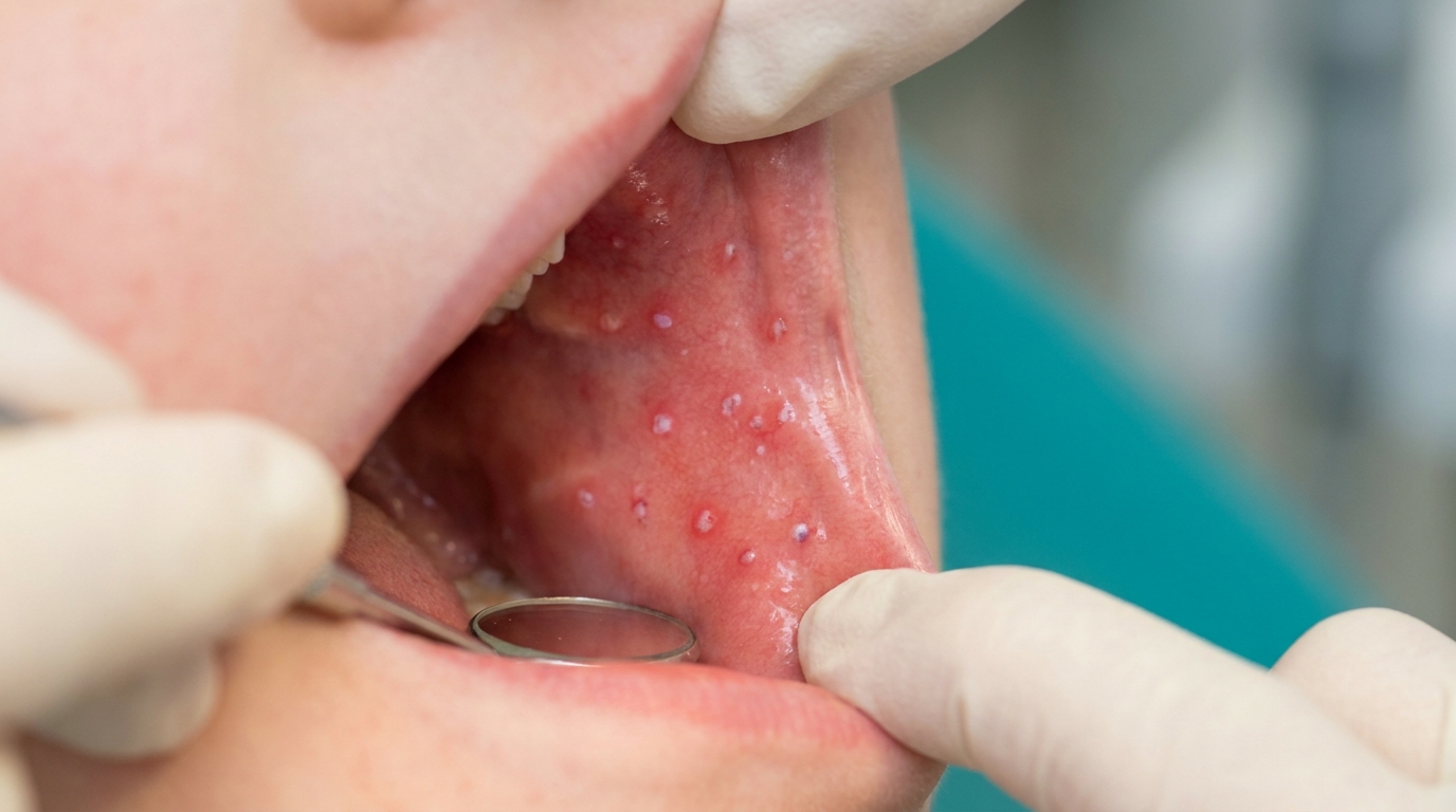
koplik spots 2n1s
rubeola (measles)
(pearly white elevated spots on erythematous background opposite to molars on buccal mucosa)
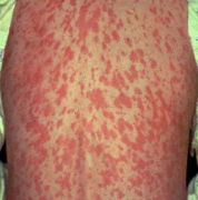
rubeola (measles) cause, prodrome, sx*
cause: morbillivirus
prodrome: cough, coryza (runny nose), conjunctivitis
blanching erythematous rash (begins on face → extremities)
koplik spots
scarlet fever cause
group A strep
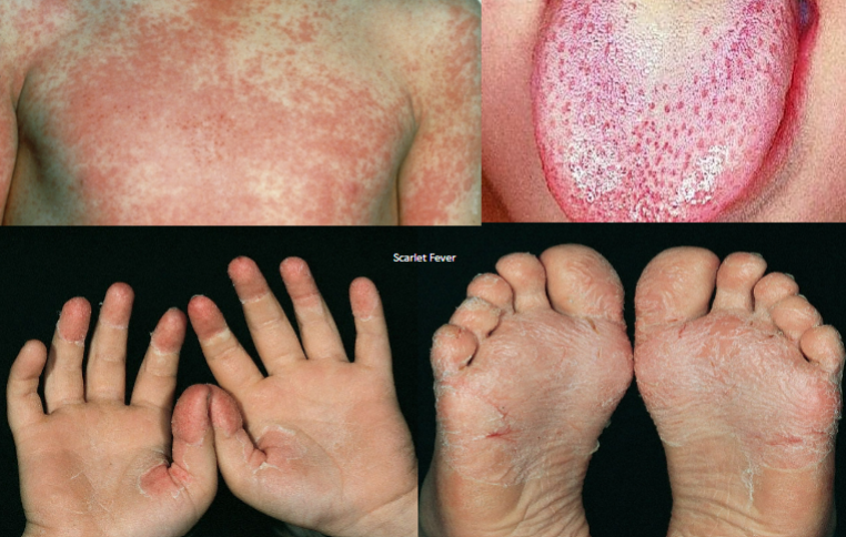
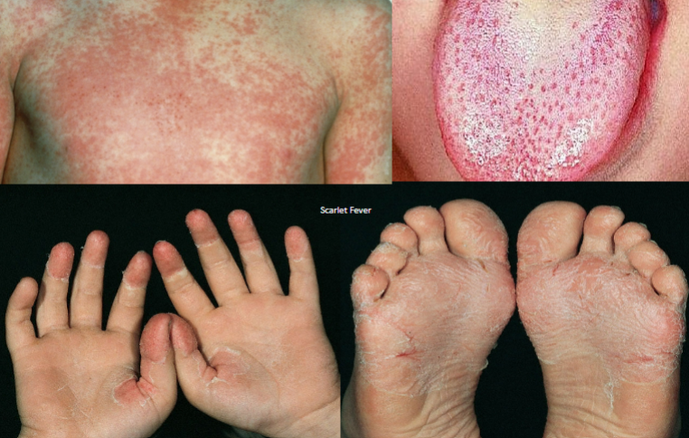
Scarlet Fever (Scarlatina) sx*
BLANCHING rash w/ erythematous macules that coalesce
sandpaper quality, starts on head/neck → trunk
strawberry tongue
pastia’s lines
circumoral pallor
skin desquamates (comes off in scales/flakes)
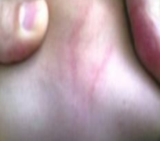
pastia’s lines definition
skin creases in the axillae, elbows, and groin might develop lines that are brighter red than the rest of the rash
rubella (german measles) cause*
togavirus
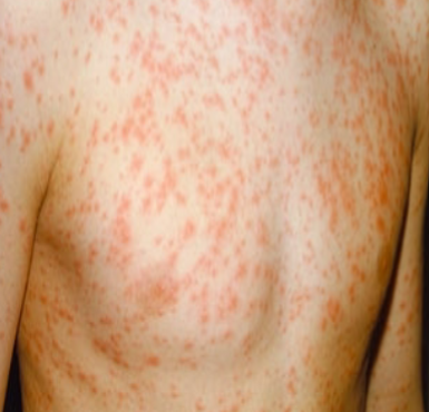
Rubella (German Measles) sx*
cervical and posterior auricular adenopathy
Forchheimer Spots → petechiae on palate
fever, rash, conjunctivitis occur simultaneously
rash starts face → down body (palpable petechiae)
duller in color than measles
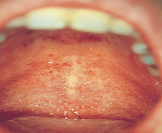
Forchheimer Spots
(rubella)
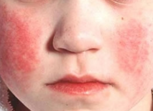
“slapped cheek” rash on face 2n1s
5th disese (erythema infectiosum)
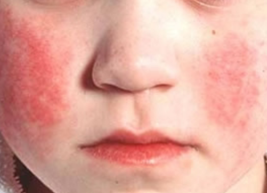
Fifth Disease (erythema infectiosum) cause*
human parvovirus (B19)
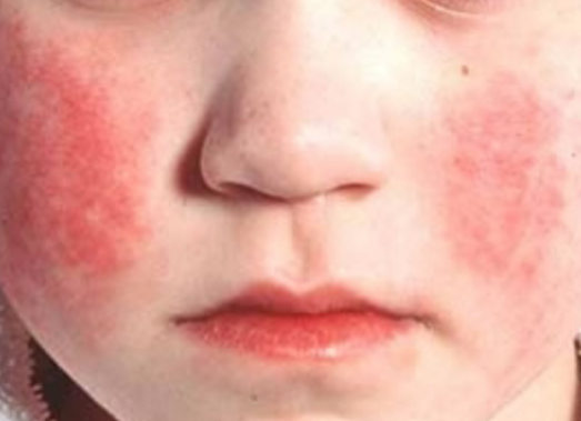
Fifth Disease (erythema infectiosum) presentation*
irritability and high fever first → slapped cheek rash and lacy rash on extremities
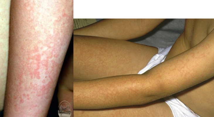
reticulating rash in 5th disease
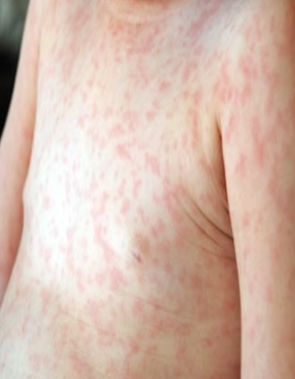
Roseola Infantum (exanthem subitum) cause*
HHV 6 and 7
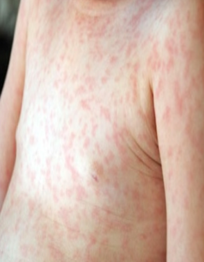
Roseola Infantum (exanthem subitum) sx*
high fever in otherwise well appearing child
rash appears as fever goes away, starts on neck/trunk → extremities
group A coxsackieviruses cause what?
herpangina
hand-foot-and-mouth disease
(picornaviridae family, genus enterovirus)
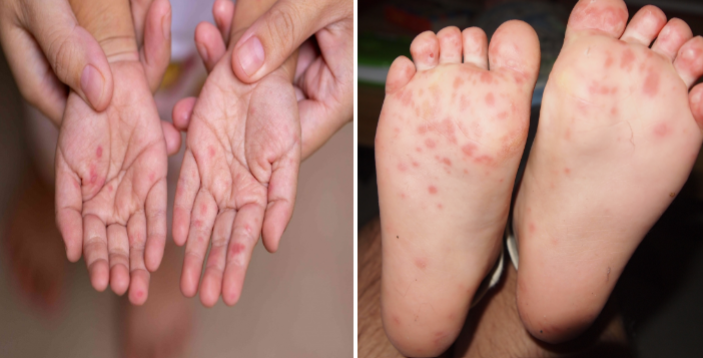
hand-foot-and-mouth (HFM) disease
severe pharyngitis, febrile, will not eat
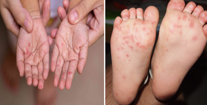
coxsackie virus disease
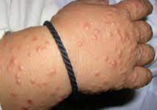
Gianotti-Crosti Syndrome (Papular acrodermatitis of childhood)
occurs after viral disease
self-limiting
lymphadenopathy in truncal area
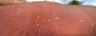
erythema toxicum
“baby acne”
harmless
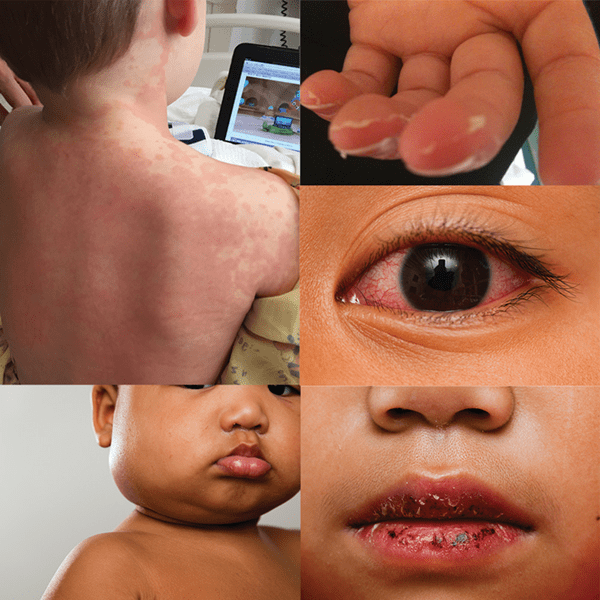
kawasaki disease sx
AKA mucocutaneous lymph node syndrome
changes to mouth/lips (cracked/erythematous lips, strawberry tongue, circumoral pallor)
polymorphous rash
conjunctivitis
erythema of palms/soles
unilateral cervical lymphadenopathy
kawasaki disease complications/sequelae
myocarditis → cardiomyopathy
complication = aseptic meningitis
tx for kawasaki’s disease*
IVIG
high dose steroids
ASA
hair growth cycle
Anagen → growing phase (2-4 years)
The normal hair shaft grows about 1 cm/month
Catagen → transitional phase (2-4 months)
Telogen → resting phase (2-4 weeks)
It is normal to lose up to ~100 hairs a day
Telogen effluvium definition and cause
stress causes hair roots to be pushed prematurely into the resting phase
cause: childbirth, stress, surgery
Alopecia Areata and types
starting early in life (children and young adults) = less likely to get better
occurs in sharply defined areas
patterned type (most common): 1+ patches → regrowth
Diffuse type (alopecia totalis) → involves entire scalp → poorer chance for permanent regrowth
Complete type (alopecia universalis) → involves entire body → poorer chance for permanent regrowth
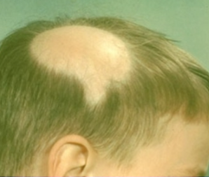
Alopecia Areata skin signs*
Non-scarring
No S/Ss of inflammation
No scale
Short “exclamation mark” hairs seen in area
Positive hair pull test
Fine “vellus” hairs seen when regrowth starts, might be pigmented
Peach Fuzz
nail pitting
→ clinical diagnosis
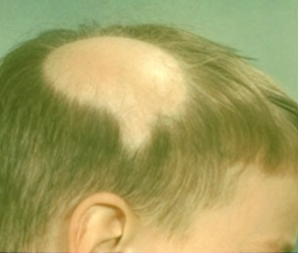
poorer prognostic factors in alopecia areata*
Onset in childhood
alopecia totalis or alopecia universalis
Duration >1 year
Ophiasis → Band-like involvement of the peripheral temporal and occipital scalp
Nail involvement
Atopy
FH
Comorbid autoimmune disease (Cicatricial alopecia)
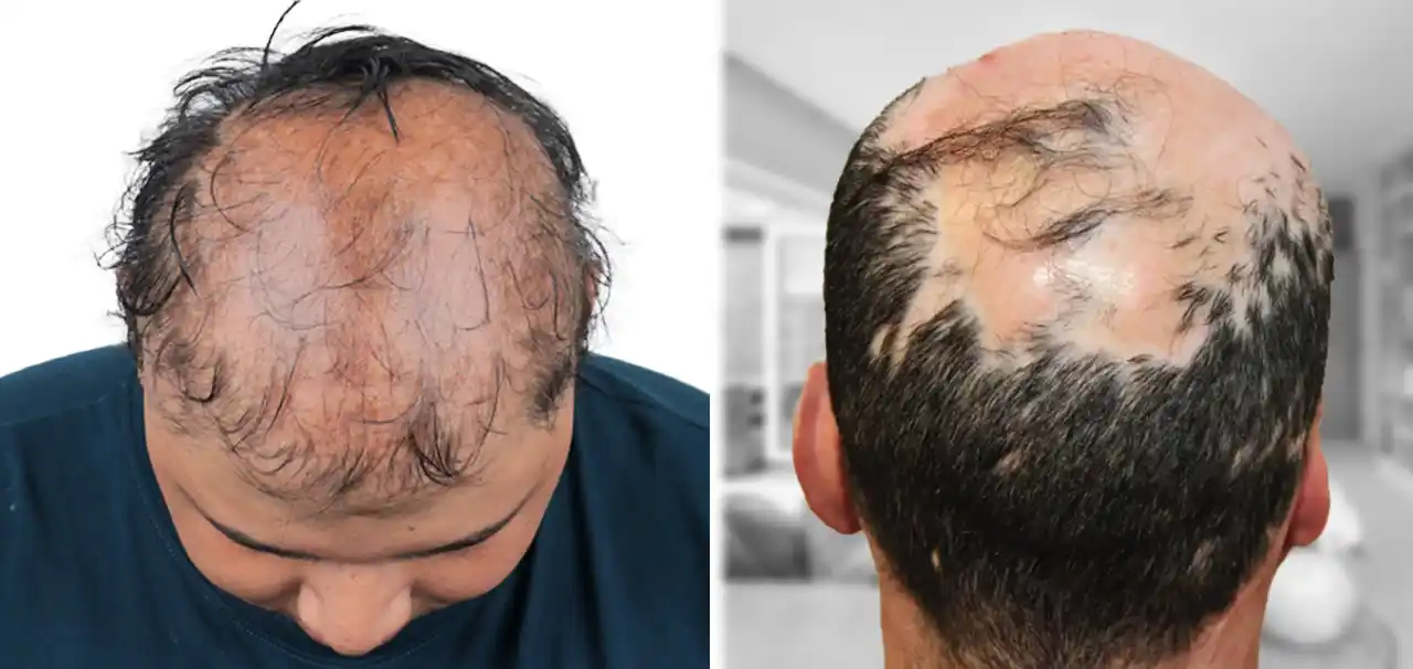
Cicatricial alopecia
scarring alopecia in discoid lupus causing total destruction of hair follicle
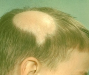
treatment for alopecia areata*
For small and solitary patches
No tx needed → Spontaneous regrowth
intralesional steroids / topical therapies
For large and numerous patches
Intralesional steroids
Minoxidil
Topical immune sensitizers
Systemic steroids x 3 months
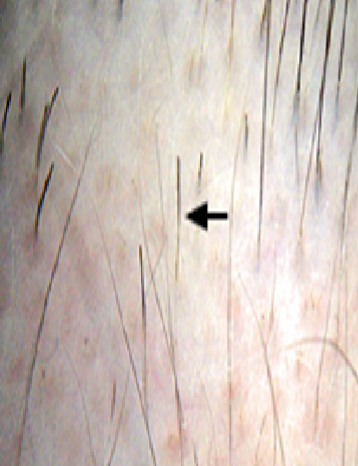
exclamation mark hairs
= alopecia areata
warts are the _ _ of the epidermis
benign lesion
verrucae → warts cause
cause: HPV
Vaccine exists for specific serotypes → 6, 11, 16, 18
transmission by simple contact
verrucae → warts types
verrucae vulgaris
verruca plantaris
verruca plana
condyloma acuminata
cervical dysplasia and neoplasia
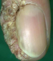
Verrucae Vulgaris
what is it
what might you find
location
common warts (HPV 1, 2, 4)
Flesh colored to grey-brown, hyperkeratotic papules/plaques w/ scale or filiform
+/- black dots (thrombosed capillaries)
hands, periungual skin, elbows/knees, face
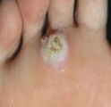
Verruca Plantaris
plantar warts (HPV 1, 2, 4)
plantar surfaces of feet
often painful
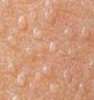
verruca plana*
flat warts (HPV 3, 10)
small (1-3), flat topped pink/tan/brown papules
forehead, legs, dorsal hands, chin, neck
condyloma acuminata*
genital warts, hpv 6 and 11
infx of anogenital skin and intra-oral issues
fleshy papules w smooth/velvety moist surfaces
can coalesce to form larger cauliflower-like mass
frequently recur after tx
low chance → malignancy
cervical dysplasia and neoplasia
HPV 16, 18, 31, 33, 45
can begin as warts that are NOT condylomata
assoc with squamous cell carcinoma of cervix, labia, penis, anus, oral cavity, tongue
bowenoid papulosis
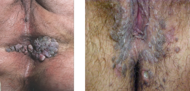
bowenoid papulosis definition
rare intermediate stage between warts caused by high risk serotypes (usually 16) and Bowen disease (squamous cell carcinoma in situ)
tx for verrucae (warts)
OTC (salicylic acid or liquid nitrogen), surgery
caustic acids (flat, plantar, common anogenital warts): cantharidin, tricholoracetic acid
chemo:
5-Fluorouracil (Efudex) → for flat warts
Imiquimod (Aldara) → for anogenital warts
Podophyllotoxin (Podofilox) → for anogenital warts
The body's immune system can destroy the warts w/o any tx; most will resolve spontaneously over weeks to months but harder if immunosuppressed
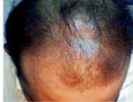
Androgenetic Alopecia (aka male pattern baldness) pop, sx*
most common form of alopecia
30-40% of M and F
non-scarring, starts at hairline and goes back
Androgenetic Alopecia (aka male pattern baldness) tx*
topical minoxidil
finasteride (more effective than minoxidil)
hair transplants
name for resting stage of hair growth cycle
telogen
if there’s stress → more hair in resting phase
telogen effluvium top 2 causes
childbirth
surgery
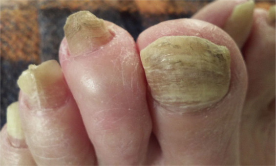
Onychomycosis (tinea unguium) definition
fungal infx of the finger/toe nail plates
15-20% of pop b/w 40-60 yo
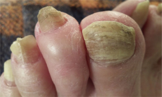
Onychomycosis (tinea unguium) etiology
dermatophytes → trichophyton rubrum and mentagrophytes most common (don’t have to know exact)
candida more often seen in fingernails
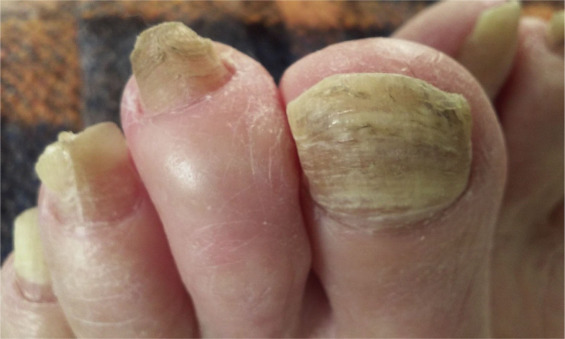
Onychomycosis (tinea unguium) diagnosis
examine all nails and skin to r/o other disease that can mimic onychomycosis
If all nails are affected, fungus is unlikely
Psoriasis, lichen planus, eczema, trauma, habitual, picking of cuticles
Scrape proximal nail bed for KOH/fungal culture
fingernails grow at a rate of ? per month, toenails grows at a rate of ? per month
3 mm/month
1 mm/monthy
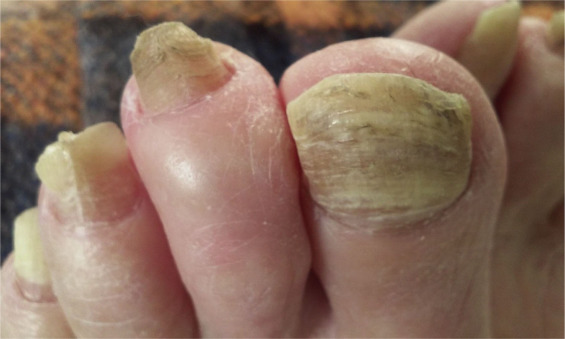
Onychomycosis (tinea unguium) tx*
what test
what is NOT effective
what IS effective
what is something to educate pts
check CBC/LFTs before, at 6 weeks and at end of tx
topical tx is NOT effective
oral therapy has highest success rate (50-80% effective but 15-20% relapse w/in 1 year)
^ terbinafine (lamisil), finger = 6 weeks, toenail = 12 weeks
itraconazole (fixed or pulse therapy)
nails do NOT appear clear for 6-12 mos after tx!!
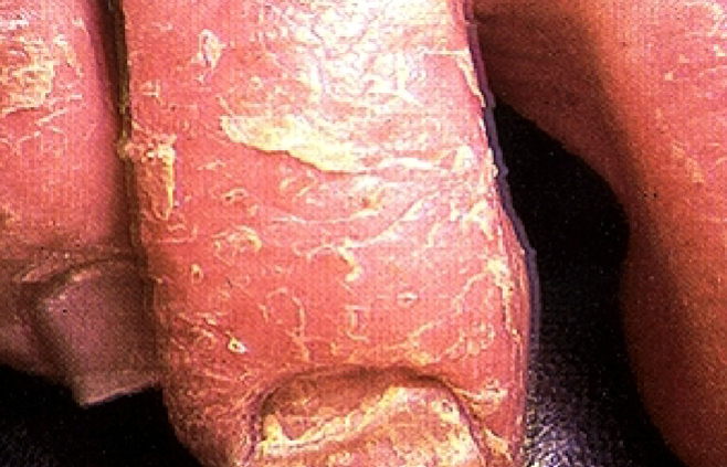
proximal subungal onychomycosis
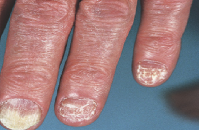
yeast onychomycosis (yeast)
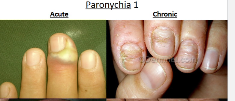
paronychia definition
Painful inflammation of the nail fold surrounding the nail plate
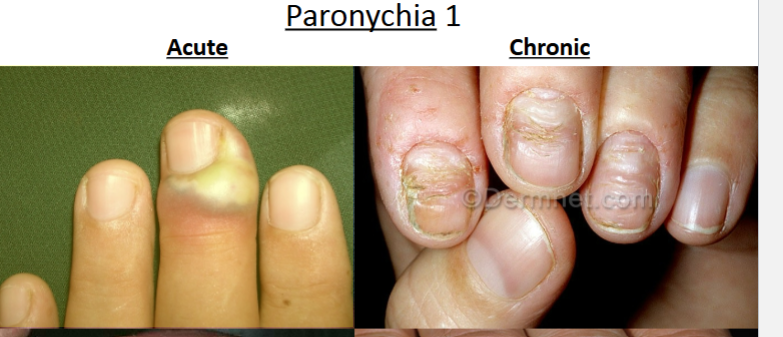
paronychia acute vs. chronic forms*
acute: bacterial (staph), precipitated by trauma or chemicals
chronic: fungal (candida), repeated wet activites, several fingers
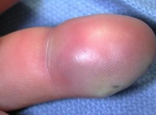
felon definition, cause, tx
bacterial infx of fleshy part of fingertip (distal)
cause: injury, extension of paronychia
tx: salt water soak, abx, i&d, do NOT incise on fleshy part, only lateral
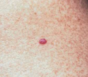
Cherry Angioma
adults, elderly
confused with spitz nevus of childhood
1-5 mm
red-purple papules
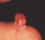
pyogenic granuloma
sudden eruption of lesion
5-10 mm, soft red papules
Arise on skin
Friable and bleed easily!!!*
common on head/neck and hands, kids/young adults
1/3 occur after local trauma
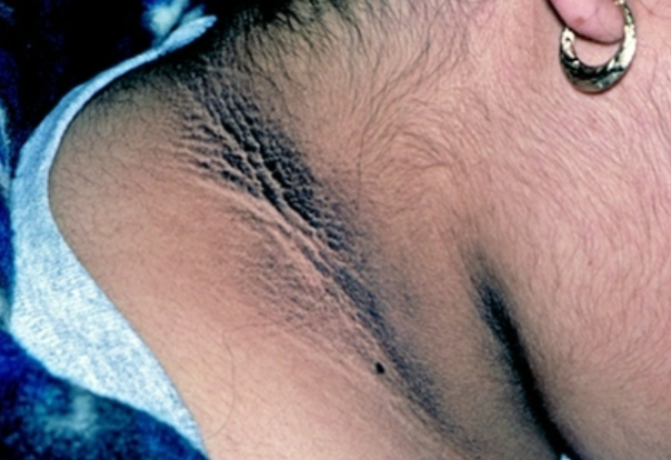
Acanthosis nigricans*
A reactive skin pattern associated w/ obesity, DM, hypothyroid, cushing’s syndrome, malignancy
Common thread in insulin resistance
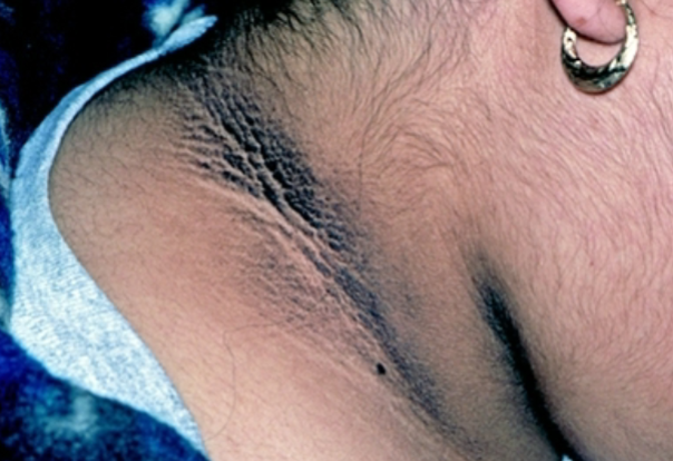
acanthosis nigricans sx*
gray-brown to black, rough to velvety, thickened plaques and prominent skin lines
occurs in flexural areas
asymptomatic
→ tx = weight loss and tx of underlying conditions
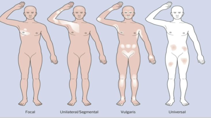
Vitiligo definition
A complete loss of skin pigmentation d/t a decrease in both number and function of melanocytes
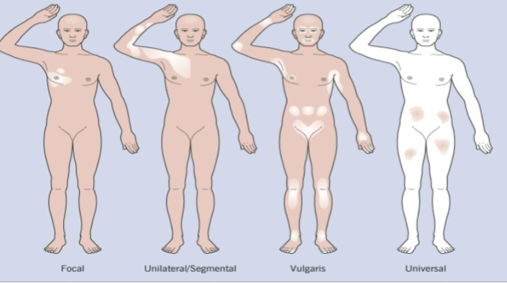
vitiligo epidemiology
age
assoc with?
examples where you see this
dark pigmented skin, can effect any race
½ of cases <20 yo
associated with high homocysteine levels (cell destruction)
Seen in graves disease, addison’s disease, hypothyroidism, pernicious anemia, T2DM, melanoma
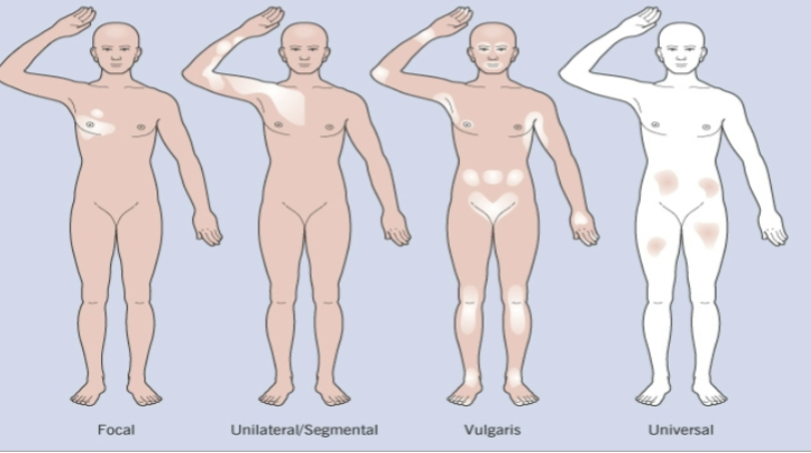
vitiligo PE*
Well demarcated borders of depigmentation
Macules/patches
dorsa of hands, face, axilla, genitalia, or around body openings
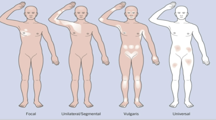
types of vitiligo*
Generalized (most common) → Widespread and symmetric distribution
Focal → Isolated distribution
Universalis → total depigmentation
Unilateral/segmental → dermatomal distribution on one side
Mucosal → MMs only
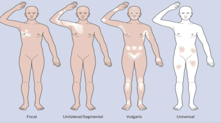
vitiligo tx*
most effective: phototherapy w/ sun protection
others: topicals, depigmentation, skin grafts, cosmetic coverup, tattooing
most common benign vascular neoplasm of childhood
hemangioma
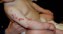
hemangioma complications
when ulcerates(diaper area) or obstruct vital functioning
→ hypopig, telang, stretched skin, scar
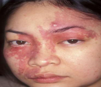
Port Wine Stain definition*
type of vascular malformation
variant of nevus flammeus
pink/red midline (unilateral) macule/patch
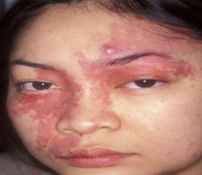
port wine stain complications*
sturge-weber syndrome**
dermal fibrosis
soft tissue/bone hypertrophy
sturge-weber syndrome definition
trigeminal nerve branches
→ glaucoma, seizures, intellectual impairment
complication of port wine stain
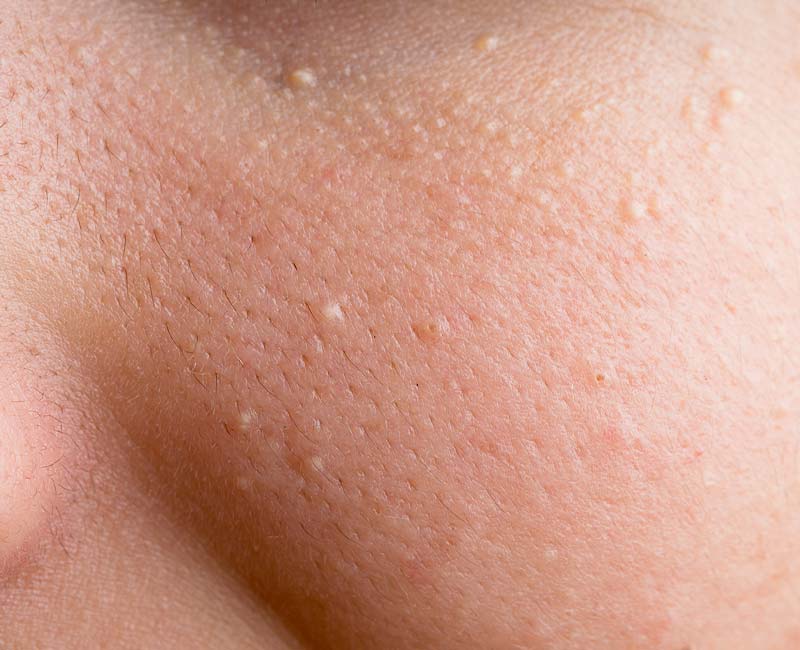
milia definition*
1-2 mm superficial white/yellow keratin containing epidural cyst
eyelids, cheeks, and forehead
Occurs at any age
No treatment necessary
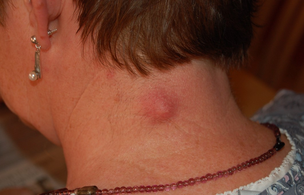
epidermal inclusion cysts*
definition
contains what
firm, mobile, painless nodules
contains KERATIN (no sebum)
epidermal cysts originate where*
epidermis
pilar cysts originate where?*
infundibulum of hair follicles
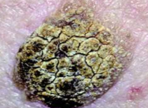
seborrheic keratosis definition, age, malignant potential*
benign epidermal growth of immature keratinocytes
>50 yo
no malignant potential
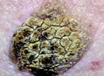
Seborrheic Keratosis features*
Warty “Stuck on” appearance
Scaly, hyperpigmented, well circumcised lesions
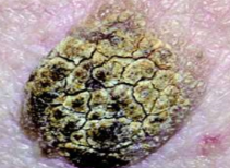
Seborrheic Keratosis tx
no tx needed unless cosmetic problems
liquid nitrogen
shave biopsy blade
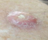
Keratoacanthoma definition*
sudden appearance of 1-2 cm skin-colored, dome-shaped papule w central keratin filled plug
can → SCC
spontaneous regression wks-mos
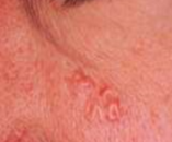
Sebaceous hyperplasia definition
unique-ish finding
what is it?
sx?
you put pressure, what do you see?
1-2 mm soft pale yellow/skin colored umbilicated papules
(scattered and enlarged sebaceous glands)
uniform telangiectasia
no sx but yields sebum w/ pressure
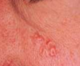
Sebaceous hyperplasia - location, pop
face
older adults
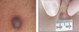
Dermatofibromas - definition, sx, what sign, location*
0.3 - 3 cm flesh to brown, firm, dome shaped or depressed, non-fixed, movable nodule
Asymptomatic
Lateral pressure → Fitzpatrick sign “dimple/retraction sign”
extremities
Skin tags - definition, location, pop, sx*
1-10 mm flesh to brown, soft pedunculated/flat/filiform or sessile
eyelids, neck, intertriginous areas
females, >30, obese
asymptomatic
Hypertrophic scar is confined to what site*
site of injury
keloid definition, cause, pop*
extends beyond site of injury
d/t inappropriate fibroblast proliferation from trauma
benign tumor, NEVER becomes malignant
darker skin types, above waist
Cutaneous horns - definition, location, appearance*
Keratinous skin tumors in the shape of conical projections above the surface of the skin
face or hands
appearance of horns, woods, coal
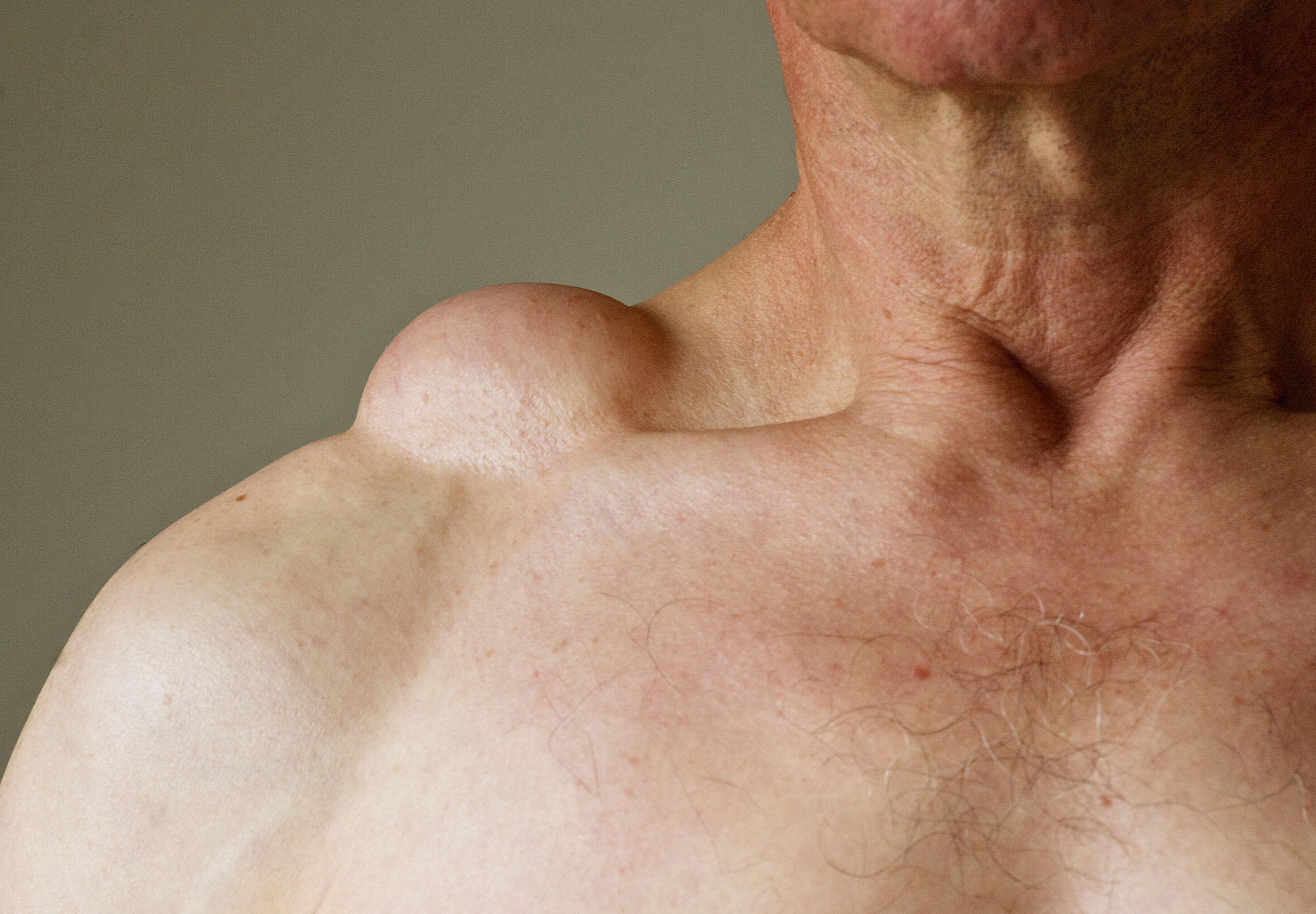
Lipomas - definition, features*
benign tumor composed of adipose tissue
Most common benign form of soft tissue tumor
Soft to the touch, moveable, generally painless
Most common benign form of soft tissue tumor
lipomas
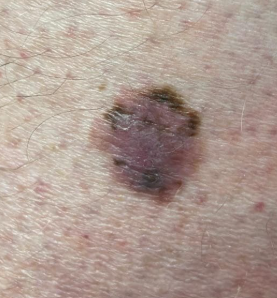
Nevi (Moles) - features, cause, sx*
cause: proliferation of melanocytes
benign BUT can → malignant melanoma
childhood-young adulthood (sus if >30)
sun-exposed skin
asymptomatic
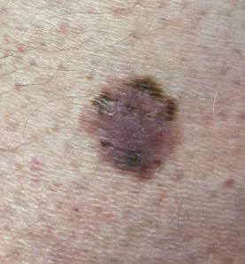
with nevi (moles), when are you suspicious?*
>30 yo
sun protected areas
sudden skin sx of itch/pain/bleeding
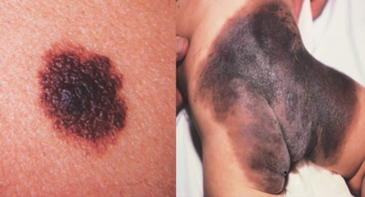
Congenital Melanocytic Nevus - inc risk of, appears when*
INC risk of melanoma!!
timing: birth or infancy
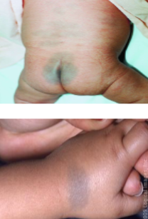
mongolian spots vs. congenital melanocytic nevi
mongolian spot (congenital dermal melanocytosis): benign, flat congenital birthmark w/ wavy borders + irregular shape (pictured above)
entrapment of melanocytes in the lower half to 2/3 of dermis
harmless and usually fade by school age
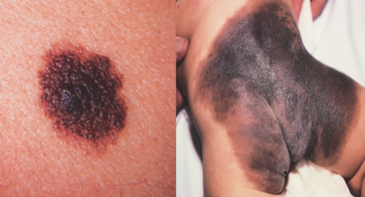

junctional common pigmented nevi*
b/w the epidermal and dermal layers of skin
pigmented (a tiny bit raised)
risk of dev cancer
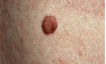
compound pigmented nevi*
features of junction and dermal nevi
pigmented and papular

dermal pigmented nevi*
deep papular lesion
appears later in life
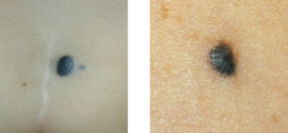
Blue Nevus
intense pigment deeper in dermis
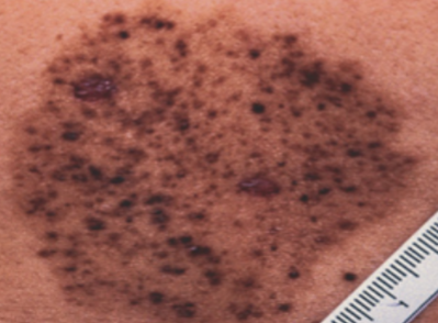
nevus spilus
black pepper appearance
tan macule w/ several smaller brown nevi
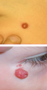
spitz nevus
dome shaped red/pink papule seen in kids
hard to distinguish from melanoma → remove
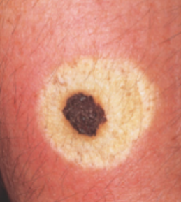
halo nevus
central placed nevus surrounded by white halo of depigmentd skin
adolescence
gradually involute and disappear → residual white patch
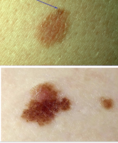
Dysplastic (Atypical) Melanocytic Nevi - definition
inc risk of ?
when do you want to excise?
benign or cancerous?
part of spectrum b/w benign nevi and melanoma
inc risk of malignancy depending on atypia severity
surgical excision of signs of dysplasia = ABCDE criteria
NOT BENIGN!
ABCDE criteria (for biopsy for cancer/dysplastic melanocytic nevi)
A = asymmetry, B = border irregularity, C = color variation, D = diameter > 6mm, E = enlarging/evolving