Chapter 3 - Bacteria & Archaea
 Call Kai
 Learn
Learn Practice Test
 Spaced Repetition
 Match
Match Flashcards
 Knowt Play
1/41

There's no tags or description
Looks like no tags are added yet.
Name | Mastery | Learn | Test | Matching | Spaced | Call with Kai | Chat |
|---|
No analytics yet
Send a link to your students to track their progress
42 Terms
Distinguishing features of Prokaryotes
The way their DNA is packaged: lack of true nucleus & histones, so DNA floats around in the cytoplasm
The makeup of their cell wall: peptidoglycan & other unique chemicals
Their internal structures: lack of membrane-bound organelles
Structures found in all bacterial cells
Cytoplasmic membrane (membrane surrounding cytoplasm)
Cytoplasm (inner most part of cell)
Ribosomes (on rough ER for protein synthesis)
Cytoskeleton (network of proteins)
Genetic material (1 chromosome, aka haploid)
Structures found in most bacterial cells
Cell wall
Glycocalyx (surface coating)
Structures found in some bacterial cells
Flagella, pili, fimbriae (hair-life appendages on surface of cell wall)
Outer membrane (part of cell wall)
Nanowires/nanotubes
Plasmids (additional DNA; can carry genes for anti-bacterial resistance)
Inclusions
Endospores (can’t be killed easily by disinfectant; hardy structures)
Not reproductive; for survival of cell!
Microcompartments
Most of these are observed in archaea as well
Bacterial shapes & arrangements
Bacteria mainly function independently as single-celled, unicellular organisms
Some act as a group in colonies or biofilms
Shapes: cocci, bacilli, vibrio, spirillum, spirochete (cocci & bacilli = most common)
Bacteria have average size of 1 micron
Pleomorphism: variations in cell wall structure caused by slight genetic or nutritional difference
So they adopt multiple shapes (shape shifters) → very rare
Cocci
Spherical/ball shaped
Cocci can be perfect spheres, but can also exist as oval & bean-shaped
Cocci = plural; Coccus = singular

Bacilli
Cylindrical/rod shaped
Rods are varied in form
Coccobacillus = when it’s short & plump
Vibrio = when it’s gently curved
Bacilli = plural; Bacillus = singular

Spirillum
Bacterium with slightly curled/spiral-shaped body
Rigid, short, few twists (resembles a corkscrew)
Spirochete
Bacterium with a spiral cell
Flexible, long, many twists (resembles a spring)
Filaments
Multiple branches produced off of a basic rod structure
Very rare
Arrangements of cocci
Single
Diplococci (pairs)
Tetrads (groups of 4)
Sarcina (cubical packet of 8, 16, or more cells → up to 64)
Streptococci (chains)
Staphylococci (irregular clusters)
Arrangements of bacilli
Single
Diplobacilli (pair of cells w/ ends attached)
Streptobacilli (chain of several cells)
Palisades (cells of a chain remain partially attached by a small hinge region at the ends)
Usually just single, not chains:
Spirilla (only sometimes in short chains)
Spriochetes: rarely remain attached after cell division
External structures
Flagella
Fimbriae
Pili
Nanotubes/nanowires
S layer
Single layer of a protein linked together like tiny chain link fences
**Only produced when bacteria are in a hostile environment**
Glycocalyx
Coating of repeating polysaccharide or glycoprotein units
Slime layer: loose, protects against loss of water & nutrients (hydrates)
Capsule: more tightly bound, denser, & thicker

Flagella - external structures/appendages
Primary function = motility (movement)
Structure (3 distinct parts):
1. Filament (outermost)
2. Hook (sheath)
3. Basal body
Immune system makes antibodies against flagella to inhibit movement

Flagella - arrangement
Polar arrangement: flagella attached at one of both ends of the cell
1. Monotrichous: single flagellum
2. Lophotrichous: small bunches/tufts of flagella emerging from same site
3. Amphitrichous: flagella at both poles of the cell
Peritrichous arrangement: flagella are dispersed randomly over surface of cell
Flagella - function
Primary function: motility (movement)
Chemotaxis: movement of bacteria in response to chemical signals
Positive vs. Negative chemotaxis
Run: rotation of flagellum counterclockwise, resulting in a smooth linear direction
Tumble: reversal of the direction of the flagellum, causing the cell to stop & change course
Fimbriae
Primary function: adhesion (attachment)
Small, bristle-like fibers sprouting off the surface of many bacterial cells
Short compared to flagella
Allow tight adhesion b/w fimbriae & epithelial cells, allowing bacteria to colonize & infect host tissues
Pili
Used in conjugation b/w bacterial cells
Well characterized in gram-negative bacteria
Type IV pilus can transfer genetic material, act like fimbriae & assist in attachment, & act like flagella & make a bacterium motile
Nanotubes (nanowires)
Very thin, long, tubular extensions of the cytoplasmic membrane
Used as channels to transfer amino acids or to harvest energy by shuttling electrons to iron-rich substances
S layer
Outside cell wall
Single layers of thousands of copies of a single protein linked together like tiny chain link fences
Only produced when bacteria are in a hostile environment

Glycocalyx
Coating of repeating polysaccharide or glycoprotein
Slime layer: loose, protects against loss of water & nutrients (also aids in attachment) → can appear as plaque
Capsule: more tightly bound, denser, & thicker; produces a sticky (mucoid) character to colonies on agar

Capsules
Formed by many pathogenic bacteria
Have greater disease-causing abilities (increased virulence)
Protects against host white blood cells called phagocytes
Biofilms: capsules can be responsible for biofilm formation
2 types of places that biofilm forms:
1. Plaque on teeth → protects bacteria from being dislodged
2. Colonization of plastic catheters, IUDs, metal pacemakers, & other implanted medical devices
The Cell Envelope
Lies outside the cytoplasm
Composed of 2 or 3 basic layers that each perform a distinct function, but together act as a single protective unit:
1. Cell wall
2. Outer membrane (part of cell wall in Gram-negative bacteria)
3. Cytoplasmic membrane
Cell wall - function
Helps determine the shape of a bacterium
Provides strong structural support to keep the bacterium from bursting or collapsing because of changes in osmotic pressure
Certain drugs target the cell wall (particularly its peptidoglycan), disrupting its integrity & causing cell lysis (disintegration or rupture) of the cell
Gains its relative rigidity from peptidoglycan (the target of antibiotics)
Cell wall - Peptidoglycan
Compound composed of a repeating framework of long glycan (sugar) chains cross-linked by short peptide (protein) fragments
Provides a strong but flexible support framework

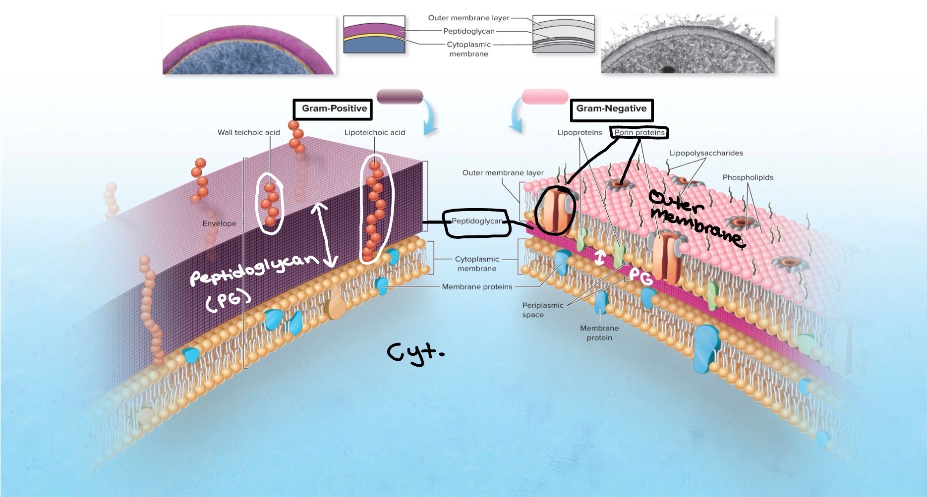
Gram-Positive cell wall
Thick, homogenous sheet of peptidoglycan
Contains teichoic acid & lipoteichoic acid
Function in cell wall maintenance & enlargement
Contribute to acidic charge on cell surface
Gram-Negative cell wall
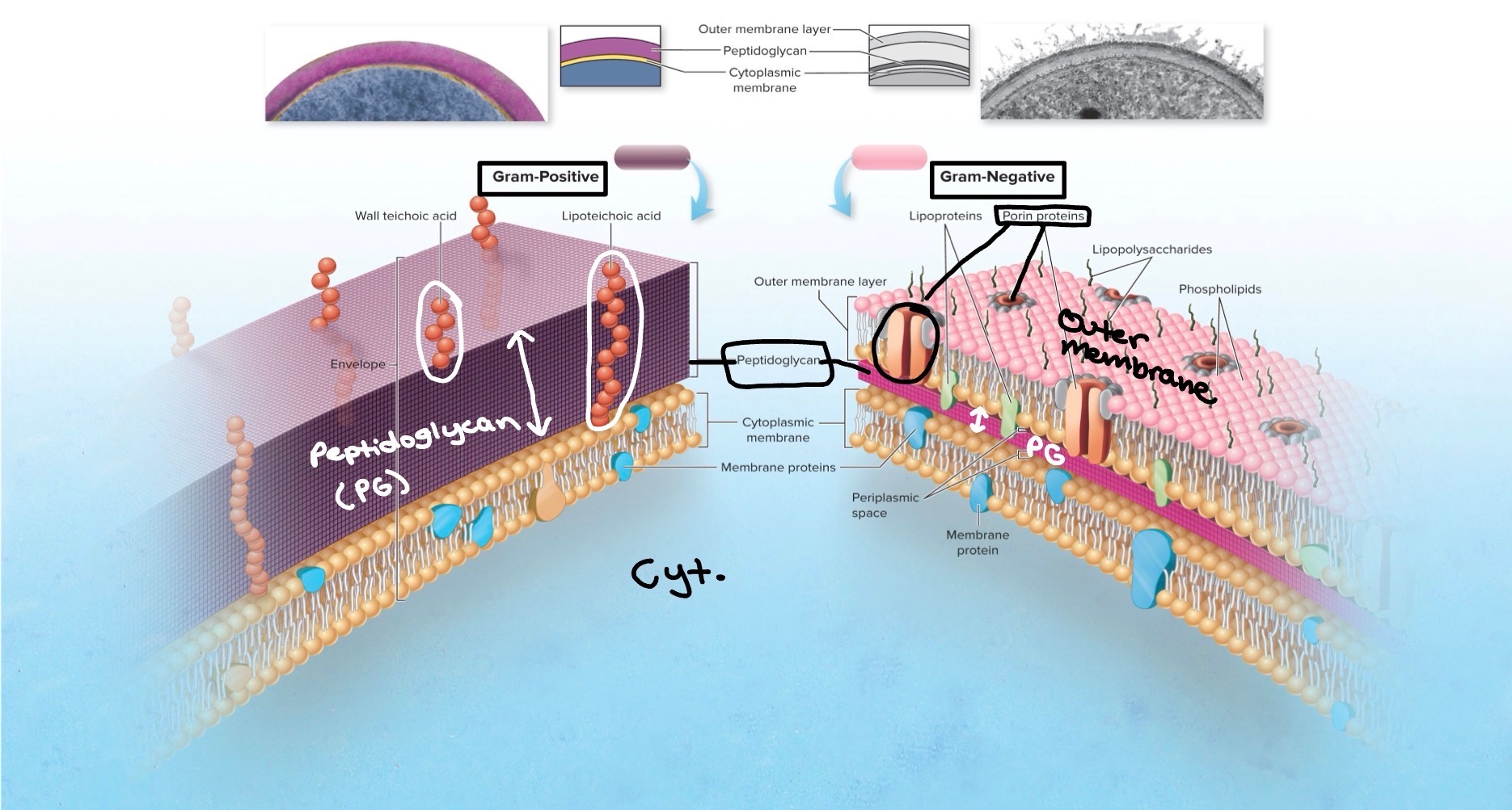
Single, thin sheet of peptidoglycan
Thinness gives gram-negative cells more susceptibility to lysis
Contains outer membrane over peptidoglycan
Outer membrane - composition
Similar in composition to most membrane, except it contains specialized polysaccharides & proteins
1. Lipopolysaccharide (LPS):
Signaling molecules & receptors
Endotoxin (toxic to body when disassociated from bacteria; leads to inflammation & septic shock)
2. Porin proteins:
Special membrane channels in outer membrane that allow certain chemical to penetrate
Affects entry of antibiotics & other chemicals

Outer membrane - functions & characteristics
Contributes an extra barrier
Resistant to certain antimicrobial chemicals
**Makes gram-negative bacteria more difficult to kill than gram-positive**
Alcohol-based compounds dissolve lipids in outer membrane & therefore damage the cells
Alcohol swabs used to cleanse skin before certain medical procedures
Treatment of infections caused by gram-negative bacteria requires drugs that can cross the outer membrane
Gram Stain
Helps determine if cell wall is Gram-positive or Gram-negative
Purple = positive
Pink = negative
Non-typical cell walls
Acid-Fast bacteria
Archaea
Acid-Fast bacteria
Not gram-positive or gram-negative
Mycobacterium & Norcardia: contain peptidoglycan & stain gram-positive, but bulk of cell wall is composed of unique lipids, so not considered gram-positive
Myolic acid:
Very long chain fatty acid
Found in cell walls of acid-fast bacteria
Contributes to the pathogenicity of the bacteria b/c it protects bacteria from getting destroyed
Makes bacteria highly resistant to certain chemicals & dyes
Archaea
Exhibit unusual & chemically distinct cell walls
Some have cell walls composed entirely of polysaccharides
Others have cell wall made of pure protein
All lack true peptidoglycan structure
Some lack a cell wall entirely
Cell-wall-deficient bacterias
Mycoplasmas
L-forms
Mycoplasmas
Naturally lack a cell wall
Sterols in the cell membranes stabilize the cell against lysis
Mycoplasma pneumoniae: “walking pneumonia”
L forms
Bacteria that naturally have a cell wall but lose it during part of their life cycle
Role in persistent infections
Resistant to antibiotics
Cytoplasmic membrane
A lipid bilayer w/ protein embedded
Functions:
Energy reaction (ATP production → happens in mitochondria in eukaryotic cells)
Nutrient processing
Synthesis
Regulates transport of nutrients & waste
Selectively permeable: special carrier mechanisms for passage of most molecules
Internal structures (in the cytoplasm)
Cytoplasm: 70-80% water plus complex mixture of sugars, amino acids, & salt
Bacterial chromosomes & plasmids (in cytoplasm):
Hereditary material of most bacteria exists in the bacterial chromosome
DNA is aggregated in the nucleoid (nucleus holds DNA in eukaryotic cells)
Plasmids: nonessential pieces of DNA (so bacteria can survive without) → confer protective traits such as drug resistance & toxin & enzyme production

Ribosomes
All bacteria have this
In the cytoplasm
Site of protein synthesis (same in eukaryotic cells)
Composed of rRNA (60%) & protein (40%)
Consist of a large & small subunit:
Large & small subunits together: 70S
Eukaryotic ribosome: 80S
Inclusion bodies
In the cytoplasm
Used for food storage
Pack gas into vesicles for buoyancy
Store crystals of iron oxide w/ magnetic properties
Cytoskeleton
In cytoplasm
Made of long polymers of protein
Contribute to cell shape
Unique to prokaryotic cells → may be a potential target for antibiotic development
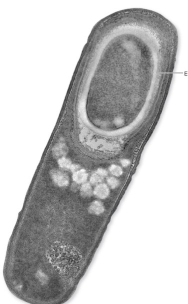
Endospores
Vegetative cell: metabolically active
When this bacterial cell dies, endospores are released, which are dormant bodies: metabolically inactive
Produced by Bacillus, Clostridium, & Sporosarcina
Sporulation: induced by environmental conditions → creation of endospore
To survive environmental stress
Endospores resist extremes of heat, drying, freezing, radiation & chemicals that would kill vegetative cells
Not meant for reproduction →