Adv EP Lesson 6 - Complications
 Call Kai
 Learn
Learn Practice Test
 Spaced Repetition
 Match
Match Flashcards
 Knowt Play
1/66

There's no tags or description
Looks like no tags are added yet.
Name | Mastery | Learn | Test | Matching | Spaced | Call with Kai | Chat |
|---|
No analytics yet
Send a link to your students to track their progress
67 Terms
layers of the heart out to in
pericardium, epicaridium, myocardium, endocardium
pericardium
Layer of connective tissue (pericardial sac) filled with lubricating fluids (serous fluids) to reduce friction and protects the heart
epicardium
Outside layer of the heart and connects to pericardium. Composed of fat and connective tissue. Vessels that run through are responsible for delivery of oxygen to the cardiac tissue
myocardium
Thick, muscular layer that generates the contractions that control the pumping of the heart
endocaridum
Lines the chambers of the heart
pericardial sac
composed of the thin visceral pericardium which consists of a single layer of cells adherent to the cardiac epicardium and the thicker, fibrous parietal pericardium composed of collagen and elastin which is adherent to the lungs, diaphragm, sternum, great vessels, and other mediastinal structures surrounding the heart.
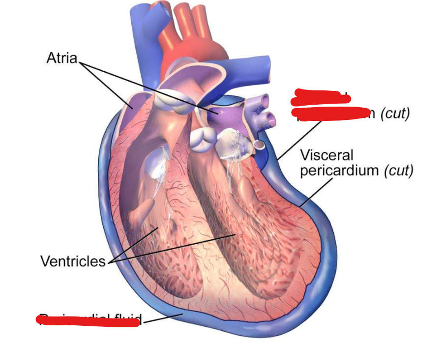

pericardial effusion
to the accumulation of fluid in the pericardial sac surrounding the heart which can lead to tampanade
tamponade
pressure on the heart due to build up of fluid in the pericardial space.
pericardial effusion side effects
pericardium stretches out to accomodate bigger volumes of fluid wihout compressing heart leading to shortness of breath, chest pain, and compression of near structures
beck’s triad
low BP
distension of jugular veins
muffled heart sounds
acute pericardial tamponade
sudden fluid acumulation and pericardium can’t adjust so a dramatic increase in pressure inside pericaridal sac
causes of acute pericardial tamponade
chest trauma, ruptured aorta, ruptured of ventricle after a heart attack
causes of pericardial effusion in EP lab
Misdirected transseptal punctures either with punctures performed too posteriorly exiting the RA into the pericardium before entering the LA or punctures exiting the LA via the roof, LAA, or the lateral LA wall
Steam pops during RF ablation, and
Mechanical perforation with catheter manipulation.
warning signs for pericardial effusion/tamponade in EP lab
Chest Pain if the patient is awake
Hypotension that could be sudden
Unexplained increase in heart rate
Still silhouette of the heart borders on fluoroscopy
Evidence of pericardial effusion on ICE, TEE or TTE
phrenic nerve injury can occur from what during PVI
RF and Cryo
what tech is most prone to phrenic nerve injury when compared to point by point ablation tech
balloon based tech
where in the heart is the risk higher for phrenic nerve injury
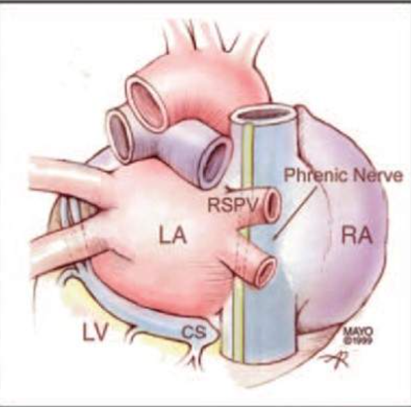
ablating the right superior pulmonary vein because of close proximity to the right sided phrenic nerve
where is the phrenic nerve
runs anterior to the right superior pulmonary vein
types of phrenic nerve injury and when it resolves
Transient Phrenic Nerve Injury (TPNI)- Resolves by end
Phrenic Nerve Palsy (PNP)- Not resolved after discharge but resolves by 12 months
Permanent Phrenic Nerve Injury (PPNI)- Does not resolve by 12 months
adjunctive methods to mitigating phrenic nerve damage
ICE, compound motor action potential, fetal heart monitor, venous pressure waveform
ICE method for mitigating
place ICE at liver to visualize diaphragm
at the first sign of decrease or loss of diaphragmatic contractions, immediately stop the ablation
compound motor action potential method for mitigating
use surface ECG leads and place along the diaphragm
immediately stop the ablation if CMAP amplitude decreases > 30-35%
fetal heart monitor method for mitigating
place across patient’s chest to detect diaphragmatic contraction
after hearing a decrescendo pitch, stop ablation
venous pressure waveform method for mitigating
femoral venous pressure waveforms are monitored using a standard pressure transducer to evaluate the strength of diaphragmatic contraction
what is a known complication of AP ablation
esophageal injury
reported rate of atrioesophageal fistula with arctic front advance
less than 1 out of 25000
strategies to minimize the risk of esophageal injury
reducing power/force/time of RF delivery on the posterior wall, using luminal esophageal temperature (LET) monitoring, use of a proton pump inhibitor, and avoiding energy delivery over the esophagus.
atrioesophageal fistula
an abnormal connection between the esophagus and the LA
thermal injury to endothelial cells of esophagus and/or damage to eophageal arteries causing ischemic necrosis of mucosal layers
symptoms of atrioesophageal fistula
Fever, rigors, fatigue, malaise, chest discomfort, dysphagia, N/V, hematemesis, neurological symptoms
diagnosis of atrioesophageal fistula
CT Scan with oral or IV contrast or MRI
treatment of atrioesophageal fistula
surgical intervention
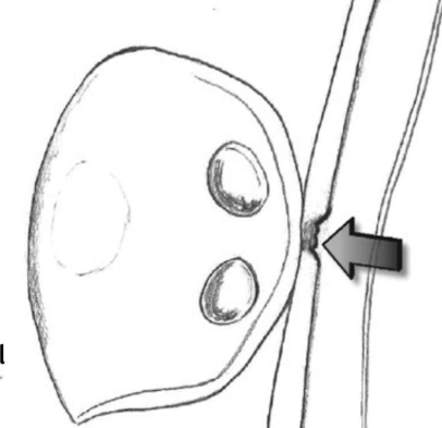
esophageal ulcer of anterior wall

esophageal pericardial fistula

atrial esophageal fistula
ways to mitigate AE fistula
Monitor esophageal temperatures during RF (ablation terminated with a 1oC rise in temperature of the esophageal temperature probe) and cryo ablation procedures (Medtronic physicians consensus suggests stopping ablation if esophageal temperature drops lower than 25oC)
Low-flow irrigation with lower RF energy delivery
Mechanical esophageal deviation
Pharmacologic prophylaxis with proton pump inhibitors (PPI) or histamine H2 receptor blockers.
stroke and transient ischemic attack
possibility due to left sided ablations
with catheter ablation the source of stroke is mainly
embolic where the stroke is outside the brain and mainly the heart
thromboembolism
obstruction of a blood vessel by a blood clot that has become dislodged from another site in the circulation
thromboembolism occurrence
occur within 24 hours of the ablation procedure
sources of thromboembolism during ablation
Stationary sheaths positioned in the left atrium
Char formation at the tip of RF ablation catheter and at the site of ablation
Disruption of thrombus located in the atrium prior to the ablation procedure and
electrical cardioversion (cardioversion is the delivery of energy that is synchronized to the QRS complex to return the heart to normal sinus rhythm)
mitigating risk of thrombus formation in the LA
Detailed pre-procedural imaging ensuring no thrombus or clot in the left atrium
Strict anticoagulation protocol- Heparin is administered before transseptal access and ACT > 300 seconds is maintained
Meticulous attention to sheath management
Careful control of RF energy to minimize char formation

clot
air embolism cause
from introduction of air via the transseptal sheath
mitigating risk of air embolism
Minimize risk of air emboli by prepping all catheters and sheaths used in the procedure properly.
It is important to remove all guide wires and sheaths slowly to minimize vacuum effect.
All infusion lines are monitored closely for any air bubbles. Sheath exchanges also can be minimized
asymptomatic cerebral emboli
an occlusion of a blood vessel in the brain due to an embolus that does not result in any acute clinical symptoms and is therefore "silent"
complication with point by point RF and cryo during PVI
PV stenosis
PV stenosis
blockage of the pulmonary veins that bring oxygenated blood from the lungs to the heart
RF risk of PV stensosi
ablation close to PV orfices and/or within the PVs, with a 5.6 fold higher incidence in comparison with antral ablation.
cryo risk with PV stenosis
Ensuring the balloon is positioned at the antrum of the vein and not deep seated is a good practice to prevent pulmonary vein stenosis from occuring.
proximal seal technique
cryoballoon technique for preventing PV stenosis, ensures antral positioning of the cryoballoon
coronary artery spasm
a temporary constriction of the muscles in the wall of the artery.
spasm can reduce or block the blood flow to part of the heart.
what were shown in PFA when delivered in proximity to the coronary arteries?
coronary artery spasms
what happened when pfa energy was applied for cavotricuspid isthmus ablation? how was it relieved?
provoked severe vasospasm
intracoronary nitroglycerin
what is subclavian access used for
implanting cardiac pacemakers
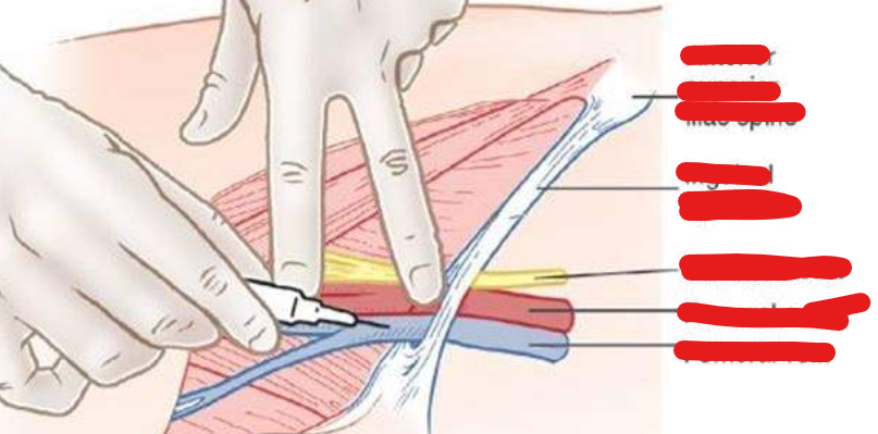

vascular access complication
includes development of a hematoma, arteriovenous (AV) fistula, or pseudoaneurysm.
causes of vascular complications
Number and size of venous sheaths used
Insertion of arterial pressure line
Intense anticoagulation management before, during and after an EP procedure
access site complications
groin hematoma, retroperitoneal bleed, Femoral Pseudoaneurysm, Arteriovenous fistula
retroperitoneal bleed treatment and symptons
When the posterior arterial wall is punctured, blood can spread into the retroperitoneal space. The retroperitoneal space is the area that lies between the sublumbar muscles and the peritoneum
Fluid resuscitation. Reversal of anticoagulation. Vascular surgery if hemodynamic instability continue
hypotension, drop in hematocrit, Flank/abdominal/back pain but won’t discern during procedure
groin hematoma and treatment
pool of mostly clotted blood that forms in the groin. A hematoma is usually caused by a broken blood vessel that was damaged at the groin site during catheter access
Ultrasound guided compression. Usually resolve spontaneously over time. Rarely surgical removal or evacuation of blood in hematoma is necessary
Arteriovenous fistula and treatments
an abnormal connection between an artery and a vein. Usually occurs as a complication when access attempt goes through the vein and artery and creates a connection between the two.
compression, possible surgery
Femoral Pseudoaneurysm
esults from an injury to your blood vessel wall (usually an artery). Blood pools in a small sac attached to one side of the artery. Unlike an aneurysm, a pseudoaneurysm only includes one or two layers of the arterial wall and usually follows interventional procedures usually follow femoral arterial access.
compression, thrombin injection, possible surgery
prevention of groin complications
Use of ultrasound during groin access
While using large sheaths predilating the veins before accessing the vein with the large sheath
Seldinger technique for venous access
Seldinger technique
to obtain safe access to central vein. The desired vessel is punctured with a sharp hollow needle, syringe is detached and guidewire is advanced through the lumen of the needle, and then the needle is withdrawn.
how do vagal responses occur
close proximity of the ganglionic plexi to the pulmonary veins. Ablating the ganglionic plexi can sometimes lead to vagal responses.
what happens when vagal responses arrise
Temporary ventricular pacing is performed