Laboratory Exam #1 Study Guide
1/70
There's no tags or description
Looks like no tags are added yet.
Name | Mastery | Learn | Test | Matching | Spaced | Call with Kai | Chat |
|---|
No analytics yet
Send a link to your students to track their progress
71 Terms
BSL-1 Organisms
Nonpathogenic
Ex. E. coli K-12
BSL-1 Safety Requirements
Standard microbiology practices
BSL-2 Organisms
Moderate-risk pathogens
Ex. Staphylococcus epidermidis
BSL-2 Safety Requirements
Gloves, eye protection, limited access
ubiquitous
microorganisms are found everywhere
ex. air, water, soil, food, human skin, plants, animals
transient microbiota
-picked up from environment
-easily removed
-more likely to include pathogens
-handwashing primarily removes transient microorganisms
resident microbiota
-normally live on human skin
-difficult to remove
-usually benificial
WHO handwashing technique
-palm to palm
-back of hands
-between fingers
-backs of fingers
-thumbs
-fingertips
-wrists
Calculate percent reduction
250 colonies before
25 colonies after
{(before-after)/before] x 100
=(225/250)x100
=90
proper vs. quick handwashing
Proper handwashing 20 sec, covers all surfaces
Quick handwashing only a few seconds

Ocular lens
number 1 on picture

Objective lenses
Objective Magnification
Scanning 4X
Low power 10X
High dry 40X
Oil immersion 100X
number 3 on picture

Stage
number 4 on picture
holds the specimen
Stage clips
holds the specimen in place

Mechanical stage controls
moves the stage left right, back forward

Condenser
gathers light from the illuminator and concentrates it into a focused cone on the specimen

Iris diaphragm
adjustable aperture located beneath the microscope stage, directly below the condenser
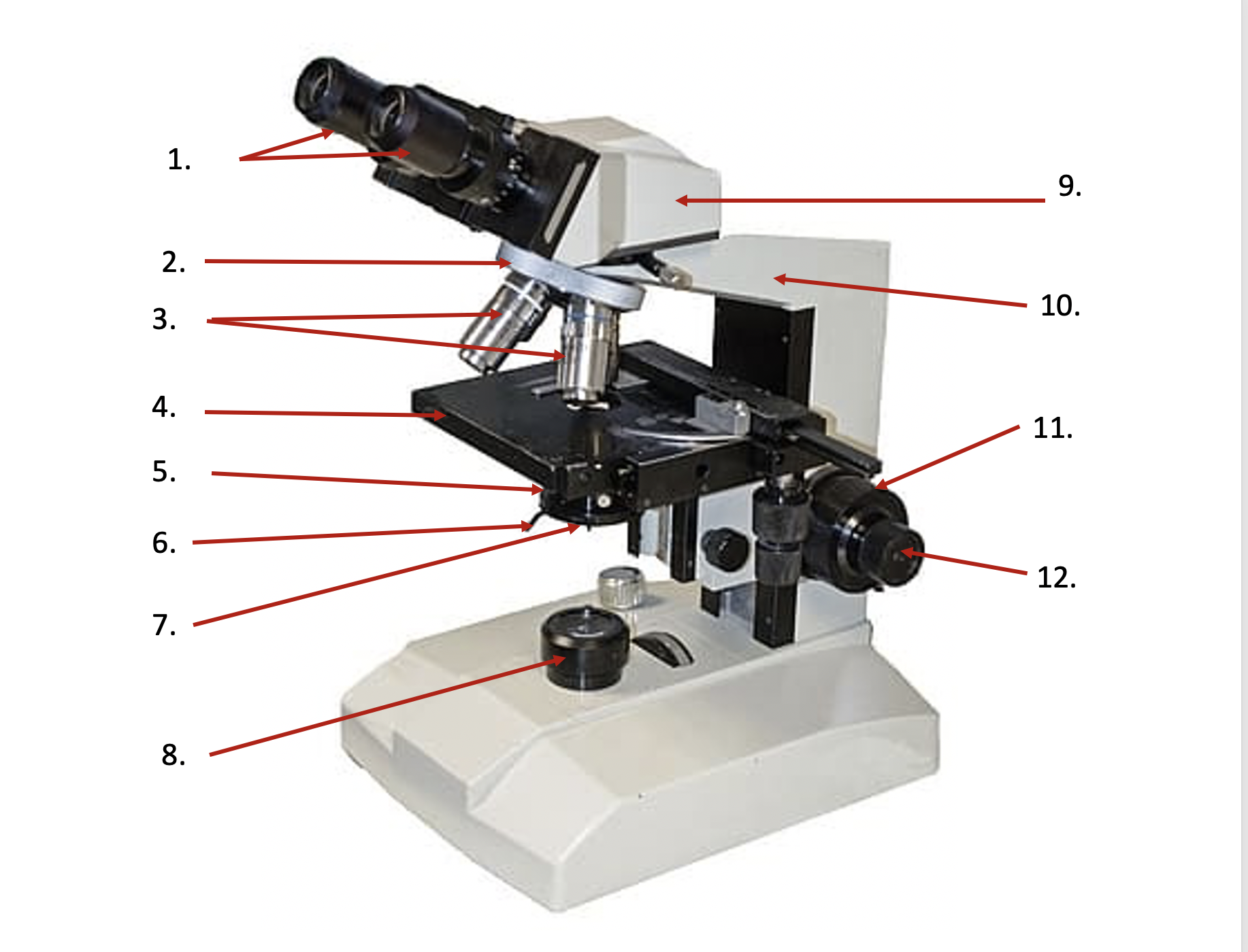
Coarse adjustment
lifts stage up and down to bring specimen in focus

Fine adjustment
focuses on the object clearly

Arm
backbone of the microscope

Base
foundation of the microscope

Light source
Provides light to view the specimens
Immersion Oil
Reduces light refraction
Improves resolution
Used ONLY with the 100X objective
Microscope Storage
• Lowest objective (4X) in position
• Stage lowered
• Oil removed
• Cord wrapped correctly
Protists
• Eukaryotic
• Mostly unicellular
• Possess membrane-bound organelles
• Have nuclei
Ex. Amoeba, paramecium, giardia, plasmodium
Trematoda Group
common name: Flukes
ex: Schistosoma
Cestoda group
common name: Tapeworms
ex: Taenia
Nematoda group
Common name: Roundworms
ex. Ascaris
Zygomycota
Produces: Zygospores
Example: Rhizopus (bread mold)
Ascomycota
Produces: Ascospores
Examples: Saccharomyces, Penicillium, Morels
Basidiomycota
Produces: Basidiospores
Examples: Mushrooms, Shelf fungi, Puffballs
Why Use Aseptic Technique
• Prevent contamination
• Maintain pure cultures
• Protect yourself
• Protect others
Sterile Transfer Steps
1. Sterilize loop
2. Cool loop
3. Flame tube
4. Transfer culture
5. Flame tube
6. Sterilize loop again
Streak Plate Purpose
Obtain isolated colonies in third or fourth quadrant
Pure culture
ne bacterial species
Mixed culture
Two or more species intentionally grown together
Contaminated culture
Unwanted microorganism introduced
Turbid
coudiness of a fluid caused by particles
Sediment
particulate matter that settles in the bottom of aquatic environments
Pellicle
thin outer protein layer that supports cell membrane in many single celled eukaryotes
Flocculent
substance that forms or consists of small woolly masses, like flocculent precipitate
Shape
distinct physical form of microorganisms
ex. coccus
Margin
edge or border of bacterial or fungal colony grown on a solid culture
Elevation
side view of a bacterial or fungal colony grown on a solid culture
ie. how much above the sutface the colony rises
Pigmentation
Natural coloration of microorganisms
Texture
physical consistency and surface charcteristics of a bacterial colony
Simple Stain
Determine: Shape, Arrangement, Size
Uses: basic dyes
ex. crystal violet, safranin, etc
SImple stain result
Cells become colored.
Background remains clear
Negative Stain
Observe: True cell size, Capsule, Delicate structures
Uses: Acidic dyes
Negative Stain result
Background becomes dark.
Cells remain unstained.
Never heat fix.
Basic Dye
Positive charge, stains cells
Acidic dye
Negative charge, stains background
Gram stain steps
1. Crystal violet
2. Iodine
3. Alcohol/acetone
4. Safranin
Gram stain results
Organism Color
Gram-positive Purple
Gram-negative Pink
Gram-positive bacteria
• Thick peptidoglycan
• Retain crystal violet-iodine complex
Gram-negative bacteria
• Thin peptidoglycan
• Lose crystal violet during decolorization
• Take up safranin
Common gram stain Errors
Over-decolorization
→ Gram-positive appears pink
Under-decolorization
→ Gram-negative appears purple
Endospore Stain organisms
Common producers:
• Bacillus
• Clostridium
Endospore Stain reagents
Primary stain: Malachite green
Counterstain: Safranin
Endospore Stain results
Endospores: Green
Vegetative cells: Pink
Acid-Fast Stain bacteria
Contain: Mycolic acids
Example: Mycobacterium
Acid-Fast Stain reagents
Primary stain: Carbol fuchsin
Decolorizer: Acid-alcohol
Counterstain: Methylene blue
Acid-Fast Stain results
Organism Color
Acid-fast Red/Pink
Non-acid-fast Blue
Problem: Gram-positive stains pink
Cause: Over-decolorization
Problem: Gram-negative stains purple
Cause: Under-decolorization
Problem: Smear washes off
Cause: Heat fixing omitted
Problem: Negative stain distorted
Cause: Heat fixed
Problem: No isolated colonies
Cause: Loop not sterilized between quadrants
Problem: Endospores pink
Cause: Malachite green not heated properly
Problem: No bacteria visible in negative stain
Cause: Too few cells or smear too thin
why laboratory safety is essential in microbiology
protect the scientist, prevent release of potentially harmful microorganisms