PCCR I: Integumentary System
 Call Kai
 Learn
Learn Practice Test
 Spaced Repetition
 Match
Match Flashcards
 Knowt Play
1/96

There's no tags or description
Looks like no tags are added yet.
Name | Mastery | Learn | Test | Matching | Spaced | Call with Kai | Chat |
|---|
No analytics yet
Send a link to your students to track their progress
97 Terms
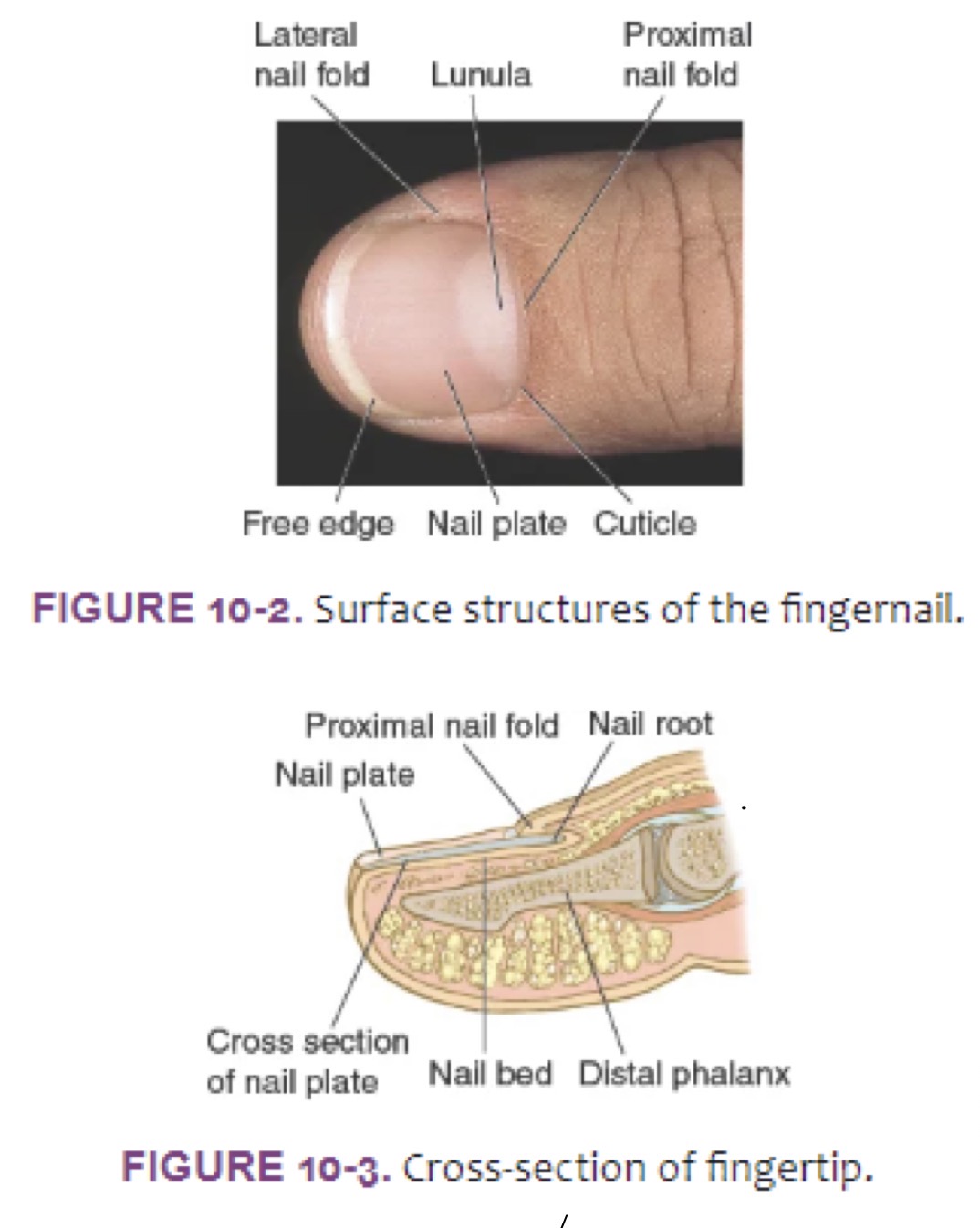
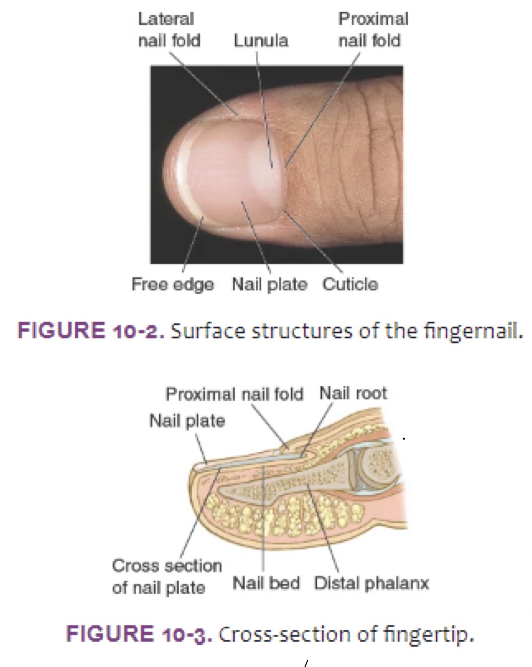
Nail Plate
Protect nail
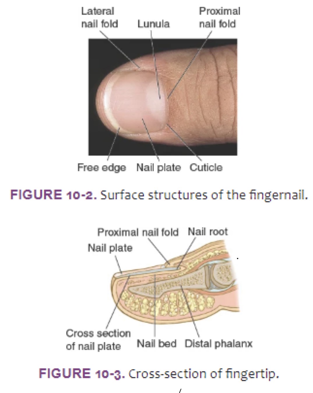
Nail Bed
vascular surface to which nail plate is attached
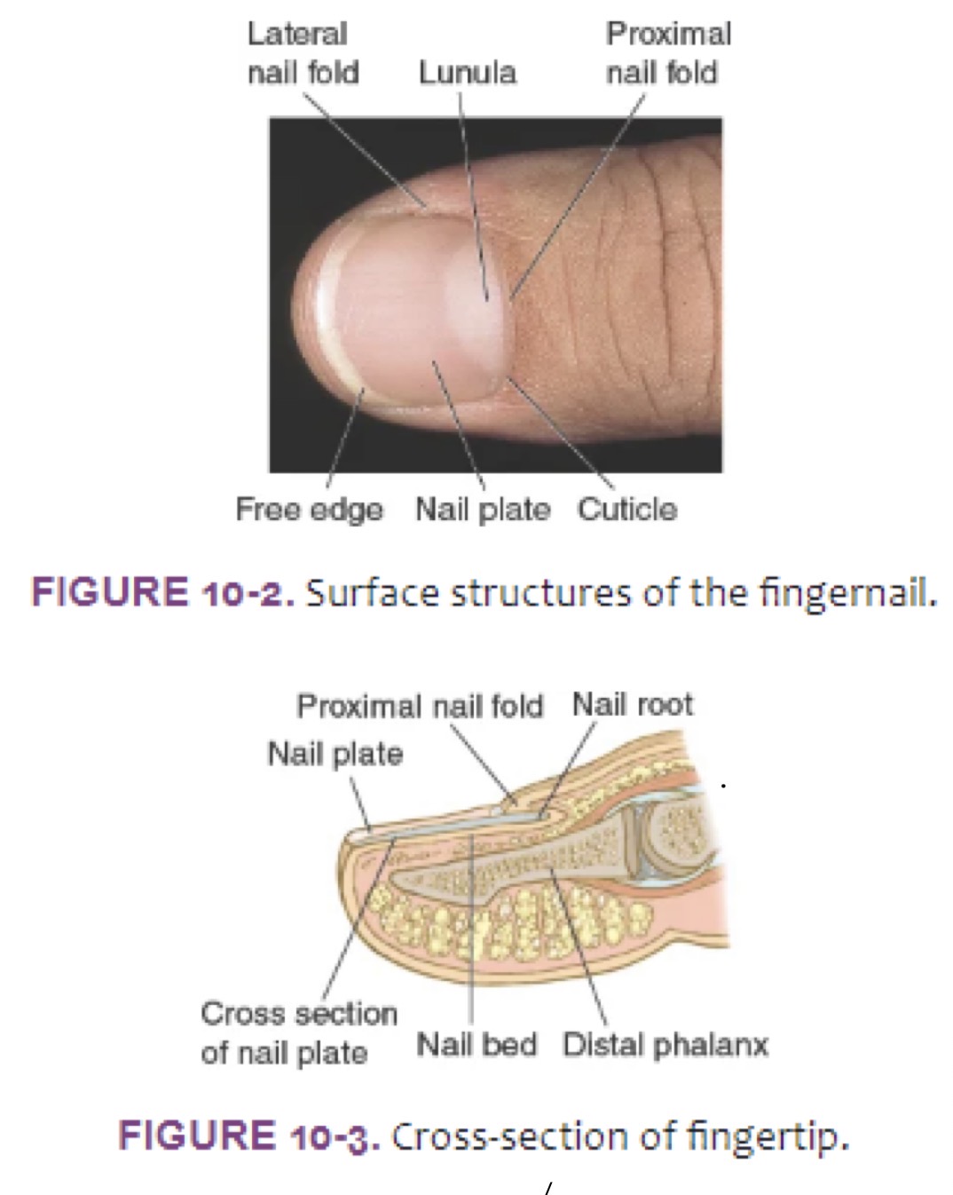
Lunula
Whitish moon at base of nail
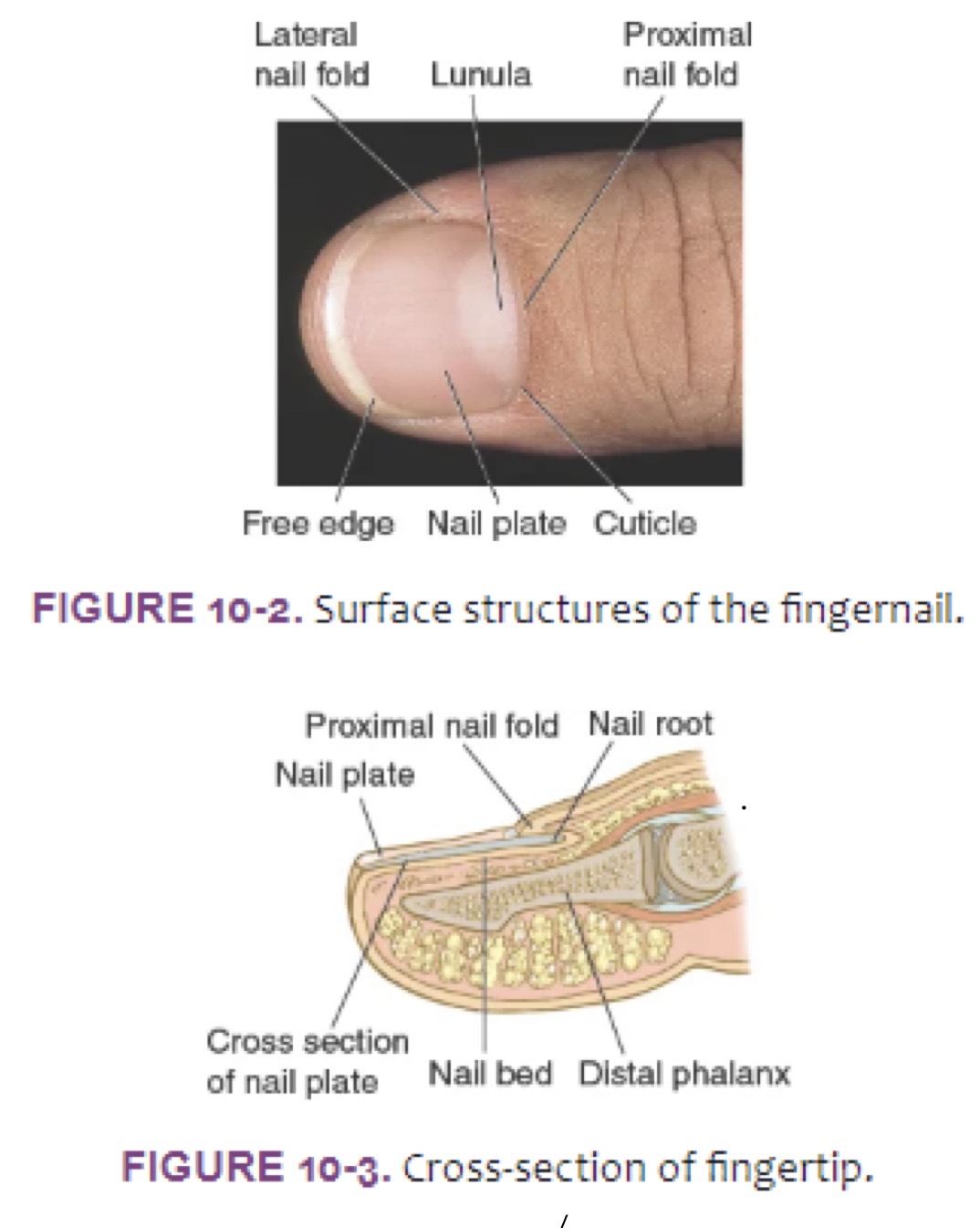
Lateral nail folds
cover the sides of the nail plate
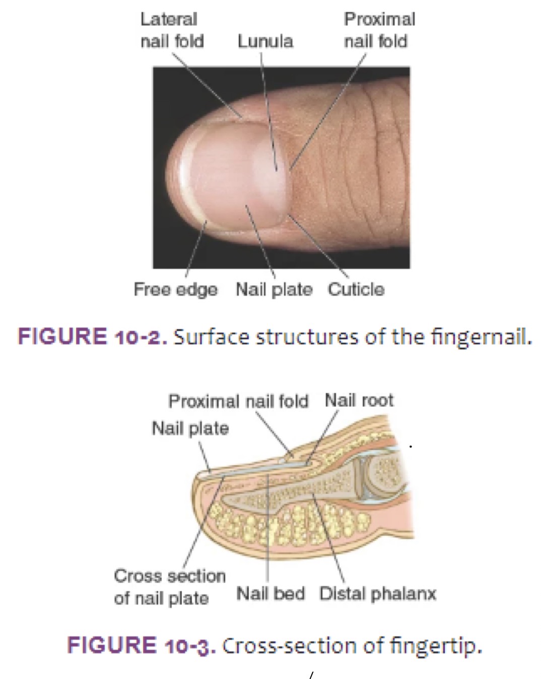
Proximal nail fold
covers nail root (proximal 1/4th of nail plate)
(normal angle b/t prox nail fold and nail plate <180deg)
Cuticle
Extends from proximal nail fold, acts as a seal
HPI
duration, evolution, periodicity, prior eps?
Any asso sympt? (itching (pruritis), pain)
ROS?
PMH: important! syst disease have cutaneous features and manifestations! Allergies?
SH: Occu (Potential exposures), diet (food changes), cosmetics, travel
ROS
Skins:
changes in color, texture, appearance
new rashes, masses, lesions, pruritis, swelling
change in size/color of moles
Hair
changes in color, texture, or appearance
Nails
changes in color, texture, or appearance
Red Flag Symptoms
recurrent or poorly healing wounds/lesions/ulcerations
often asso w/ hx of DTM, autoimmune, endocrine
WORRISOME: DTM, autoimmune - HIV, Endocrine - Cushing Disease, Venous insufficiency
HPI - Lesions
any concern abt new lseion?
PMH of skin cancer? (y - type/loca/date/self-skin exam/sunscreen)
FH of skin cancer? (y - types)
SH of self-skin exams, sunscreen?
HPI - Rashes & Pruritis
HPI of palliative/provocative factors (moisturizers or OTC products)
Asc/ROS: Pruritis (itching?) → IMPORTANT! is it b4 or after?;;; any dry skin?
PMH of season allergies, asthma, atopic dermatitis (AKA eczema)
HPI - Hair Loss and Nail Changes
HPI - any thinning or shedding (y - where: @roots or break shaft?)
SH of hair care practices (freq of shampooing, hair dyes, chem relaxers, heating appliances)
Examination of Skin - TOOLS
adequate lighting
measuring tape/small ruler
small magnifying glass/ dermoscope (dep on user skill/ knowledge)
pt gowned (unclothed expect underwear, open back)
Examination - Seated (HEAD TO TOE)
inspect hair and scalp (distribution, texture, and quantity)
sep hair using fingers/ cotton-tip applicator
inspect head and neck, including forehead, eyebrows, eyelids, eyelashes, conjunctivae, sclerae, nose, ears, cheeks, lips, oral cavity, chin, and beard
including terminal hair of eyelashes, eyebrows, beard
inspect upper back
ask to lean forward and for permission to open back of gown
inspect shoulders, arms, and hands including palpation of fingernails
including color shape, and lesion on fingernails
inspect chest and abdomen
have pt raise/lower gown to expose area (SOLELY) you are examining (may do axilla now or w/ breasts)
inspect anterior thighs and legs
include web0spaces b/tw toes, soles of feet, and toenails
inspect feet and toes including soles, interdigital areas and toenails
same as above
Examination - standing
Inspect lower back
may need to ask pt to lean forward slightly
inspect posterior thighs and legs
(only expose buttock for exam, otherwise covered)
inspect breasts, axillae, and genitalia (axillary and pubic hair)
ensure chaperone for safety
Directed Exam for Hair Loss
Overall pattern of loss/thinning?
Any bogginess, pustules, scaling, erythema, scarring?
PALPATE:
hair pull test: 50-60 hairs b/tw thumb and index/middle fingers and firmly pull from scalp
tug test: group of hairs and pull along hair shaft, abnormal for breakage
ABCDE-EFG Method for Melanoma
A - asymmetry?
B - border irregularities
C- color variation? (2+ not good)
D- Diameter >6mm? (~size of pencil eraser)
E- Evolving/ rapid changing
E- Elevation
F- FIRM on palpation
G - Growing progressively over sev weeks
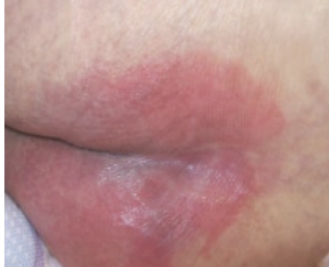
Pressure Injury Stg I
Intact skin w/ localized area of nonblanchable erythema, MAY appear DIFFERENT in darkly pigmented skin
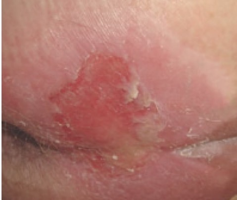
Pressure Injury Stg 2
Partial-thickness skin loss of skin w/ exposed dermis
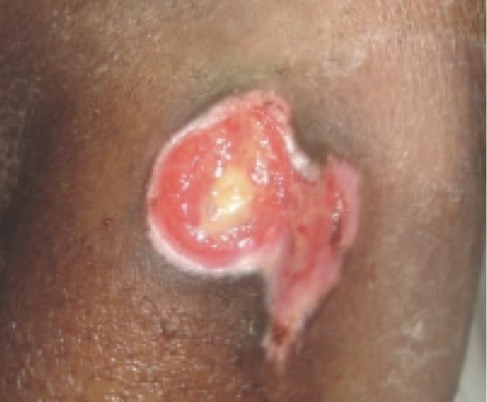
Pressure Injury Stg 3
Full-thickness skin loss, in which adipose is visible in ulcer and granulation tissue and rolled wound edges is often present
Unstageable Pressure Injury
Full-thickness skin and tissue loss in which the extent of tissue damage w/in ulcer cannot be confirmed b/c it is obscured by slough/eschar
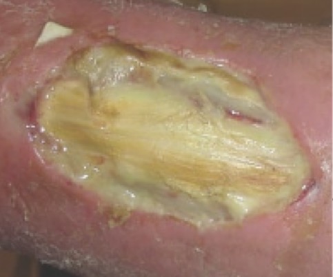
Pressure Injury Stg 4
Full-thickness skin and tissue loss in w/ exposed or directly palpable fascia, muscle, tendon, ligament, cartilage, or bone in ulcer
Deep Tissue Pressure Injury
Persistent nonblanchable deep red/maroon/purple discoloration
can be intact/non-intact skin!!
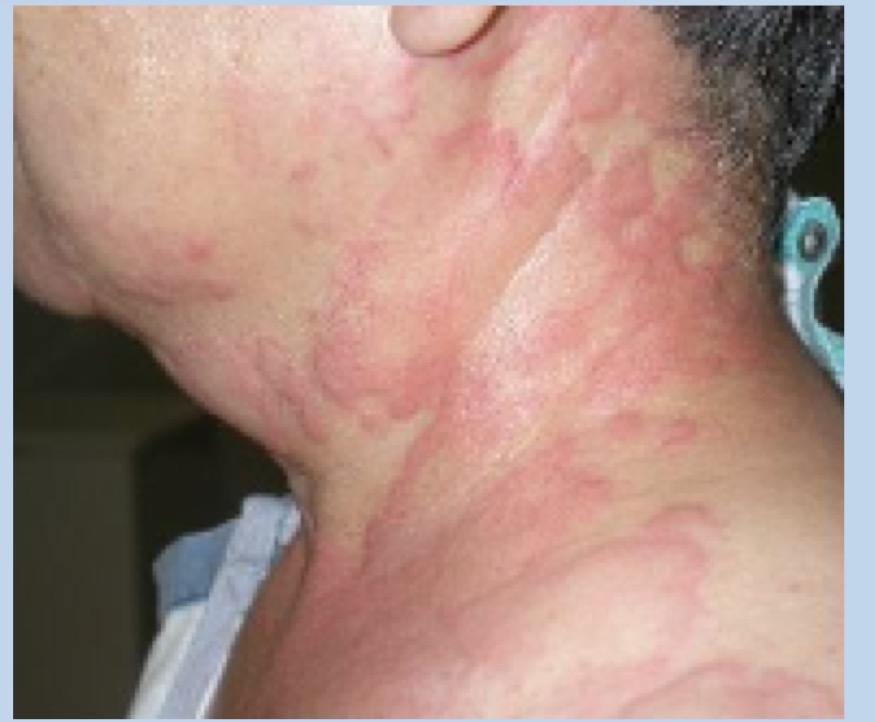
Wheal (aka hives/urticaria): circumscribed area of dermal edema
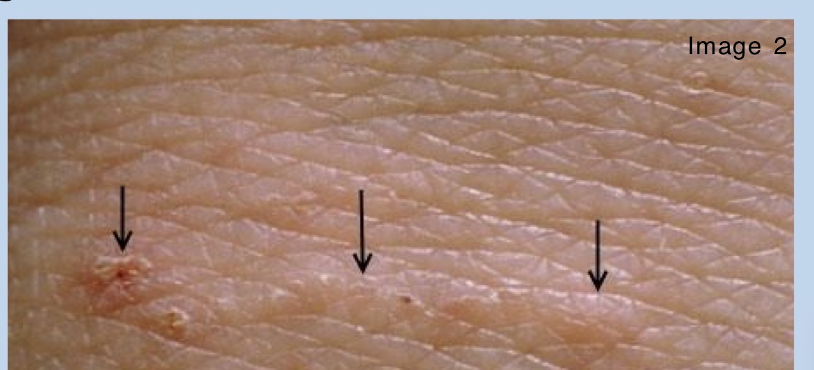
Burrow: small, linear/serpiginous (snake-like/curved/branching) paths in epidermis
created bhy scabies mite!!!
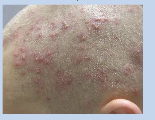
Pustule: small circumscribed elevated epidermis w/ purulent fluid
EX:
bacterial folliculitis (erythematous, pus0filled lesions around hair follicles)
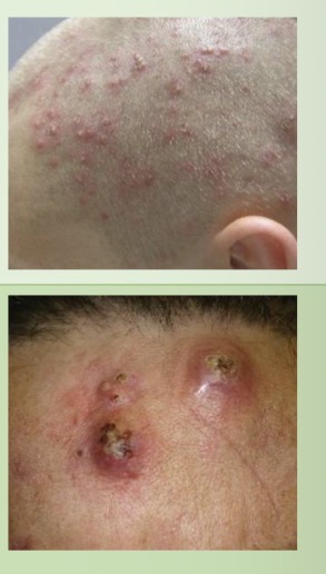
abscesses (fluctuant deep infections, can arise from furuncles, folliculities, or other primary lesions)
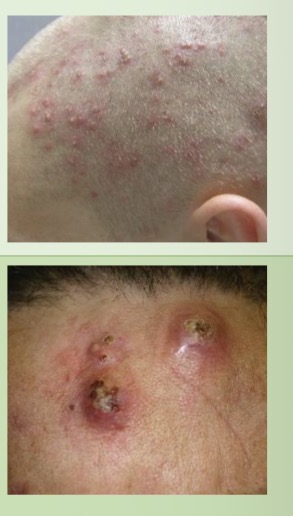
Bacterial Folliculitis: erythematous, puss-filled lesions around hair follicles
Abscesses: fluctuant deep infections, can arise for furuncles (inflamed hair follicles), folliculitis (bacterial folliculitis), or other primary lesions
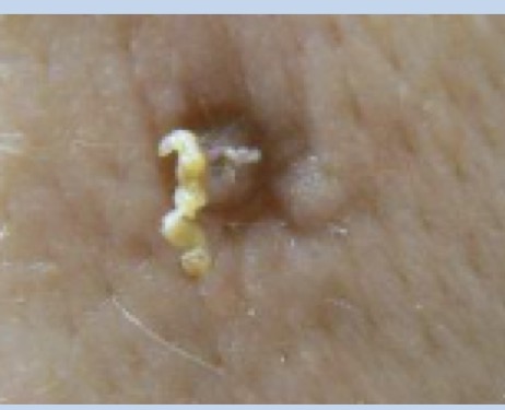
Cyst: encapsulated collection of fluid or semisolid substance
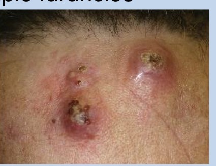
Furuncle (Inflamed hair follicle!!!)
mult = carbuncle
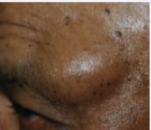
Nodule: raised area of skin → large, solid elevation extending DEEPER into skin layers (>1cm)
ref papule (<1cm) and plaque (flat, >1cm)
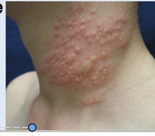
Vesicle: raised and fluid filled (<1cm)
ref bullae (>1cm)
EX:
bullous drug reaction: dusky patch w/ inner violaceous (purple) patch and central tense bulla
herpes simples: vesicles (sometimes pustules) grouped together
herpes zoster (AKA shingles): grouped vesicles in single dermatomal distribution that DOESN’T cross midline
allergic dermatitis: often papules (solid elevation of skin <1cm) and vesicles due to allergic trigger
poison ivy (may have transudative crust)
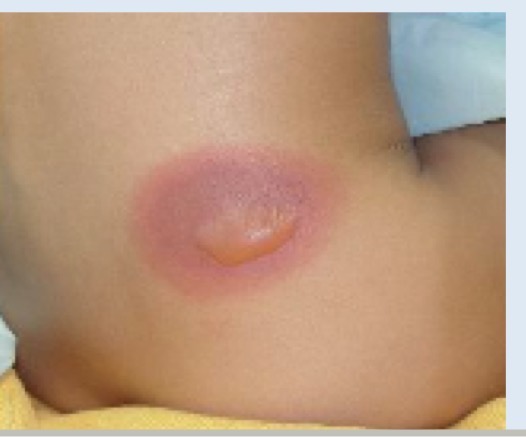
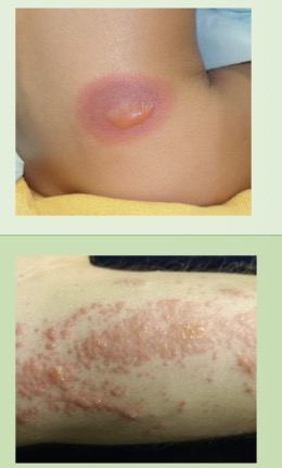
Bullous Drug Reaction: dusky patch w/ inner violaceous patch and central tense bulla (raised and fluid-filled >1cm)
Allergic Dermatitis: often papules (solid elev of skin <1cm) and vesicles (raised and fluid-filled <1cm) due to allergic trigger
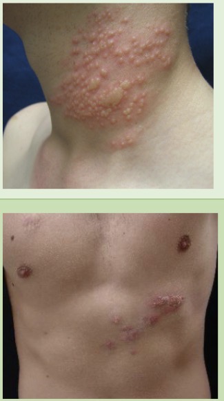
Herpes Simplex: vesicles (sometimes pustules -small circumscribed elev epidermis w/ purulent fluid) grouped together
Herpes Zoster (AKA shingles): grouped vsicles in single dermatomal distribution that DOESN’T cross midline
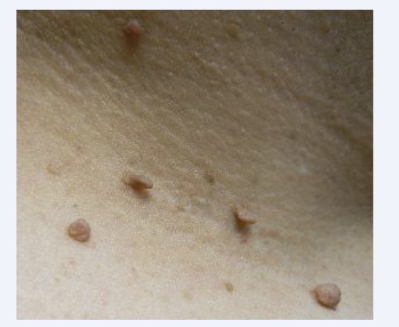
Papule: raised area of skin → solid elevation <1cm
EX:
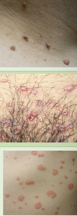
skin tags: fleshy skin-colored to light brown papules, often located in skin folds (Lateral neck, axillae)
molluscum contagiosum: firm, smooth-domed papules w/ central unbilications
guttae psoriasis: scatted erthematous, well circumscribed scaling papules (+plauques too!)
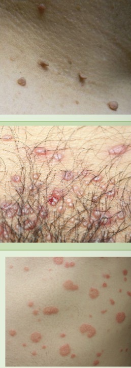
Skin tags: should know
Molluscum contagiosum: firm smooth-domed papules w/ central umbilications (indents in middle!)
guttae psoriasis: scatted erythematous, well-circumscribed scaling papules (+Plaques! - large flat elev of skin >1cm)
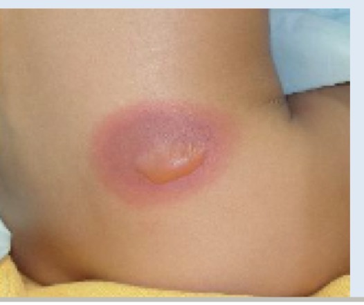
Bullae: raised and fluid-filled (>1cm)
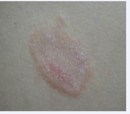
Plaque: raised area of skin with large, flat elevation >1cm
EX:
plaque psoriasis: scattered, erythematous or bright pink, flat-tooped plaques w/ overlying silvery scale MC on extensor surfaces in childhood (eg. elbow crease)
atopic dermatitis: bilateral eczamatous plaques, poorly circumscribed and often lichenified (thickened skin from rubbing/scratching) MC of flexor surfaces (eg. knees/elbows)
nummular dermatitis: mult, round coin-like exzematous plaques w/ overlying transudative crust
pityriasis rosea rash: mult, round, violaceous plaques
herald patch of pityriasis rosea: single oval, raised but flat-topped plaque
initial sign of pityriasis rosea
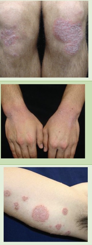
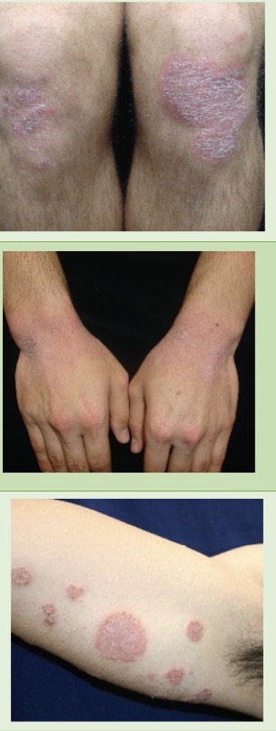
plaque psoriasis: scattered, erythematous or bright pink, flat-tooped plaques w/ overlying silvery scale MC on extensor surfaces in childhood (eg. elbow crease)
atopic dermatitis: bilateral eczamatous plaques, poorly circumscribed and often lichenified (thickened skin from rubbing/scratching) MC of flexor surfaces (eg. knees/elbows)
nummular dermatitis: mult, round coin-like exzematous plaques w/ overlying transudative crust
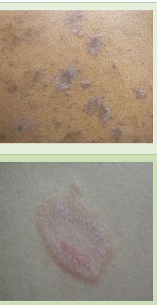
pityriasis rosea rash: mult, round, violaceous plaques
herald patch of pityriasis rosea: single oval, raised but flat-topped plaque
initial sign of pityriasis rosea
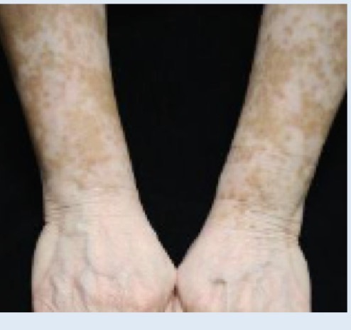
Patch: flat area of color change of skin >1cm
EX:
vitiligo: confluent, depigmented patches
tinea cruris: erythematous, geographic patches w/ peripheral scaling
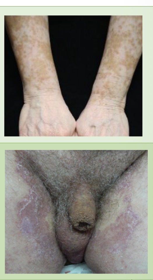
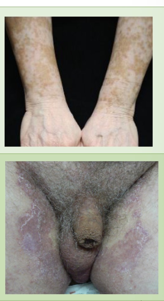
vitiligo: confluent, depigmented patches
tinea cruris: erythematous, geographic patches w/ peripheral scaling
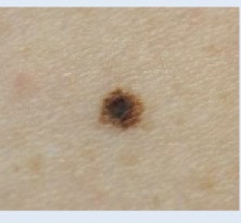
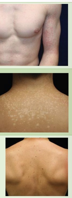
Macule: flat area of color change <1cm
EX:
morbilliform drug reactions: erythematous, confluent round cacules
tinea versicolor: mult. hypopigmented or hyperpigmented or tan macule
benign melanocytic nevi: symmetrical pigmented, round/oval, brown macules
Benign Skin Lesions
Seborrheic Keratosis
Solar Lentigines
Benign Melanocytic Nevi (macule)
NODULES:
Dermatofibroma
keloids
epidermal inclusion cyst
pilar cysts
lipomas
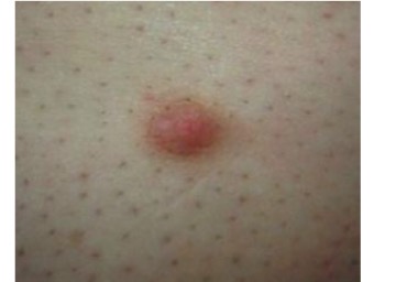
Dermatofibroma: firm nodule, often w/ hyperpig
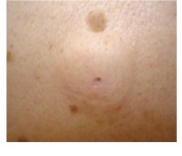
Epidermal Inclusion Cysts: tethered subcut cyts, often w/ malodorous whitish-yellow substance/discharge
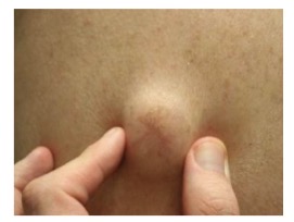
Lipomas: rubbery, may be mass like BUT freely mobile
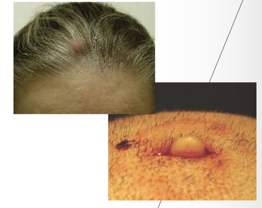
Pilar Cyst: MOBILE cysts that on excision reveals pearly-white balls
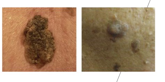
Seborrheic Keratosis
verrucous (“warty lesions”) texture
appear like sutck on/flattened ball of wax
erytheme (if inflamed), may crumble/bleed if picked
mimic for squamous cell carcinoma
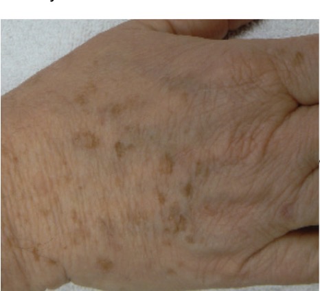
Solar Lentigo: light brown, uniform color BUT may be asymmetric
mimic for melanoma in situ
Cancerous/Pre-cancerous skin lesions
Actinic keratosis
cutaneous horn/keratotic scale
squamous cell carcinoma (SCC)
basal cell carcinoma (BCC)
superficial
nodular
ulcerated
Melanoma
amelanotic
melanoma in situ
acral melanoma
blue-black melanoma
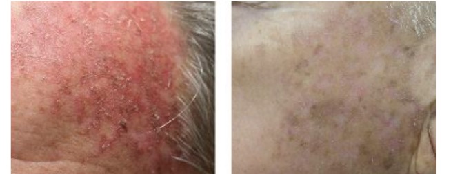
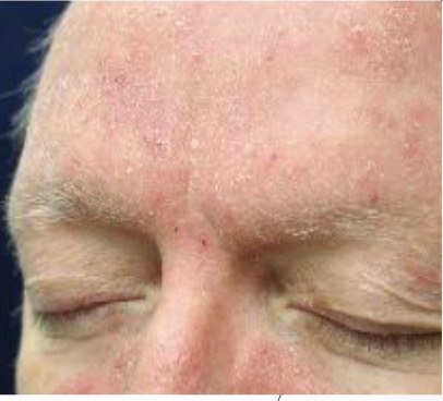
Actinic Keratosis (precursor to SCC): superficial keratotic papules
“come and go” on sun damaged skin
easier to feel than see
MIMIC: superficial xerosis/ seborrheic dermatitis
scale is less keratotic (more dry)
improves w/ moisturizers or mild topical steroids
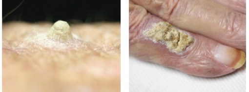
Cutaneous horn/keratotic scale
scale of keratin
occurs w/ actinic keratoses and SCC
cutaneous horn should be biopsied to r/o SCC
MIMIC: warts
skin-colored/pink
often have hemorrhagic punctae (tiny, pinpt spot of bleeding <3mm, appear like small red/purple dots)
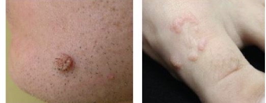
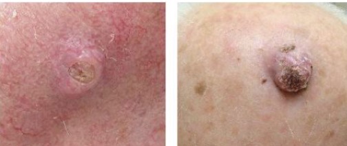
Squamous Cell Carcinoma (SCC)
keratocanthomas are rapidly arising SCC w/ crateriform center w/ smooth, firm border
Highest areas of metastasis: lips/ears/scalp
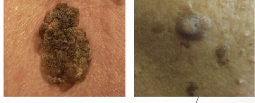
MIMIC: Seborrheic Keratosis (benign skin lesions)
stuck on/flattened ball of wax
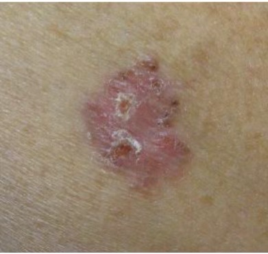
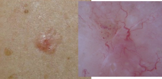
Superficial Basal Cell Carcinoma
pink patch, often w/ focal scaling (localized, distinct area of dry, flaking skin cells)
will NOT heal
MIMIC: Actinic Keratosis (precursor SCC) & SCC in Situ
due to keratotic scaling of SCC
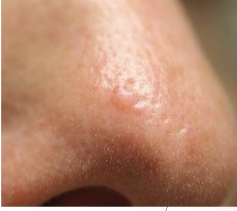
Nodular Basal Cell Carcinoma
pink papule w/ translucent or pearly white appearance (maybe focal pig)
usually w/ overlying telangiectasias
MIMIC: Sebaceous Hyperplasia
yelllowish globular papules, usually w/ central depression
telangeictasis go AROUND
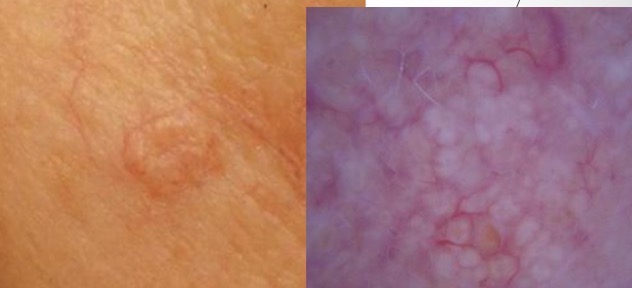
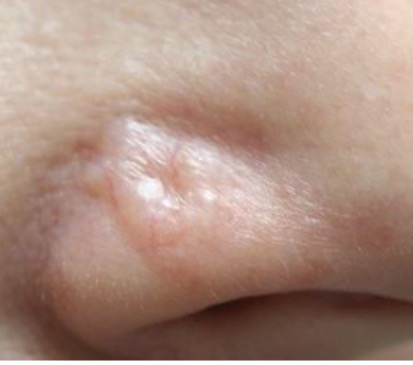
Basal Cell Carcinoma (BCC): pearly pink plaque w/ central depression and TELANGIESTASIAS
MIMIC: Fibrous Papule
skin-colored NO telangeictasias
may become excoriated (loss of skin’s surface)
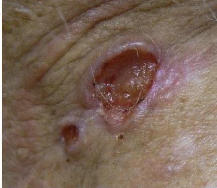
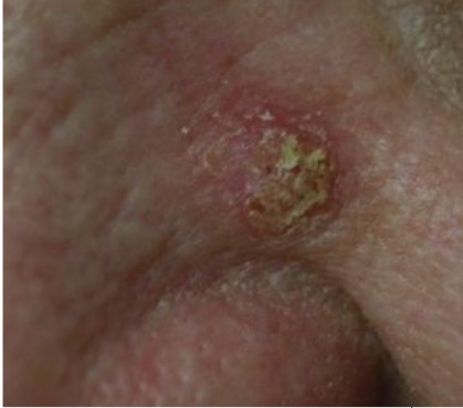
Ulcerated Basal Cell Carcinoma: non-healing ulcer, resulting in rolled border/edge
MIMIC: SCC
may be ulcerated
on exam edges FIRMER to palpation than BCC
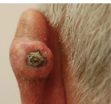
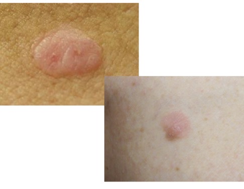
Amelanotic Melanoma:
MC in very fair-skin
evolution or rapid change #1 FEATURE
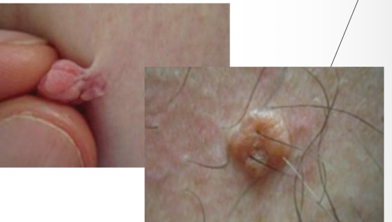
MIMIC: Skin tags/Intradermal Nevi
soft, fleshy, typ around neck/axillae/back
may have hint of brown pig
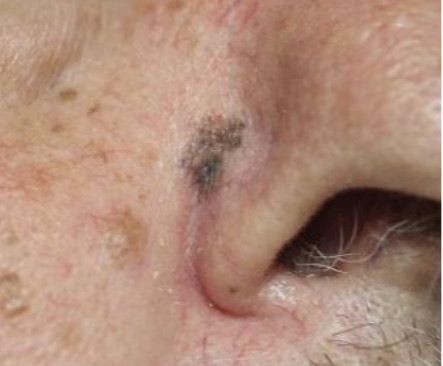
Melanoma in Situ
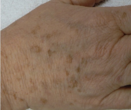
MIMIC: Solar Lentigo (benign skin lesion)
light brown, uniform color BUT may be asymmetric
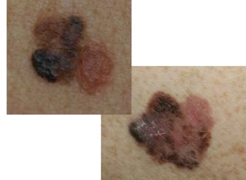
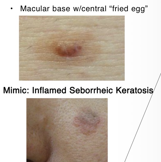
Melanoma:
can arise from existing nevi (benign)
varigated color (brown/reds)
MIMIC: Dysplastic Nevus
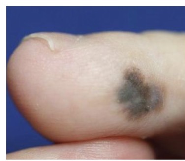
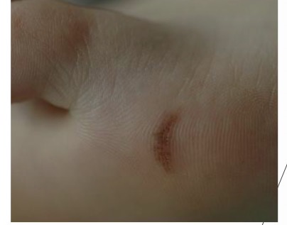
Acral Melanoma:
rapid change/evolution
consider biopsy >7mm, rapidly growing, or concerning features on dermoscopy
MIMIC: Acral Nevus
likely benign <7mm; dermoscopy reassuring (parallel fullow/lattice pattern)
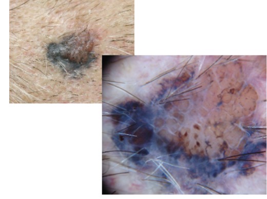
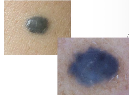
Melanoma w/ blue-black areas:
blue-black areas very concerning ESP if asymmetric
MIMIC: Blue Nevus
homogenous blue-gray appearance both clinically and dermoscopy
Vascular/Purpuric Skin Lesions
Spider Angioma
Spider Vein
Cherry Angioma
Petechia/purpura
Ecchymosis
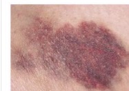
Ecchymosis: blood outside vessels; often secondary to bruising/trauma (seen in bleeding disorders)
Color/size: purple/purplish blue, fading green, yellow, and brown w/ TIME.; variable size, >petechiae mm
shape: rounded/oval/irregular; maybe subcut flat nodules (HEMATOMA)
Pulsatility and Effect of Pressure: NONE
Distribution variable
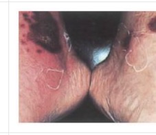
Petechia/Purpura: blood outside vessels possible suggestive of bleeding disorder.
Petechia → emboli to skin
palpable purpura in vasculitis
Color/size: DEEP red/ reddish purple; FADE over time
petechia 1-3 mm; purpura larger
Shape: rounded/ smt irregular; flat
Pulsatility and Effect of Pressure: NONE
Distribution Variable
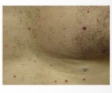
Cherry Angioma: NO SIG, increases in size and numbers w/ age
Color/SIze: bright/ruby red (w/ age purplish) 1-3mm
Shaped: Round/flat/smt riase; may have purple halo
Pulsatility and Effect of Pressure: NONE (partial blanching)
Distribution around TRUNK and extremities
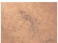
Spider Vein: accom increased pressure in SUPerficial veins (AKA varicose veins)
Color/Size: Bluish w/ variable size
Shape: variable ~spider/linear/irregular/cascading
Pulsatility and Effect of Pressure: NONE (no blanching, but diffuse blanches vein)
Distribution often ON LEGS near veins. also ant chest
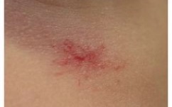
Spider Angioma: singles are normal and common on face/chest. Typ of preg and liver disease
Color/Size: Fiery Red; v small-2cm
Shape: central body (smt raised) surrounded by erythema and radiating legs
Pulsatility and Effect of Pressure: often seen in center of spider when pressure with a glass slide is applied; pressure causes blanching of spider
Distribution around face/neck/arms/upper trunk almost never below waist
Acne Vulgaris Primary Lesions
Mild Acne: comedonal, occasional papules (solid raised elev of skin <1cm)
Moderate Acne: Inflammatory; comedones, papules and pustules (small circumscrib elev of purulent fluid)
Severe Cystic Acne: nodulocystic (deep, painful, inflamed lumps/nodules under skin)
Acne Vulgaris Secondary Lesions
Pitting and Scarring
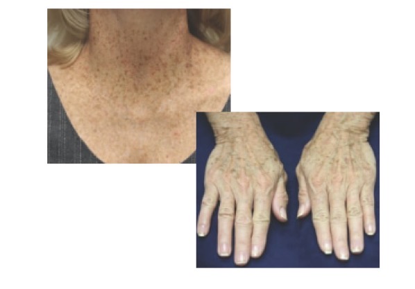
Sun Damage: Solar Lentigo
Bilat Symmetric; brown macule (<1cm flat area of color change) on sun exposed skin
not freckle
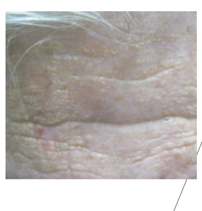
Sun Damage: Solar Elastosis
Yellowish white macules (<1cm flat skin color change) on sun-exposed skin, ESP forehead
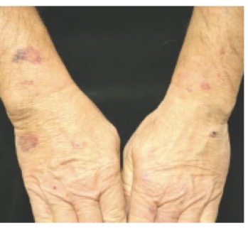
Sun Damage: Actinic Purpura (skin lesion)
Ecchymosis (bruise) to dorsal forearms and hands
doesn’t extend about “shirt-sleeve” on upper arm
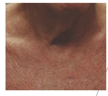
Sun Damage: Poikiloderma
Red patches in sun-damaged areas w/ fine telangiectasias and area of BOTH hyperpig and hypopig
Sun Damage: Wrinkles
Start earlier age
Sun Damage: Cutis Rhomboidalis Nuchae
Deep wrinkles on POSTerior neck in “crisscross” pattern
Male Pattern Hair Loss
Frontal hairline regression
thinning on posterior vertex
Female Hair Pattern Loss
Thinning from crown down hairline w/o hairline regression
Alopecia Areata
Sudden onset of clearly demarcated hair loss
SMOOTH skin w/o hair
NO visible scaling or erythema
Scarring Alopecia
Shiny skin w/ complete loss of hair follicles
Discoloration
Tinea Capitis
Round scaling patches of alopecia
Dermoscopy: broken hairs/corkscrew hair visible
maybe boggy on palpation
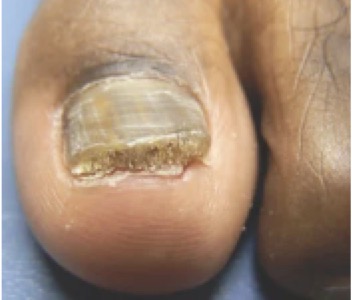
Onchomhycosis: nail thickening and subungal debris
usually from dermatophyte but also molds (athletes ft)
tinea pedia precursor
typ in older folk, unable to keep toe hygeine
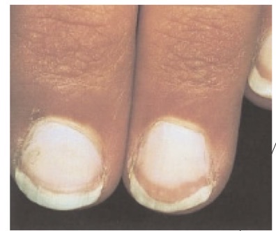
Terry Nails: nail plate turn white w/ ground-glass appearance, distal reddish brown, and obliteration of lunula
typ affects ALL nails
asso w: liver diease, heart failure, DTM
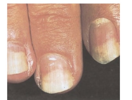
Onycholysis: painless separation of whitened nail plate from nail bed (=enlarged free edge)
usually result of trauma (distal → prox)
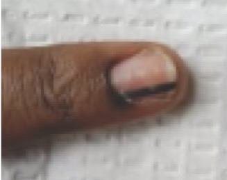
Melanonychia: linear streak formed as the nail grows out, caused by increased pigmentation in nail matrix
NEVUS: thin and uniform
SUBUNGAL MELANOMA: wide and/or irregular
in mult nails? may be from ethnic variation
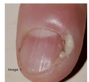
Paronychia: superficial infection of proximal and/or lateral nail folds
S/Sxs: red, swollen, tender
caused by localized trauma: nail biting, manicure, freq immersion in water (hand washing!)
Clubbing of Fingers
Bulbous swelling of soft tissue at base of nail ( angle >180deg b/tw nail and prox fold)
Asso w: congenital heart disease, interstitial lung disease, lung cancer, IBD, malignancies
related to vasodilation (maybe hypoxia? not largely understood)
Pitting of Nail
punctate depression of nail plate (defective layering by prox nail matrix)
asso w: reactive arthritis, sarcoidosis, alopecia areata, atopic/chemical dermatitis
Transverse Linear Depressions (Beau Lines) of Nail
Transverse depression of nail plate usually bilaterally
result of: severe illness, trauma (manicure), cold exposure
Self Examination
USE: mirror in well lit room
Steps
1: Examine full body front/back then left/right
2: Check elbows, forearms, underarms and palms
3: Look at backs of legs, feet, webspaces b/t toes and soles of feet
4: Examine neck and scalp w/ hand mirror, separate hair
5: Check back and buttock using hand mirror
Patient Education
Avoid UV radiation and tanning beds
Regular use of suncreen: SPF 30+ w/ reapp every 2 hrs outside/in water
USPSTF
United States Preventative Services Task Force
Counsel adults 24yr+ w/ fair skin types regarding minimizing exposure
AAD
American Academy of Dermatology
High risk persons should follow with dermatology for regular clinical skin exmas
ACS
Doesn’t have specific guidelines!
Recommends self-exams for persons at increased risk
Point-of-Care Ultrasound (POCUS)
Purpose?
Help visualize structures of the skin, differentiate skin lesions
Helps determine need for non-invasive vs invasive management
Ex: cellulitis vs abscess
POCUS: Cellulitis
Red Swollen area w/..
Tenderness, warmth, pain
Possible fever if severe
Induration to palpation
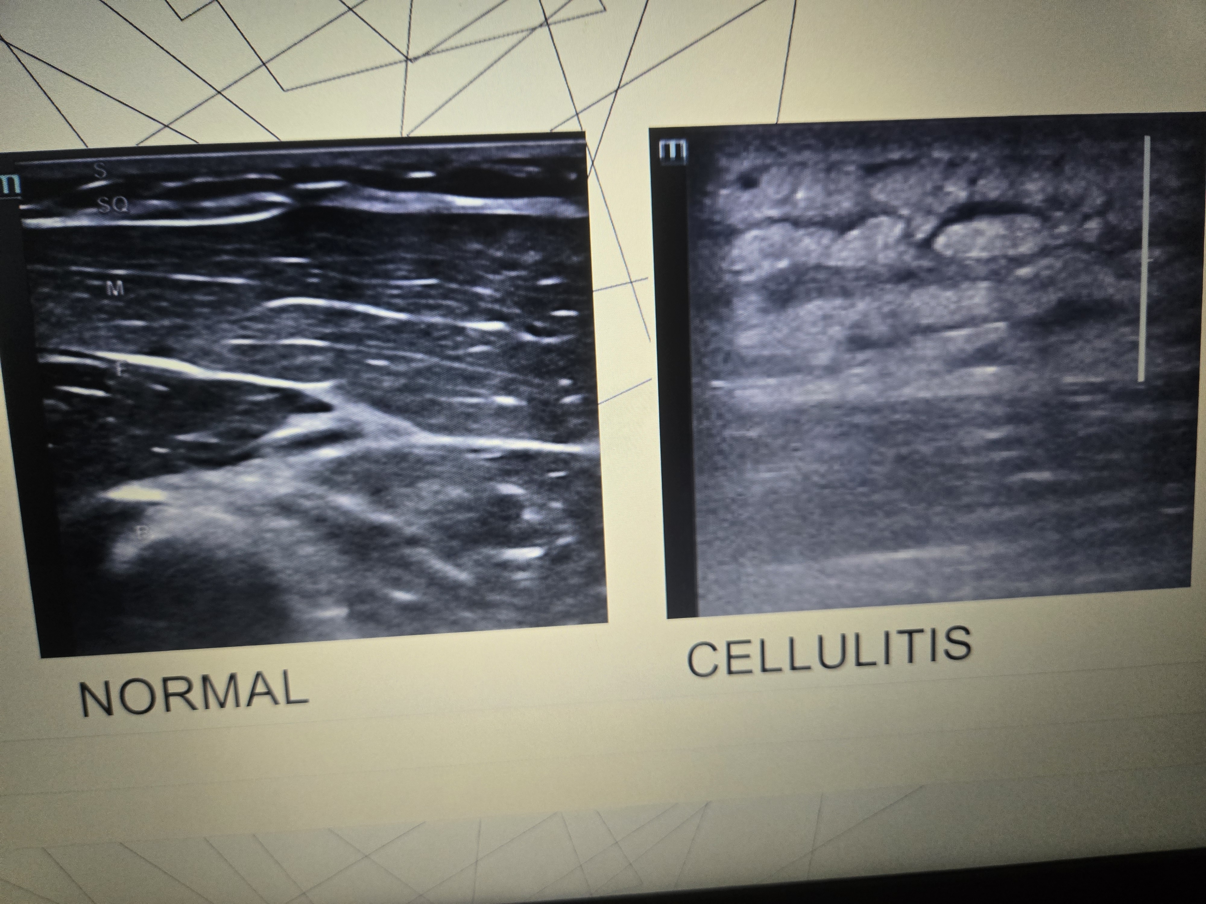
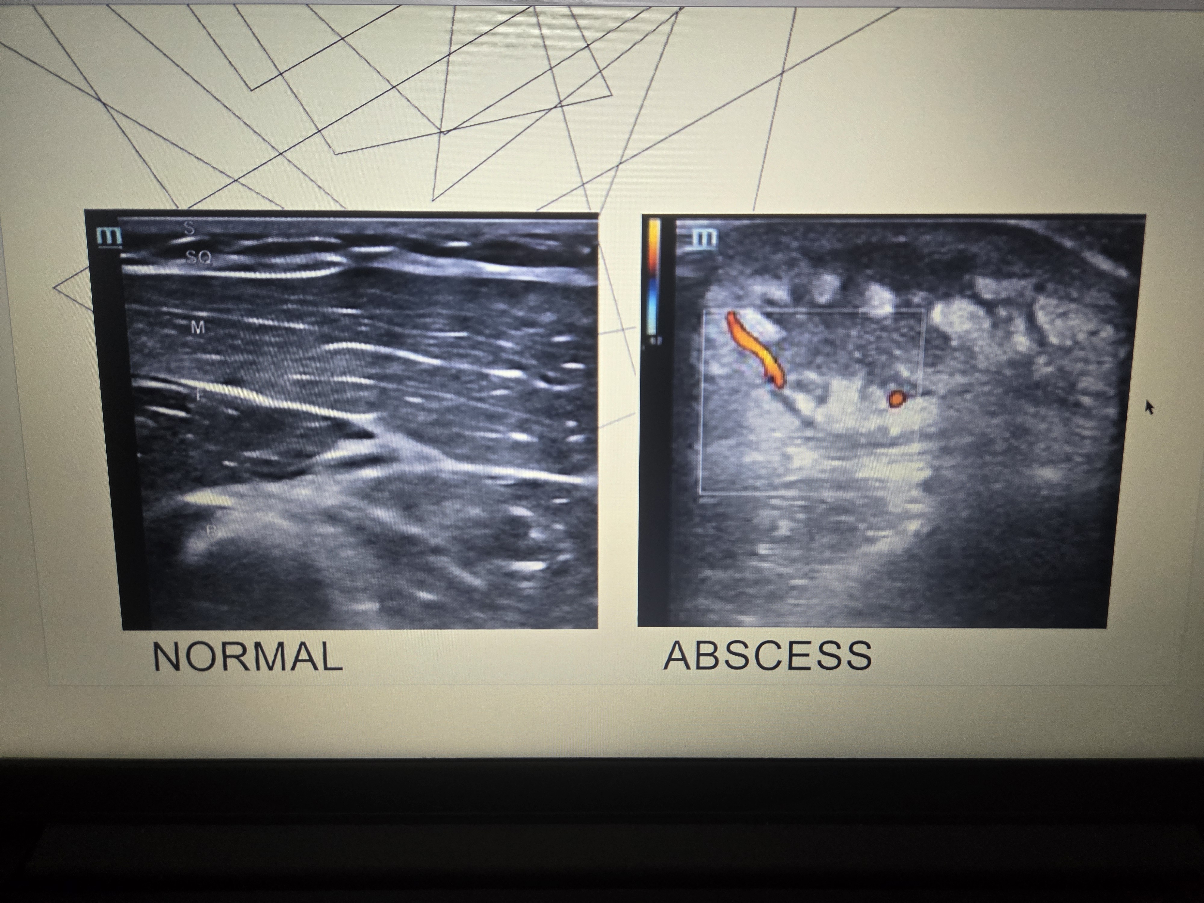
POCUS Abscess
Red swollen lump w/… tenderness, warmth, often more intense pain, and central pocket of pus
Possible fever (esp not drained)
Will feel induration (hardening) esp around lump and fluctuance (liquidy under pressure)