Lecture 17+18- damage, regeneration and repair in the audio-vestibular (AV) system
 Call Kai
 Learn
Learn Practice Test
 Spaced Repetition
 Match
Match Flashcards
 Knowt Play
1/20

There's no tags or description
Looks like no tags are added yet.
Name | Mastery | Learn | Test | Matching | Spaced | Call with Kai | Chat |
|---|
No analytics yet
Send a link to your students to track their progress
21 Terms
What are the main types and causes of hearing loss?
Conductive hearing loss: problem transmitting sound through the external/middle ear.
Causes: earwax/blockage, otitis externa/media(infections), glue ear, middle-ear bone problems, ruptured eardrum.
Treatment can include wax removal, antibiotics or grommets.
Sensorineural hearing loss: damage to the cochlea, auditory nerve or brain; cochlear hair cells are the main target.
Mixed hearing loss: combination of conductive + sensorineural loss.
Severity: mild → moderate → severe → profound.
Severe/profound loss may require cochlear implants.
Hearing loss prevalence increases with ageing.
What roles do inner and outer hair cells play?
Inner hair cells: provide ~95% of auditory nerve input → transmit sound information to the brain.
Loss of inner hair cells can cause profound hearing loss.
Outer hair cells: amplify cochlear vibrations → improve sensitivity and frequency selectivity.
Outer hair-cell loss contributes to mild–moderate hearing loss, often treated with hearing aids.
Ageing is a major cause; ~50% of people >75 have significant hearing loss.
How does noise exposure damage the cochlea?
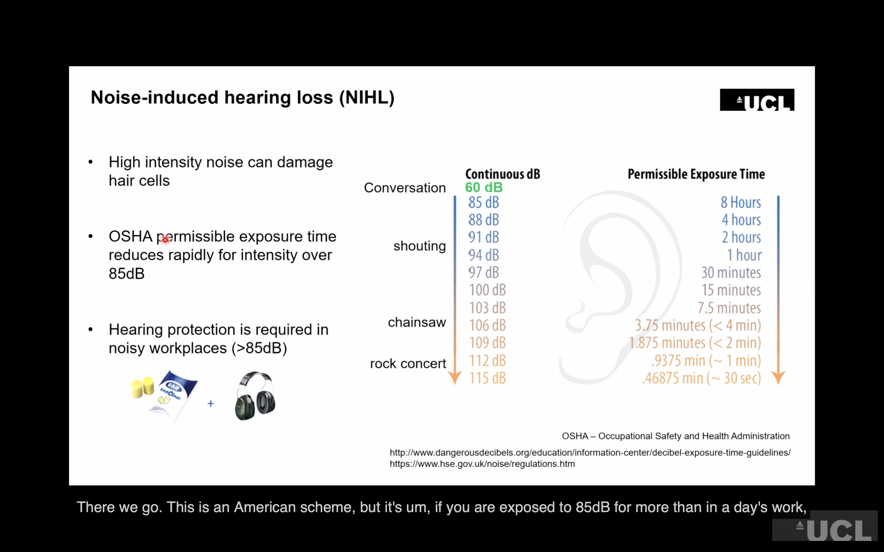
Major risk factors: noise, ageing, genetics and ototoxic drugs.
Increasing noise exposure causes:
Tip-link breakage → early damage that can repair.
Stereocilia damage/splaying with greater exposure.
Synaptic damage at pre- and postsynaptic levels.
Severe exposure → cell membrane and reticular-lamina disruption.
Hair-cell death through apoptosis or necrosis.
Damage is frequency-specific because the cochlea is tonotopic:
Base = high frequencies
Apex = low frequencies
Repeated exposure can produce frequency-specific notches on an audiogram.
Prolonged loud noise can eventually cause permanent hearing loss.
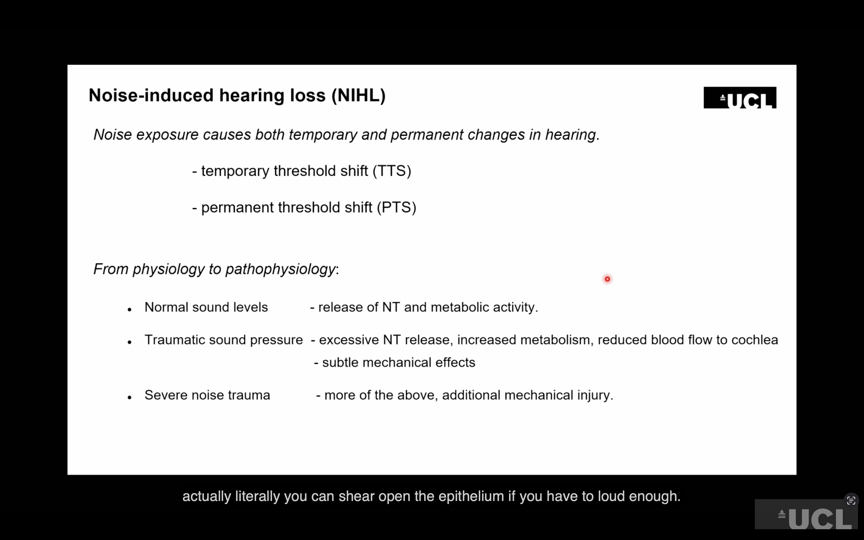
What is a temporary threshold shift (TTS), and why can hearing appear normal afterwards?
TTS: temporary increase in the hearing threshold after noise exposure → hearing later recovers.
Noise increases neurotransmitter release and metabolic demand; high levels can also reduce blood flow through vasoconstriction.
Severe noise can cause mechanical damage to the cochlea.
Example: mice exposed to 100 dB noise for 2 hours developed up to a 40 dB threshold shift, which largely recovered over subsequent weeks.
Important: recovery of the hearing threshold does not necessarily mean the cochlea is undamaged.
Permanent synaptic damage can remain, causing difficulty hearing particularly in noisy environments.
How is cochlear damage measured and related to hearing?
Audiogram: measures hearing thresholds; damage to a particular cochlear frequency region produces corresponding hearing loss.
Auditory Brainstem Response (ABR): electrodes measure neural responses to sound without requiring behavioural responses.
ABR waves reflect activity progressing through the auditory pathway:
Wave I: cochlea/auditory nerve output.
Later waves reflect activity further through the brainstem and auditory pathway.
Severe cochlear damage can cause loss of hair cells, organ of Corti and nerve fibres.
Anatomical damage can match the frequency-specific pattern of the audiogram.
A recovered threshold with reduced Wave I can reveal persistent cochlear/synaptic damage that a standard audiogram misses.
What did the noise-exposure experiment in mice show?
Mice were exposed to 100 dB, 8–16 kHz band noise for 2 hours
This caused a temporary threshold shift (TTS) of up to 40 dB, particularly at high frequencies
Auditory brainstem responses (ABRs) were measured using electrodes to record brain activity in response to sound
ABR waves show activity travelling from the cochlea/auditory nerve → cochlear nucleus → inferior colliculus → cortex
Hearing thresholds recovered to approximately normal within 2–8 weeks
However, Wave I amplitude remained reduced
Wave I represents output from the cochlea through the auditory nerve
This showed that cochlear output remained permanently reduced despite recovery of hearing thresholds
What is “hidden hearing loss” and what causes it?
Noise exposure can permanently damage auditory nerve fibres/synapses even when a standard audiogram appears normal.
After noise exposure there is a reduction in synaptic ribbons and auditory nerve fibres connecting to inner hair cells/audiotry nerve fibres get disconnected, reducing cochlear output = hidden hearing loss because conventional hearing tests may show normal thresholds.
The lost nerve fibres normally oversample information from inner hair cells, helping the brain understand sounds in complex/background noise.
hidden hearing loss can cause particular difficulty understanding speech in noisy environments despite a normal audiogram.
What is age-related hearing loss (presbycusis), and what happens to the cochlea?
Most common sensory deficit; affects approximately 25% of people aged 65–75+ and >70% of people over 75.
Usually progressive, bilateral, symmetrical and sensorineural.
Strongly influenced by genetics, which affect susceptibility and rate of progression.
Outer hair cells are particularly vulnerable and are progressively lost , leading to reduced sensitivity and frequency discrimination; inner hair cells and auditory nerve fibres can also be lost.
Hair cells do not regenerate in humans, so damage is permanent.
Hearing loss is not caused by ageing alone: noise, toxic medications/chemicals, smoking, diet and chronic health conditions can contribute.
Ageing + noise exposure have additive effects, meaning noise can accelerate age-related hearing loss.
What are the 4 main pathological types of age-related hearing loss?
Sensory: mainly loss of outer hair cells → typically high-frequency hearing loss.
Metabolic/strial: damage to the stria vascularis/transport epithelium, disrupting the ionic environment of the cochlea → broader-frequency hearing loss.
Neural: loss of spiral ganglion cells/neuronal connections → generally broad-frequency but potentially severe hearing loss.
Mechanical/conductive: stiffening of the basilar membrane/cochlear mechanics, reducing its movement.
Later evidence suggests outer hair-cell loss is a major component, while auditory nerve fibre loss/hidden hearing loss may also contribute.
What is ototoxicity and what can cause it?
Ototoxicity: damage to the sensory organs of hearing and/or balance; can be cochleotoxic and/or vestibulotoxic.
It can occur as a side effect of drugs taken for another condition or through environmental/workplace chemical exposure.
Important examples:
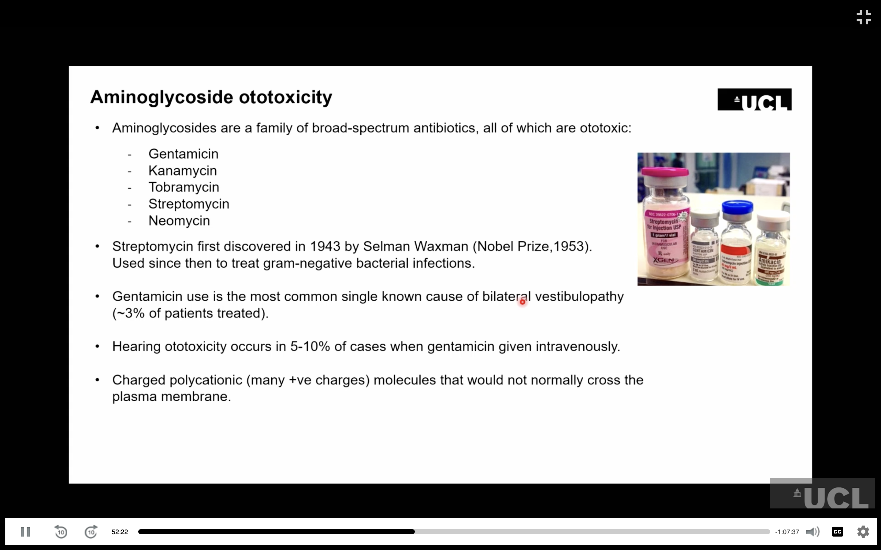
Aminoglycoside antibiotics such as gentamicin and streptomycin → hearing and/or vestibular damage.
Chemotherapy, especially cisplatin/carboplatin → often permanent, high-frequency hearing loss.
Quinine/antimalarials and aspirin overdose can also cause hearing problems.
More than 200 medications are potentially ototoxic.
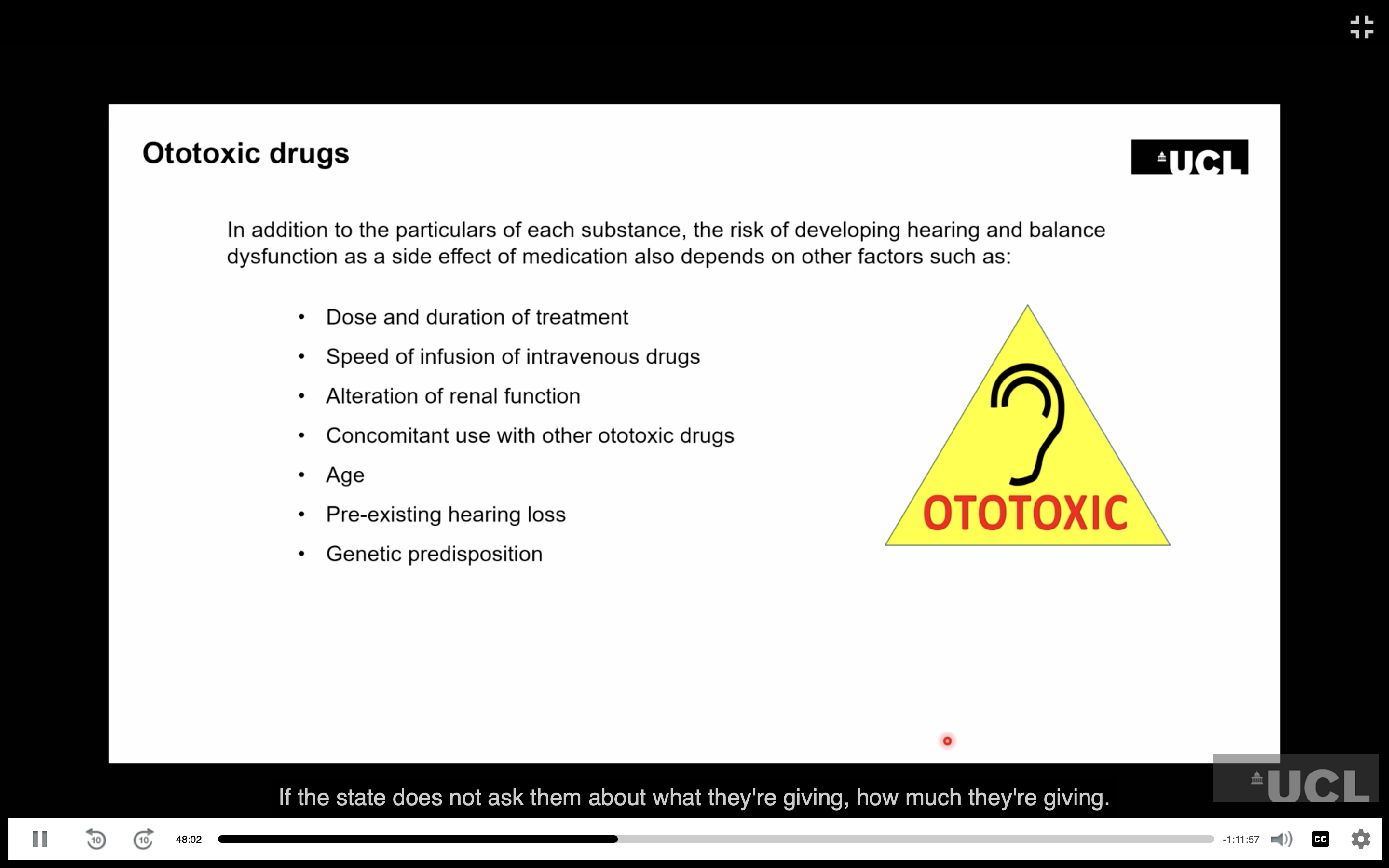
Risk depends on dose, duration, route of administration and combinations with other ototoxic drugs.
Loop diuretics can greatly worsen ototoxicity when combined with other ototoxic drugs.
Early signs include tinnitus, hearing loss, dizziness/vertigo and oscillopsia.
Ototoxic drugs can also affect the kidneys and neuromuscular junction.
How do ototoxic drugs enter and damage cochlear hair cells?
Drugs mainly reach the cochlea through the blood/vasculature, particularly via the stria vascularis, then enter the inner-ear fluid and hair cells.
Aminoglycosides are positively charged and can enter hair cells mainly through mechanotransduction channels, which are partly open even at rest.
Increased sound can increase drug entry through these channels; other channels and endocytosis may also contribute.
Aminoglycosides can damage mitochondria/ribosomal machinery, causing:
Reactive oxygen species (ROS)
Cytochrome-c release
Activation of apoptosis/programmed cell death
Cisplatin cross-links DNA to kill cancer cells but is also taken up by hair cells, particularly outer hair cells, causing permanent, often high-frequency hearing loss.
Cisplatin entry may involve mechanotransduction channels, diffusion or other pathways.
What are the current strategies for restoring hearing, and why is regeneration important?
Hearing aids: microphone + sound processor amplify sound; less effective in noisy environments.
Cochlear implants: electrodes directly stimulate auditory nerve fibres.
Auditory brainstem implants: stimulate the auditory brainstem when cochlear nerve fibres are unavailable.
These are devices, not drugs.
Regeneration is the major goal because restoring lost hair cells/neurons could treat the underlying damage rather than compensate for it.
Future approaches include gene therapy, pharmacological therapies and stem cells.
How do cochlear and auditory brainstem implants work?
Cochlear implant:
Electrode array inserted into the cochlea, typically covering ~1.5 of its 2.5 turns.
Usually ~12 electrodes, with up to 22 possible.
Electrodes stimulate auditory nerve fibres along the tonotopic axis.
~12 frequency channels replace the information normally carried by ~30,000 nerve fibres.
Allows speech and communication despite reduced frequency resolution.
Sound is often perceived as electronic/squeaky rather than natural.
Auditory brainstem implant:
Used when there are insufficient auditory nerve fibres to stimulate.
Electrode placed on the auditory brainstem.
Less effective and has poorer frequency tuning than a cochlear implant.
Why is hearing loss in humans considered irreversible?
Humans are born with essentially their full complement of hair cells.
Mammalian hair cells are not naturally replaced after they are lost.
Progressive loss:
Outer hair-cell loss → hearing threshold increases.
Complete loss of hair cells in a region can produce ~40 dB hearing loss.
More extensive loss can produce 70–80 dB+ loss.
Severe loss can cause sound to be heard through the opposite ear by bone conduction.
Extensive hair-cell loss produces a permanent threshold shift.
Animal models show the same lack of regeneration in mammals.
How do hair cells die, and why does the mechanism matter for treatment?
Hair cells can die by both:
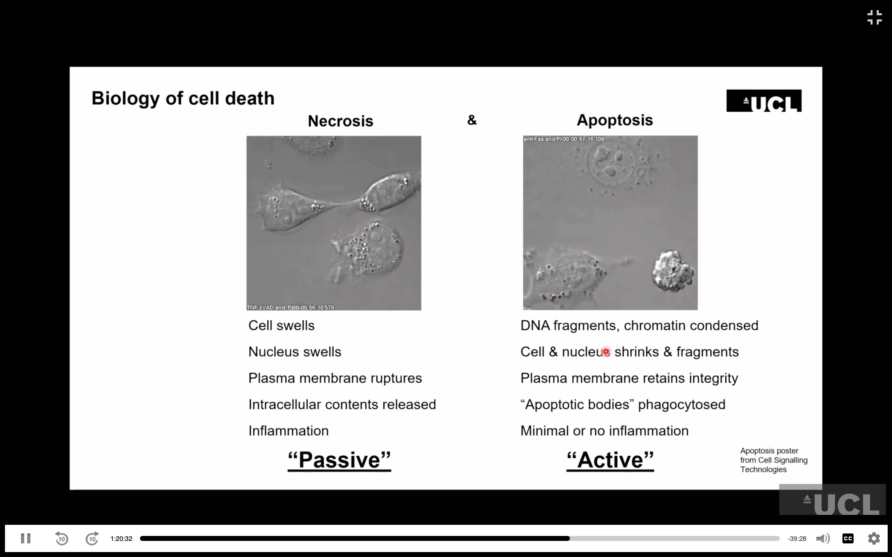
Apoptosis: controlled cell death; cell contents remain contained and are subsequently cleared.
Necrosis: cell swells and membrane breaks down → cellular contents are released.
Necrosis:
Causes inflammation.
Can damage neighbouring cells through a bystander effect.
Therefore, preventing the initial damage may be particularly important.
Hair-cell death depends on factors such as:
Ageing
Noise exposure
Ototoxic drugs
There are also intermediate forms of cell death, creating potential pharmacological targets.
In guinea pigs after severe ototoxic damage, hair cells can progressively disappear over 8–12 weeks, producing permanent hearing loss.
Which animals can regenerate hair cells, and what can we learn from them?
Chickens, frogs, fish and other non-mammalian vertebrates can regenerate hair cells.
Many cold-blooded vertebrates can produce hair cells throughout life.
Continuous hair-cell turnover depends on ongoing cell division/stem-cell activity.
Birds normally have ongoing hair-cell production in some auditory/vestibular systems.
The avian basilar papilla is tonotopic and mechanically detects sound, although it differs structurally from the mammalian cochlea.
In chickens, hair-cell production is normally stopped after development but can be reactivated following noise or damage.
What happens when a chicken's basilar papilla is damaged by noise?
Chickens exposed to 120 dB noise for 48 hours develop hair-cell damage, particularly around the 1.5 kHz region.
Damaged hair cells disappear, but new hair cells appear within ~10 days.
New cells develop new hair bundles, recapitulating aspects of embryonic development.
Hair-cell numbers subsequently recover.
Hearing thresholds also recover substantially over time, although not always completely to baseline.
This demonstrates that non-mammalian auditory systems can achieve both hair-cell regeneration and functional hearing recovery.
What are the three endogenous mechanisms of hair-cell recovery?
1. Supporting-cell division
Supporting cells re-enter the cell cycle.
Divide to produce new cells, including hair cells and supporting cells.
2. Phenotypic conversion
A supporting cell directly converts into a hair cell.
Possible because supporting cells and hair cells originate from the same developmental progenitors.
3. Repair
Existing damaged hair cells can repair structures such as stereocilia bundles and tip links.
These mechanisms are not mutually exclusive.
Phenotypic conversion may provide faster recovery, while cell turnover/division may be slower.
What evidence supports supporting-cell division and phenotypic conversion in hair-cell regeneration?
Cell-cycle markers can identify cells that have re-entered the cell cycle:
Historically tritiated thymidine.
More recently BrdU and other chemical/antibody-based markers.
In damaged chick basilar papilla:
Supporting cells take up cell-cycle markers.
New hair cells and supporting cells appear together.
Increased pairs/doublets suggest supporting-cell division.
Cell-cycle activation occurs locally around the damaged region.
Phenotypic conversion is also demonstrated:
After hair cells are completely removed in vitro, new hair cells appear.
Many new hair cells do not contain cell-cycle markers.
Therefore, they cannot have arisen through cell division alone.
This supports supporting-cell → hair-cell phenotypic conversion.
Both mechanisms can operate in the same tissue.
Can damaged hair-cell structures repair themselves?
Tip links between stereocilia can be repaired.
Removing calcium from the surrounding solution breaks tip links.
Returning the cells to normal calcium allows tip links to recover within ~24 hours.
Similar recovery has been observed in chicken and mouse hair cells.
In mouse cochlea, recovery of dye uptake through mechanotransduction channels occurs over ~24 hours, supporting restoration of transduction.
Repair may therefore be another potential therapeutic target, although it appears to be a low-probability event and is difficult to study.
What approaches are being investigated to induce hair-cell regeneration?
Gene therapy
Targets Atoh1, a master transcription factor required for hair-cell formation.
Removing Atoh1 prevents hair-cell production.
Overexpressing Atoh1 can induce hair-cell formation from suitable inner-ear cells.
First human gene-therapy trial used an adenovirus injected into the cochlea to target supporting cells.
Trial was not successful and some hearing loss occurred, but it demonstrated that the approach was feasible and largely safe.
More recent gene therapy has successfully replaced missing genes in some specific genetic hearing disorders, particularly in young children.
Pharmacological therapy
Targets Notch signalling, which normally prevents neighbouring supporting cells from becoming hair cells through lateral inhibition.
Blocking γ-secretase reduces Notch signalling → removes the inhibition → promotes hair-cell formation.
Worked particularly well in immature systems.
Adult noise-damaged mice showed some functional recovery, but the subsequent clinical trial was not effective.
The trial was nevertheless important for developing future inner-ear drug-delivery approaches.
Stem cells/organoids
Stem cells can be pluripotent or multipotent.
Adult cells can be reprogrammed into induced pluripotent stem cells using stem-cell factors.
3D organoids can reproduce aspects of inner-ear development.
Mouse organoids can contain hair cells and supporting cells arranged in an inner-ear-like epithelium.
Human induced pluripotent stem cells can also generate inner-ear organoids.
Stem-cell-derived neuronal progenitors are being investigated to replace lost cochlear neurons, potentially followed by cochlear implantation