a+p: skin
 Call Kai
 Learn
Learn Practice Test
 Spaced Repetition
 Match
Match Flashcards
 Knowt Play
1/58

There's no tags or description
Looks like no tags are added yet.
Name | Mastery | Learn | Test | Matching | Spaced | Call with Kai | Chat |
|---|
No analytics yet
Send a link to your students to track their progress
59 Terms
skin (aka cutaneous membrane)
made up of epithelial tissue and connective tissue
largest organ in the body - 7% of total body weight

epidermis
epithelial tissue - keratinized stratified squamous epithelium
keratin makes the cell very durable and protects epidermis
avascular (no directly blood supply)
regenerates/replaces every 35-45 days

cells of the epidermis
keratinocytes
melanocytes
dendritic cells
tactile cells
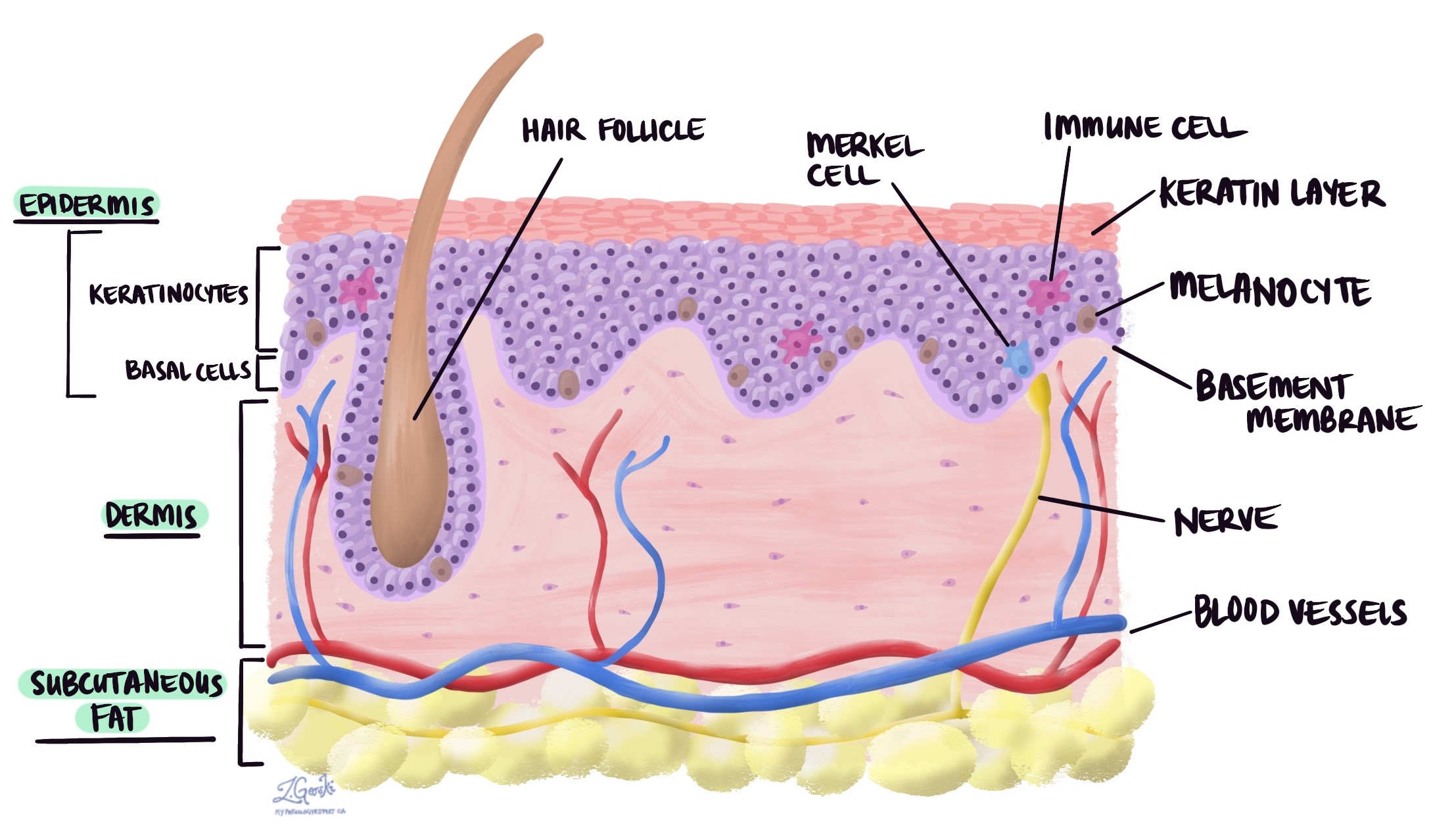
keratinocytes
produce keratin - provides skin with durability
allows epidermis to function as a protective layer
primary type of cell found in the epidermis and is incorporated to the plasma membrane
produce vitamin D precursor
melanocytes
produce melanin
help protect keratinocytes from UV damage
dendritic cells
layer of immune protection
tactile cells
sensory reception
sense touch, makes epidermis sensitive to touch
stratum basale (stratum germinativum)
deepest layer - is attached to the dermis (where we find blood vessels)
single row of constantly dividing stem cells
constantly making new cells - new cells get pushed to the top by old cells
cells reach skin surface in 35-45 days - die when they move up because they’re too far away from the dermis
cells in the stratum basale are closest to the dermis (oxygen and blood supply) which supports their high metabolic processes

the _______ ________ is where our epithelial tissue transitions to connective tissue
stratum basale
melanocytes are found in the _____ ________ and produce ________ _______ in the skin
stratum basale, melanin pigment
melanin production
melanocytes release melanin-filled melanosomes
taken up/used by keratinocytes to protect their nuclei from UV damage
UV exposure increases melanin production
layers of the epidermis
stratum corneum
stratum lucidum
stratum granulosum
stratum spinosum
stratum germinativum (basale)

stratum granulosum
is made up of cells that used to be in the stratum spinosum but got pushed up
start of keratinization - increase friction = increase keratinization (callus on hands)
cells start to die off because they’re too far from dermal capillaries to survive
includes lamellar granules
lamellar granules in stratum granulosum
promote water retention - creates a layer to prevent water from escaping
topical medications
topical = polar
can’t penetrate lamellar coating because it is repelled by the hydrophobic heads
unable to enter blood vessels in dermis
since these medications can’t pass all the way through to the blood stream it only has a local effect (affects where you applied it on surface of skin)
transdermal medications
transdermal = nonpolar
able to cross epidermis - passes through bilayer
potential for systemic absorption - able to be absorbed into the bloodstream
ex: birth control patch
stratum lucidum
only present in thick skin → extra protection
ex: palms of hand and soles of feet
not found in all regions of the skin
thick skin
palms, soles, fingers, toes - areas of the body where we encounter friction, abrasion, and mechanical functions/stress
contains stratum lucidum - extra layer
thicker stratum corneum
sweat glands but no hair follicles or sebaceous oil glands
thicker layer of cells in all layers
thin skin
areas of the body under less mechanical stress
fewer layers of cells at all levels - doesn’t require extra layers of protection
contains sweat/oil glands, hair follicles, and sebaceous glands
thick or thin skin refers to the _________
epidermis
stratum corneum
thickest layer of skin - ¾ of thickness of epidermis
protects deeper cells from damage or pathogens entering
prevents water loss - maintains hydration
dead keratinocytes at surface shed regularly - losing keratinocytes very frequently
melanin
produced by melanocytes
protects skin from UV damage and affects skin color
eumelanin
pheomelanin
freckles/moles
more melanin granules = darker skin
how do melanocytes affect skin color?
all humans have roughly the same number of melanocytes but their activity varies which causes skin color differences
eumelanin
brown/black
pheomelanin
yellow/red (lips)
freckles/moles
local accumulations of melanin
skin color and UV light
excessive UV light exposure damages skin
damages keratinocyte DNA —> increased cancer risk
exposure to ______ is essential for ________ ___ production and required for ______ _________
sunlight
vitamin D
calcium absorption
melanin _______ UV light _________
reduce, penetration
darker skin is more protective
individual and population level effects on skin color and UV light
ancestry:
if you are a descendant from those who did not live in light areas or were exposed to sunlight, then their bodies were adapted to produce lower amounts of melanin in the melanocytes
less active melanocytes on a daily basis
skin color
melanin
carotene
hemoglobin
carotene
yellow/orange pigment
acquired from fruits/vegetables (ex carrots)
converted to vitamin A
promotes healing in the skin and recovery from tissue damage
hemoglobin
less melanin → keratinocytes more transparent (can see the blood from outside of the skin - pinker skin and red lips)
color of blood sometimes visible through epidermis
skin color alterations
cyanosis
pallor
erythema
jaundice
bruises
cyanosis
low oxygenation of hemoglobin (low oxygen level of blood)
peripheral cyanosis
hemoglobin carrying less oxygen may blood look more blue or purple than red, affecting skin color
peripheral cyanosis
reduced blood flow in extremities - blueish tint in fingers or toes
pallor
anemia, low blood pressure
unusual paleness in our skin - not supplying with enough blood or blood doesn’t have enough hemoglobin
erythema
fever, hypertension (high blood pressure), inflammation, allergy
increase in blood supply - any immune response sending more blood to areas
jaundice
liver disorders - issue with liver functioning
causes yellow ish skin
bruises
clotted blood beneath skin
color changes over time as blotted blood changes
damage to blood vessels but not the skin
dermis
is deep to epidermis
papillary layer
reticular layer

papillary layer
attacked to overlying epidermis
“proper connective tissue”
areolar tissue
dermal papillae

areolar tissue
loose connective tissue
lots of ground substance → retains water/maintains hydration
dermal papillae
capillaries, nerves, touch receptors
blood supply that epidermis relies on
push up to epidermis
increased surface layer - helps keep them attached
friction ridges
dermal papillae and overlying epidermis in areas of thick skin
epidermal ridges that lie on top of dermal ridges
sweat from pores leave unique patterns - fingerprint
found on anterior surfaces of fingers and toes

friction ridges enhance _____ and ______ _________
grip, tactile sensation (sensitivity)
reticular layer
nerves, blood vessels, lymphatic vessels - leather is made from reticular layer
dense irregular tissue

dense irregular tissue
dense connective tissue
provides multidirectional strength and support - lots of collagen protein fibers that help give it that multidirectional strength and support
helps give out skin strength and support while interacting with other forces
dermal trauma
stria (stretch marks)
blisters
striae
tears to dermis caused by extreme stretching
epidermis is more able to withstand stretching than dermis
occurs during weight gain and pregnancy
blisters
fluid-filled pockets caused by repetitive trauma - repeated mechanical stress
separation of epidermal and dermal layers
the layers break and are disrupted - fluid then fills the space between the 2 layers, forming the blister
hypodermis
adipose and areolar tissue
highly vascular
extensive blood supply - sight of subcutaneous injection (absorbed by blood and sent to rest of the body)
anchors skin to muscle while allow movement
allows skin to move over out muscles
provides insulation
adipose tissue retains heat very well - maintains body heat
ground substance gives it a looser structure

wrinkles
reduction in protein fibers, ground substance, and adipose tissue in dermis/hypodermis - they decrease as we age
skin loses its support and flexibility, causing it to sag
deeper in more mobile regions - wrinkles are prevalent in areas around areas that move the most (eyes and mouth)
what accelerates the formation of wrinkles?
UV exposure
cigarette smoke
burns
tissue damage caused by heat, electricity, radiation, and chemicals - breakdown/denature proteins and cells in our body
first degree burns
second degree burns
third degree burns
first degree burn
only epidermis impacted
localized redness, edema
swollen, red, and painful
temporary, heals quickly
ex: sunburn
second degree burn
all of epidermis damaged - some of dermis impacted
blisters
potential for infection
the loss of the epidermis layer makes the skin more likely to get infected
third degree burn
epidermis and dermis destroyed, extends into hypodermis
nerve damage
causes it to be less damaged because our sense receptors are damaged
require medical intervention - too severe for body to heal itself
medical interventions for third degree burns
debridement (removal of damaged area)
antibiotics
temporary coverings (bcs we lost the protective layer and hydration)
skin grafts