4A. 1: Pelvis + LL
5.0(1)
Studied by 2 peopleCard Sorting
1/47
Earn XP
Description and Tags
Last updated 5:32 PM on 4/18/23
Name | Mastery | Learn | Test | Matching | Spaced | Call with Kai |
|---|
No analytics yet
Send a link to your students to track their progress
48 Terms
1
New cards
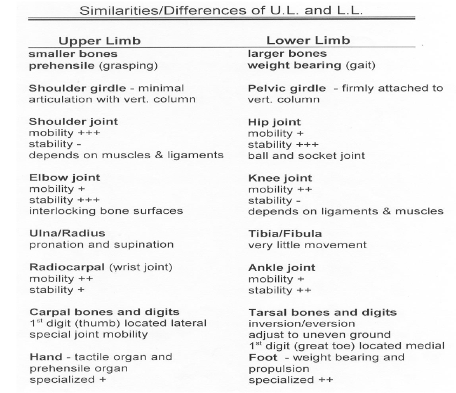
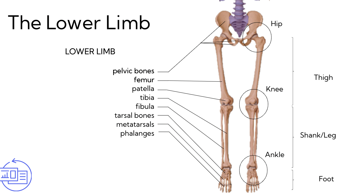
Upper Limb vs Lower limb
\
2
New cards
Why does extension happen on the posterior side in the lower limb?
Consequencive embryonic development
3
New cards
Consequencive embryonic development
around 8 weeks: limb rotation
* before that, arms and legs grow as little buds
arm: supinates
leg: pronates
* before that, arms and legs grow as little buds
arm: supinates
leg: pronates
4
New cards
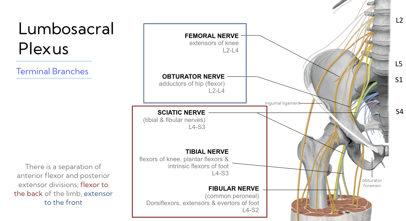
Lumbosacral plexus
\
5
New cards
All of the anterior rami that recombine to form these peripheral nerves come off of the ________ and ______ regions
lumbar and sacral
6
New cards
the lumbosacral plexus extends from __ to __
L2-S4
7
New cards
Femoral nerve
extensors of the knee
* L2-L4
* L2-L4
8
New cards
Obturator Nerve
adductors of hip (flexor)
* L2-L4
* L2-L4
9
New cards
Sciatic nerve
a combo of Tibial & fibular nerves (everything posterior)
* L4 - S3
* L4 - S3
10
New cards
Tibial nerve
flexors of knee, plantar flexors & intrinsic flexors of foot
* 4-S3
* 4-S3
11
New cards
Fibular nerve
(common peroneal)
Dorsiflexors, extensors, evertors of foot
* L4-S2
Dorsiflexors, extensors, evertors of foot
* L4-S2
12
New cards
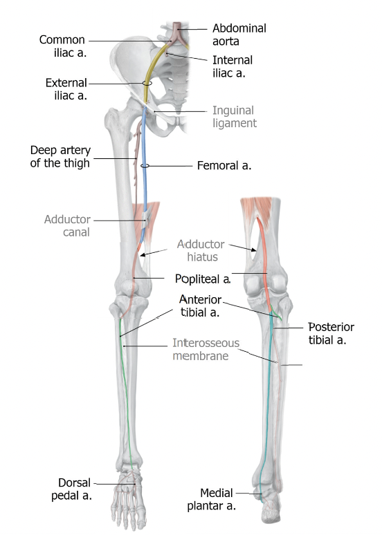
arterial supply
ALL blood starts of in the:
* ==Abdominal Aorta==
AA then bifurcates to form the:
* ^^L and R common Iliac arteries^^
These bifurcate again to form:
* Internal and external Iliac artery
external becomes:
* %%Femoral artery%%
Femoral goes through the Adductor canal, then through a hole called adductor hiatus to become:
* @@popliteal artery@@ (back of the knee)
popliteal artery bifurcates to form:
* ==anterior tibial artery==
This sneaks through interosseous membrane and comes round to the anterior aspect and becomes:
* ^^the dorsal pedal artery^^ (top of foot)
The other branch off popliteal is the:
* posterior tibial artery
which runs along the interosseous membrane to become:
* %%the medial plantar artery%% (bottom of foot)
This gives off a branch called:
* @@fibular artery@@ (supplies the lateral aspect the shank)
13
New cards
Internal and external iliac arteries
Internal IA:
* supplies musculature and viscera of pelvis
\
External IA:
* supplies lower limb
* supplies musculature and viscera of pelvis
\
External IA:
* supplies lower limb
14
New cards
When the external artery passes through the ______ it becomes the femoral artery
Inguinal ligament
\
\*Deep artery of the thigh comes off of femoral artery
\
\*Deep artery of the thigh comes off of femoral artery
15
New cards
there are _ routes of venus blood
2
((deep veins - blue) - same as arteries)
(superficial veins - green)
((deep veins - blue) - same as arteries)
(superficial veins - green)
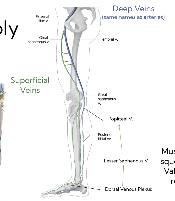
16
New cards
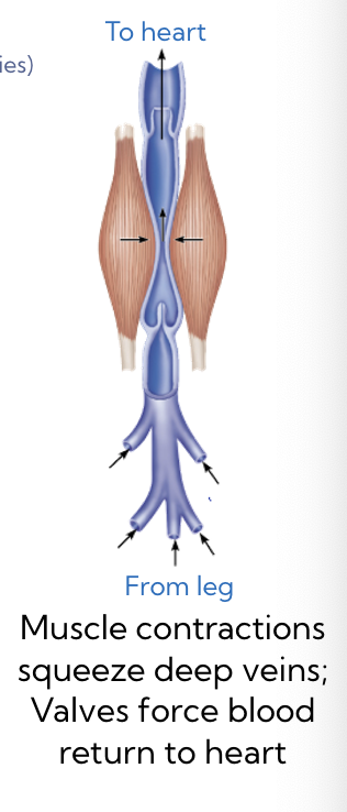
deep veins are responsible for
returning blood during exercise
17
New cards
superficial veins are responsible for
returning blood at rest
18
New cards
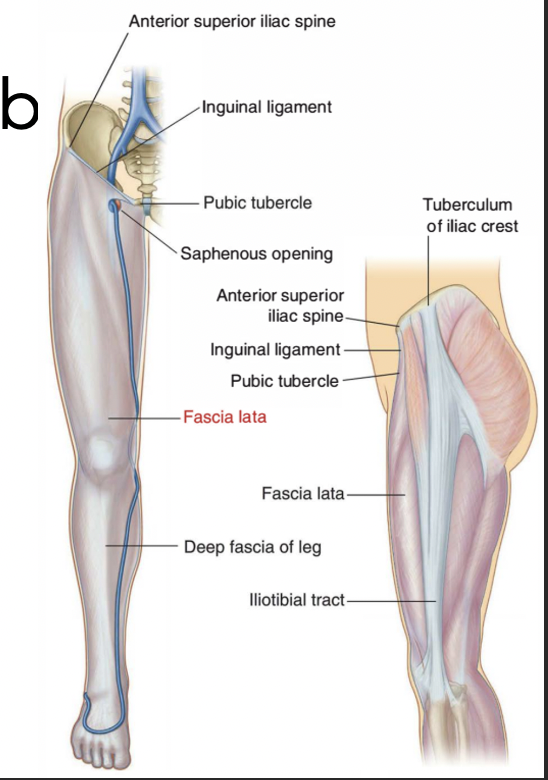
Fascia lata
fascial sleeve, continuous with both the:
* i**nguinal ligament**
* **inferior abdominal wall**
It covers the whole leg (think of a pair of tights)
* **thickens on the Iliotibial tract**
* i**nguinal ligament**
* **inferior abdominal wall**
It covers the whole leg (think of a pair of tights)
* **thickens on the Iliotibial tract**
19
New cards
Deep fascia of the shank
“crural fascia”
* divides shank into 3 compartments:
* Anterior, lateral, posterior
* remember compartment syndrome
* divides shank into 3 compartments:
* Anterior, lateral, posterior
* remember compartment syndrome
20
New cards
The lower limb
21
New cards
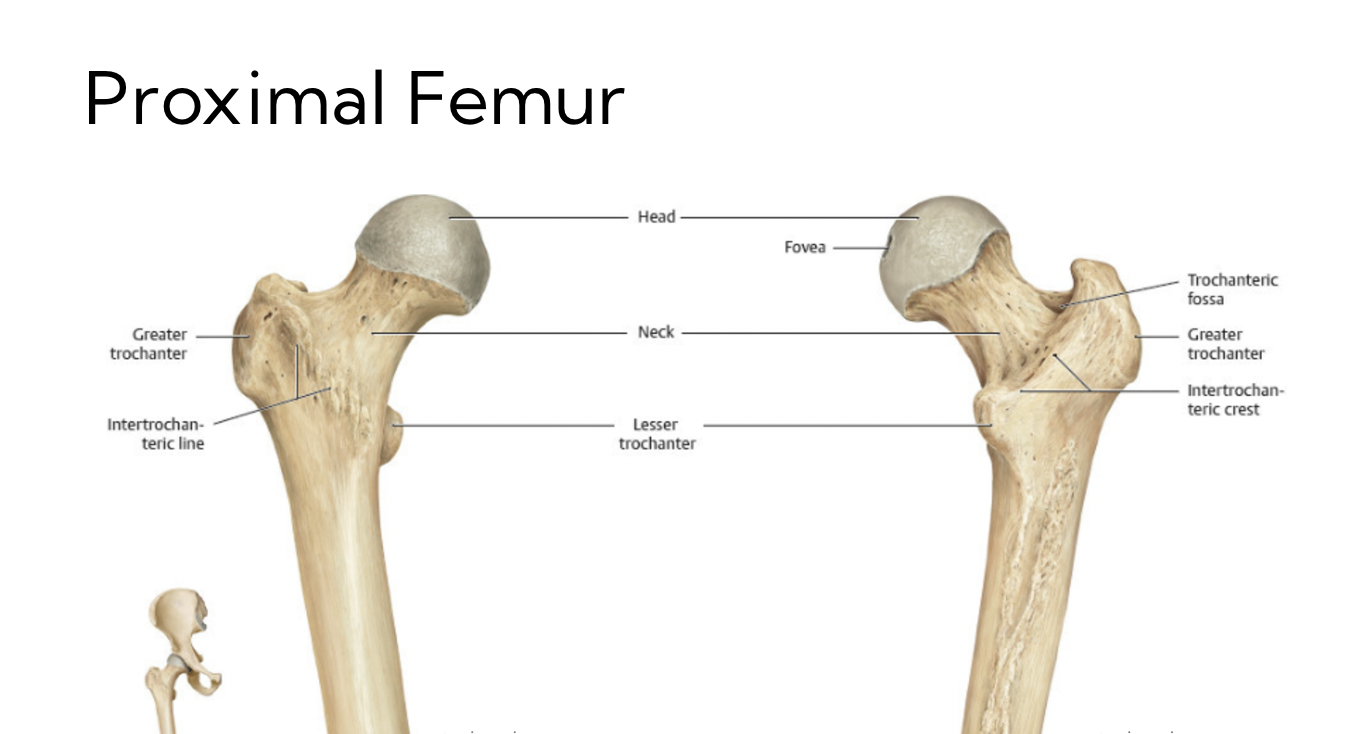
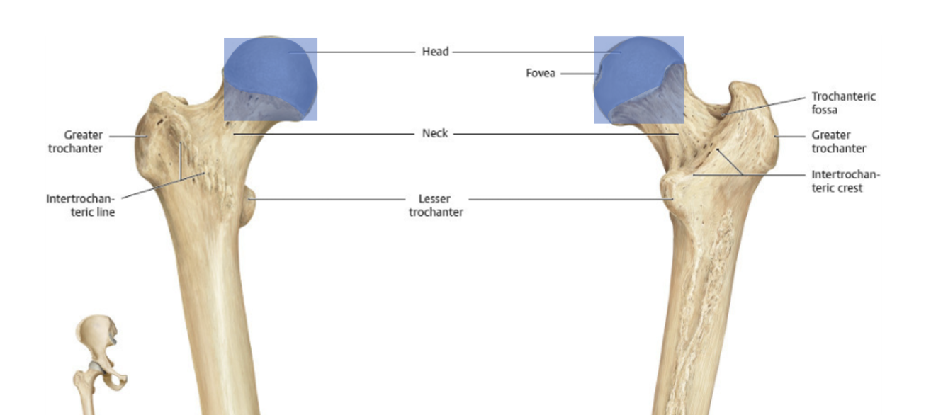
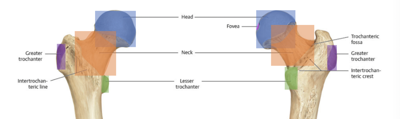
Proximal femur
22
New cards
Femoral head
23
New cards
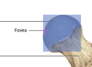
Indentation of the femoral head
Fovea
* where the ligament of the head of the femur attaches
* contains an artery
* where the ligament of the head of the femur attaches
* contains an artery
24
New cards
greater and lesser Trochanter
25
New cards
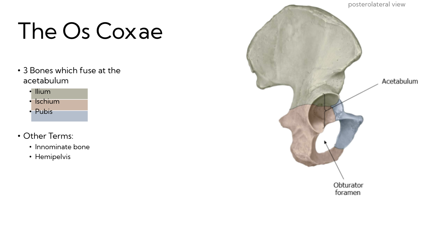
The pelvis
3 fused bones (plus sacrum)
→ The Ox Coxae
* Ilium
* Ischium
* Pubis
\
Or…
* Innominate bone
* Hemipelvis
→ The Ox Coxae
* Ilium
* Ischium
* Pubis
\
Or…
* Innominate bone
* Hemipelvis
26
New cards
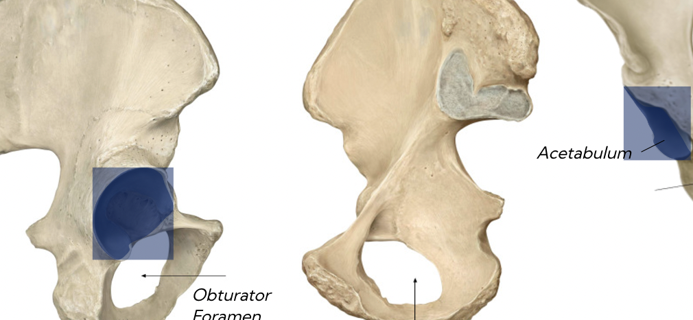
Acetabulum
The area where these come together to form the hip joint
27
New cards
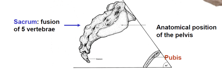
In anatomical position, the pubis is:
Inferior to sacrum
(pelvis is tilted forward)
(pelvis is tilted forward)
28
New cards
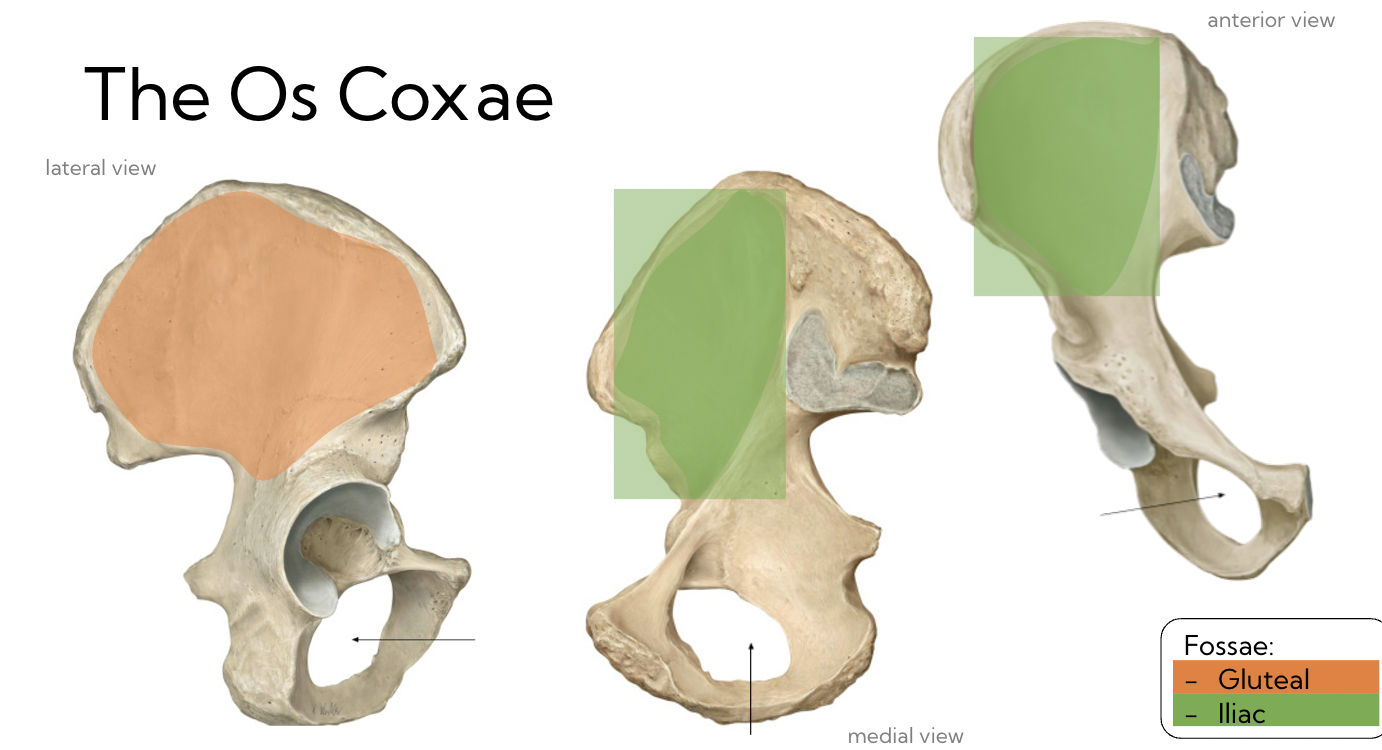
The Os Coxae Fossa
Fossae
* Gluteal (orange)
* Iliac (green)
* Gluteal (orange)
* Iliac (green)
29
New cards
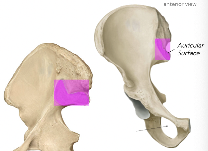
Auricular surface
The area where the sacrum articulates with the Os Coxae
‘ear shaped”
‘ear shaped”
30
New cards
Acetabulum
socket for hip joint
31
New cards
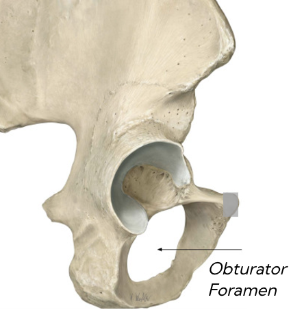
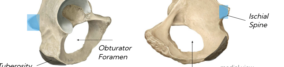
Obturator foramen
hole in inferior aspect
32
New cards
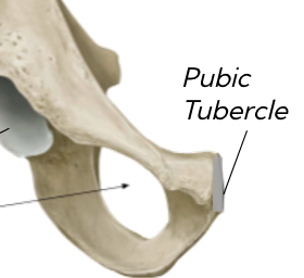
Pubic tubercle
where it joins with the other half
33
New cards
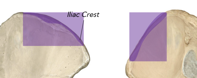
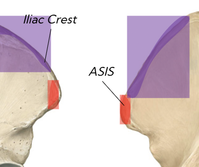
Iliac crest
where your hands go on your hips
34
New cards
Anterior superior Iliac spine (ASIS)
bony point right on anterior aspect
35
New cards
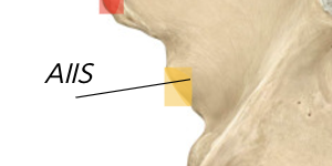
anterior inferior iliac spine (AIIS)
36
New cards
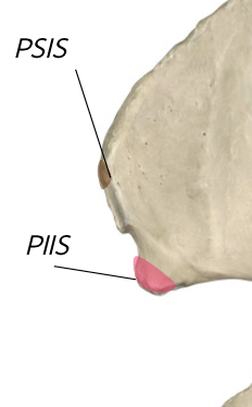
Posterior Superior & Posterior Inferior Iliac spine (PSIS) (PIIS)
37
New cards
Ischial spine and tuberosity
\*sit on your isch
38
New cards
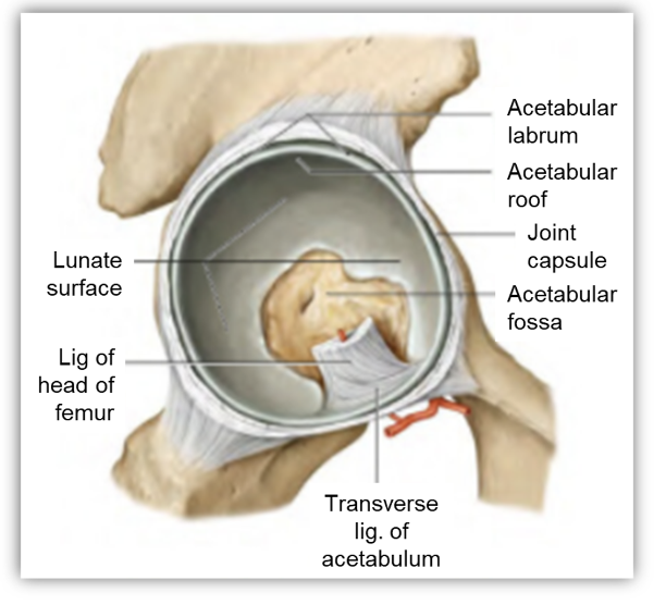
Lateral view of Acetabulum
The socket of the hip joint
* labrum
* lunate surface (covered by articular cartilage)
* ligament of the head of the femur (attaches to fovea)
* labrum
* lunate surface (covered by articular cartilage)
* ligament of the head of the femur (attaches to fovea)
39
New cards
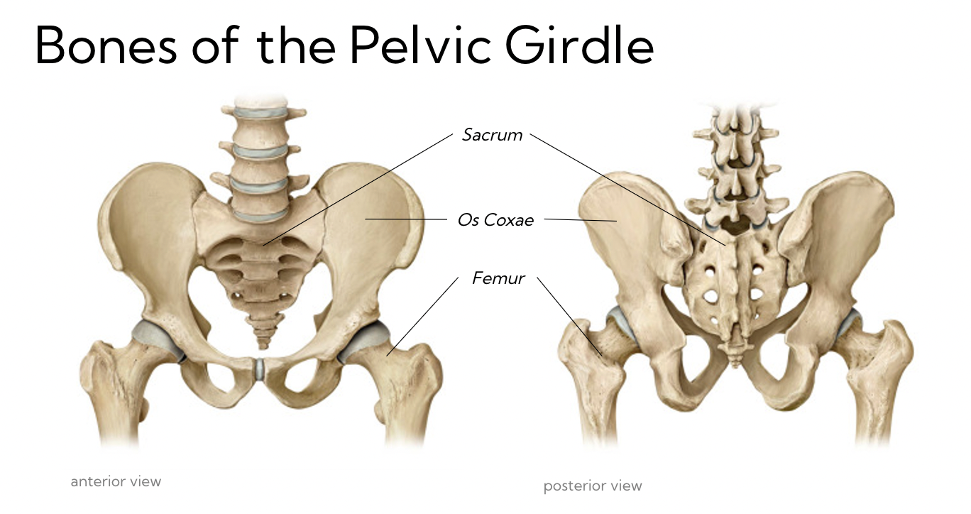
Bones of the pelvic girdle
40
New cards
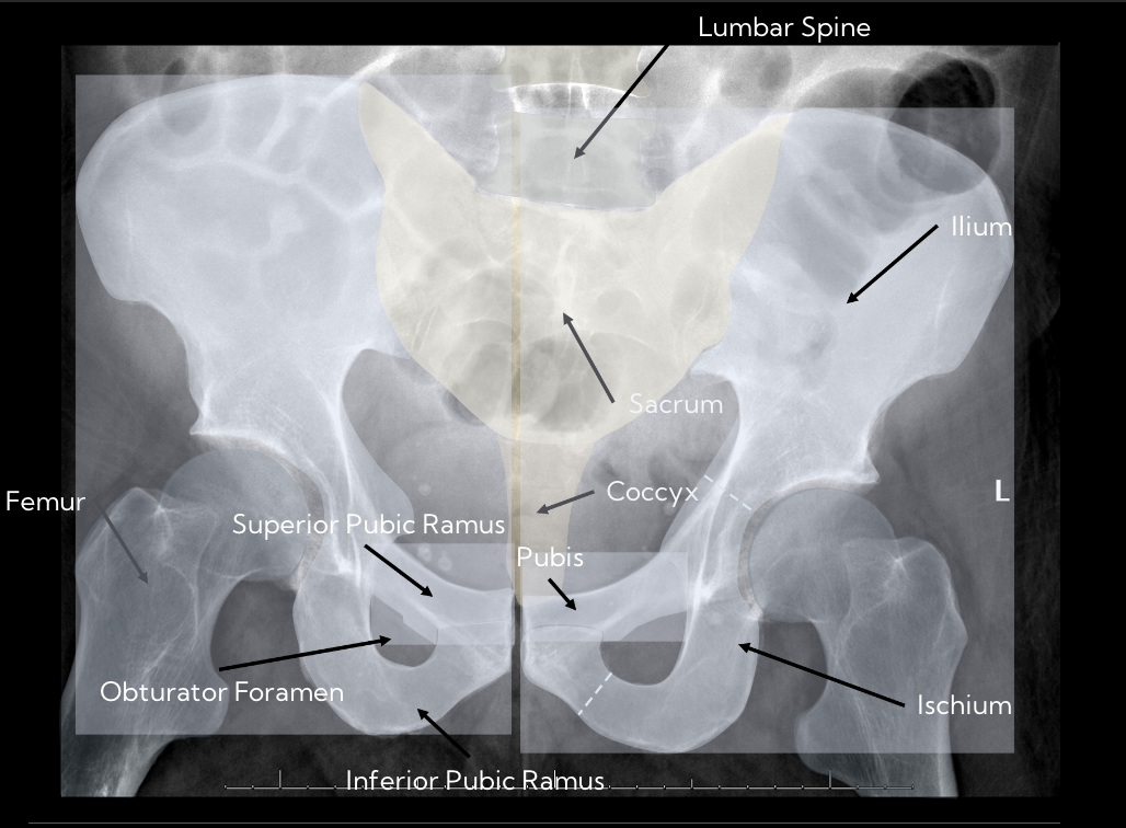
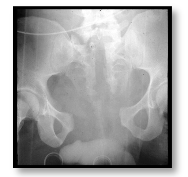
radiograph of pelvis
41
New cards
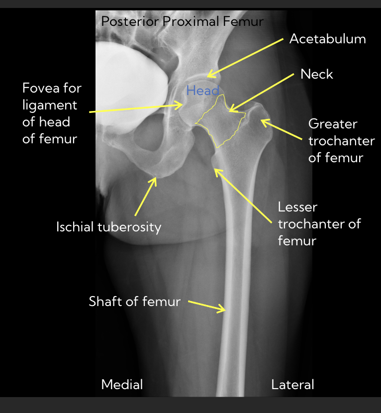
Radiograph of femur
42
New cards
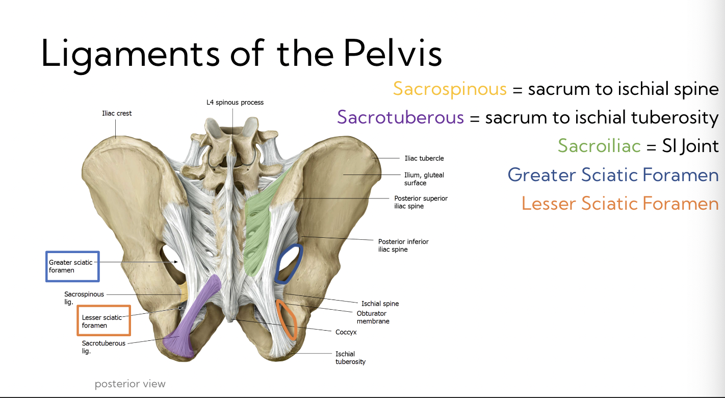
Ligaments of the pelvis
Sacrospinous
* sacrum to ischial spine
Sacrotuberous
* sacrum to ischial tuberosity
Sacroiliac
* SI joint
Greater Sciatic foramen
Lesser Sciatic foramen
→ both formed from ligaments
* sacrum to ischial spine
Sacrotuberous
* sacrum to ischial tuberosity
Sacroiliac
* SI joint
Greater Sciatic foramen
Lesser Sciatic foramen
→ both formed from ligaments
43
New cards
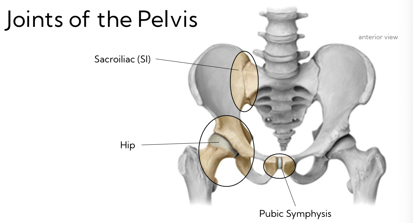
Joints of the pelvis
3 joints
44
New cards
What kinds of of joints are the 3 joints of the pelvis?
Sacroiliac
* synovial, Bilateral
\
Pubic Symphysis
* cartilaginous (fibrocartilaginous disc)
\
Hip
* Synovial, Bilateral
* synovial, Bilateral
\
Pubic Symphysis
* cartilaginous (fibrocartilaginous disc)
\
Hip
* Synovial, Bilateral
45
New cards
Sacroiliac joint
Sacrum + ilium joint
* Bilateral
* Synovial
* Immobile (due to strong ligaments)
* Anterior/ posterior sacroiliac
* Bilateral
* Synovial
* Immobile (due to strong ligaments)
* Anterior/ posterior sacroiliac
46
New cards
Pubic Symphysis
L + R pubic Rami
* Cartilaginous joint - symphysis
* Hyaline Cartilage on ends of bones, fibrocartilage disc in-between
* Relatively immobile
* during pregnancy, these joints become more mobile
* Cartilaginous joint - symphysis
* Hyaline Cartilage on ends of bones, fibrocartilage disc in-between
* Relatively immobile
* during pregnancy, these joints become more mobile
47
New cards
Open book fracture
**Separation of pubic symphysis**
* normal = 4-5mm
* Pregnancy = 8-9mm
\
**2 main causes:**
* diastasis symphysis pubis (during childbirth)
* Traumatic injury
\
**Complications:**
* substantial blood loss in pelvic cavity
* Infection & hemorrhage
* normal = 4-5mm
* Pregnancy = 8-9mm
\
**2 main causes:**
* diastasis symphysis pubis (during childbirth)
* Traumatic injury
\
**Complications:**
* substantial blood loss in pelvic cavity
* Infection & hemorrhage
48
New cards
Summary