Veterinary Systems Exam 1
 Call Kai
 Learn
Learn Practice Test
 Spaced Repetition
 Match
Match Flashcards
 Knowt Play
1/1003

There's no tags or description
Looks like no tags are added yet.
Name | Mastery | Learn | Test | Matching | Spaced | Call with Kai | Chat |
|---|
No analytics yet
Send a link to your students to track their progress
1004 Terms
Why are stains used in histology?
To provide contrast
How does plane of section alter the 2D appearance of 3D structures?
It reduces 3D objects to thin, flat slices
What is histology?
The study of microscopic tissues and cells
What is stain in histology?
Dyes used to create contrast for tissue/cellular recognition
What is hematoxylin?
A plant dye combined with a metal ion to become a blue, positively charged (cationic) dye
What is eosin?
A fluorescent red dye derived from fluorescein (negatively charged)
What is H&E?
Hematoxylin and eosin stain
What is basophilic?
Loves base (binds to basic dyes)
What is eosinophilic?
Loves eosin (binds to eosin)
What is the purpose of staining?
Provide contrast to introduce color, shape, and texture cues
(T/F) Staining allows for visual pattern recognition
True
What are the chemical properties of hematoxylin stain?
Plant dye combined with a metal ion to become a blue positive charged (cationic) dye
What are the chemical properties of eosin stain?
A fluorescent red dye derived from fluorescein (negatively charged)
Hematoxylin is _______, while Eosin is _______
Cationic, Anionic
The combination of visual cues to recognize patterns and identify structures is a process within _________
histology
Why is it difficult for us to recognize structures on an unstained slide?
Little contrast makes it difficult to distinguish cellular and tissue structures
(T/F) Histologic stains bind differently to tissue components
True
Which two stains allow for the recognition of general tissue architecture?
Hematoxylin and eosin
Hematoxylin is ______ in color
blue
Eosin is _______ in color
pink
Hematoxylin binds to _______
acids
Eosin binds to _______
proteins
Which stain is dark blue in color, basic (cationic), binds to acidic/negative-charged structures, and often stains nucleic acids used to visualize the nucleus and ribosomes?
Hematoxylin
Which stain is basic and binds to acidic/negatively-charged strcutures?
Hematoxylin
Which stain is pink/red in color, acidic (anionic), binds to basic/positively-charged structures, and often stains cytoplasmic proteins/collagen/muscle fibers to visualize the cytoplasm and matrix?
Eosin
Nuclei, nucleoli, ribosome-rich cytoplasm, rough ER, and hyaline cartilage matrix are _________ structures
basophilic
Protein-rich cytoplasm, muscle fibers, collagen fibers, and red blood cells are _________ structures
Eosinophilic
DNA/chromatin, abundant rRNA, membrane-bound ribosomes, and sulfated GAGs and proteoglycans are _________ structures
basophilic
Abundant cytoplasmic proteins, actin and myosin, extracellular protiens, and hemoglobin are _________ structures
eosinophilic
(T/F) Tissue sections allow us to study microanatomy, but they have limitations
True
Routine light microscopy requires tissue to be cut into thin sections so that ______ can pass through
light
_________ converts a three-dimensional structure into a two-dimensional structure
Sectioning
The ______ of section describes the orientation of the cut
plane
The ______ of section describes where along the structure the cut is made
level
What is a longitudinal section?
Cut parallel to the long axis of the structure

What is a transverse section (cross-section)?
Cut perpendicular to the long axis
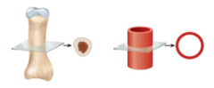
What is an oblique section?
Cut at an angle between longitudinal and cross sections

What is a tangential section?
Cut barely passes through the edge of a structure

The nucleus, nucleolus, chromatin, euchromatin, heterochromatin, RER, SER, Golgi apparatus, perinuclear halo, mitochondria, cytoskeleton, and secretory granules are...
major cellular structures visible in H&E sections
Which cellular structure is responsible for storing DNA and regulating gene expression?
Nucleus
Which cellular structure is responsible for producing rRNA and assembling ribosomes?
Nucleolus
Which cellular structure is responsible for synthesizing proteins from mRNA?
Ribosomes
Which cellular structure is responsible for synthesizing secreted and membrane proteins?
Rough endoplasmic reticulum (RER)
Which cellular structure is responsible for modifying, sorting, and packaging proteins?
Golgi apparatus
Which cellular structure is responsible for storing and releasing secreted products?
Secretory granules
Which cellular structure is responsible for controlling transport and cellular signaling?
Plasma membrane
Which cellular structure is responsible for synthesizing lipids, detoxification, and storing calcium?
Smooth endoplasmic reticulum (SER)
Which cellular structure is responsible for generating ATP through oxidative phosphorylation?
Mitochondria
Which cellular structure is responsible for maintaining the shape and enabling cellular movement?
Cytoskeleton
Which cellular structure is responsible for digesting and recycling cellular materials?
Lysosomes
Which cellular structure is responsible for storing lipids for energy?
Lipid droplet
What are 4 basophilic structures in H&E?
Nucleus, nucleolus, high concentrations of ribosomes (RER), and secretory granules
Which cellular structure is usually the most prominent basophilic structure in H&E sections?
Nuceli
Nuclei can be described by their ________ pattern
chromatin
Which chromatin type is described as dispersed chromatin and appears to have lighter staining?
Euchromatin
Which chromatin type is described as condensed chromatin and appears to have darker staining?
Heterochromatin
(T/F) Most nuclei will rarely contain both euchromatin and heterochromatin
False (most nuclei WILL contain both euchromatin and heterochromatin)
Chromatin patter often reflects _________ activity
transcriptional
Which chromatin type is generally accessible and transcriptionally active?
Euchromatin
Which chromatin type is generally condensed and transcriptionally inactive?
Heterochromatin
What are 4 eosinophilic structures in H&E?
Mitochondria, cytoskeleton, ECM proteins (collagen & elastin), many secretory granules, and most proteins
Lipid droplets, golgi, glycogen, mucus, and the plasma membrane are _____ stained structures in the H&E
poorly
Why are lipid droplets poorly stained structures?
They are extracted during routine paraffin processing
Why are Golgi poorly stained structures?
They are mostly membrane and hydrophobic
Why does glycogen have poor staining properties?
It is often extracted or poorly visualized
Why does mucus have poor staining properties?
Often pale or poorly preserved (acidic mucins may be basophilic)
Why does the plasma membrane have poor staining properties?
It is too thin to resolve directly
How might fixation create histologic artifacts?
May pull cells and extracellular matrix away from one another, creating artificial clear/white spaces
How might processing create histologic artifacts?
Processing may extract material, creating blank/white gaps
How might sectioning create histologic artifacts?
Sectioning may create tears, folds, or knife/chatter marks, which alter the physical composition of tissues on slides
How might staining create histologic artifacts?
Unfiltered stain or acid-formalin reacting with blood may leave crystaline, black/brown, or deep purple granular debris on tissue sections
How might mounting steps create histologic artifacts?
Tears, folds, chatter/knife marks, shrinkage, precipitates, and dissolution/extraction are examples of common _________
artifacts
Thin tissue sections are prone to _________ during cutting or placement on the slide
tearing
Thin tissue sections are prone to _______ over during placement on the slide, causing a lack of uniformity and an uneven appearance
folding
Which artifact type is caused by microtome blade vibration, causing fine, parallel thick-and-thin bands across tissue?
Chatter marks
Which artifact type is caused by dull or knicked blades, leaving straight vertical scratches?
Knife marks
Fixation and dehydration pulls cells and the extracellular matrix away from one another, creating artifical clear or white artifacts/spaces, otherwise known as _________
shrinkage
Which artifact type is defined by an unfiltered stain or acid-formalin reacting with blood, leaving crystalline, brown/black, or deep purple granular debris resting on the tissue section?
Precipitates
Which artifact type is defined by fats and poorly fixed structures that are prone to dissolving or washing off the slide?
Dissolution/extraction
(T/F) When staining, real structures often follow predictable patterns
True
________ appear irregular or out of place and may cause hypo/hyper-staining
Artifacts
What is connective tissue?
One of the four primary tissue types
What are the functions of connective tissue?
Supports, connects, protects, stores, transports, and participates in tissue repair and defense
What is the difference between the stroma and parenchyma?
The stroma is the structural framework of an organ, and the parenchyma is the functional component of an organ
How many primary tissue types are there?
4
The connective tissue forms the _____ and supporting framework of most organs
stroma
Bone, adipose tissue, cartilage, and blood are specialized __________ tissues
connective tissues
What two components make up connective tissues?
Cells + Extracellular Matrix (ECM)
What two components make up the Extracellular Matrix (ECM)?
Fibers + Ground Substance
What are fibers?
Filamentous structural proteins that provide strength
Collagen, reticular fibers, and elastic fibers are examples of ______
fibers
What is ground substance?
Gelatinous material
Tissue fluid, glycosaminoglycans, proteoglycans, and adhesion proteins are examples of ________
ground substance
Which connective tissue component is responsible for ECM synthesis, remodeling, repair, immune surveillance/defense, energy storage, and insulation?
Cells
Which connective tissue component is responsible for providing strength, resistance, a supportive framework, stretch, and recoil?
Fibers
Which connective tissue component is responsible for binding water, maintaining hydration, resisting compressive forces, regulating diffusion of wastes/nutrients, and supporting cell adhesion/migration/signaling?
Ground substance
How can we differentiate loose and dense connective tissues?
Cellularity, fiber density, ground substance abundance, and mechanical properties
What are the 3 types of connective tissues?
Connective tissue proper, specialized connective tissue, and embryonic connective tissue
What is the most common connective tissue type?
Connective tissue proper